The Brain Anatomy Coloring Book
Learn the Structure and Function of Your Brain Through Interactive Illustrations
Many people struggle to visualize and retain complex brain anatomy despite its fundamental importance to understanding human health and behavior.
191 pages8 chaptersJan 5, 2026Anatomy
About this book
This interactive coloring book transforms passive learning into an engaging, multi-sensory experience that makes neurological structures memorable and meaningful. By combining detailed, scientifically accurate illustrations with guided coloring activities, you'll develop a genuine understanding of how your brain is organized and how each region contributes to your thoughts, emotions, and physical functions. Whether you're a student preparing for exams, a healthcare professional seeking to refresh your knowledge, or simply curious about the organ that makes you who you are, this book provides the perfect balance of educational depth and creative engagement. Each chapter focuses on a specific brain region or system, with clear labels, functional descriptions, and coloring exercises designed to reinforce learning. You'll progress from basic brain organization to complex neural networks, building confidence and competence with every page you complete.
Table of contents
- IntroductionWhy Brain Anatomy Matters, How Coloring Enhances Learning, How to Use This Book, Essential Anatomical Terms and DirectionsRead below
- Chapter 1: Brain Organization and OverviewThe Three Major Divisions of the Brain, Protective Structures: Meninges and Cerebrospinal Fluid, Blood Supply and the Blood-Brain Barrier
- Chapter 2: The Cerebral Cortex and LobesStructure and Organization of the Cerebral Cortex, The Frontal Lobe: Executive Function and Motor Control, The Parietal Lobe: Sensation and Spatial Processing, The Temporal Lobe: Hearing and Memory, The Occipital Lobe: Vision and Visual Processing
- Chapter 3: Subcortical Structures and the Limbic SystemThe Thalamus and Hypothalamus: Sensory Relay and Regulation, The Hippocampus: Memory Formation and Consolidation, The Amygdala: Emotion and Fear Processing, Limbic System Connections and Emotional Regulation
- Chapter 4: The Brainstem and CerebellumThe Midbrain: Sensory and Motor Integration, The Pons and Medulla: Vital Life Functions, The Cerebellum: Balance, Coordination, and Motor Learning
- Chapter 5: White Matter and Neural PathwaysUnderstanding White Matter and Axonal Connections, Major White Matter Tracts and Communication Highways, Hemispheric Communication: The Corpus Callosum
- Chapter 6: Neurons and NeurotransmittersNeuron Structure and Types, Synaptic Transmission and Neural Communication, Major Neurotransmitters and Their Functions
- Chapter 7: Sensory and Motor SystemsSensory Pathways: From Receptors to Cortex, Motor Pathways: From Brain to Muscles, Sensorimotor Integration and Coordination
- Chapter 8: Brain Plasticity and AdaptationNeuroplasticity: The Brain's Ability to Change, Learning and Memory Formation at the Cellular Level, Recovery and Adaptation After Brain Injury
- ConclusionIntegrating Your Brain Anatomy Knowledge, Next Steps in Your Learning Journey
- Glossary of Brain Anatomy Terms
- Further Reading and Resources
Introduction
The human brain represents one of the most complex and fascinating structures in the known universe. Weighing approximately three pounds and containing roughly 86 billion neurons, this remarkable organ controls every aspect of your existence—from the automatic beating of your heart to your most abstract thoughts and creative ideas. Despite its fundamental importance to everything you experience, think, and do, the brain remains mysterious to many people. Understanding its anatomy provides a foundation for comprehending how you perceive the world, form memories, regulate emotions, and coordinate movement. This knowledge isn't just academic; it empowers you to make informed decisions about your health, understand neurological conditions, and appreciate the incredible biological machinery that makes you uniquely human.
Traditional approaches to learning brain anatomy often rely on passive reading and memorization of complex terminology, which can feel overwhelming and disconnected from practical understanding. Many students and curious learners struggle to visualize three-dimensional structures from two-dimensional diagrams, and the sheer volume of anatomical terms can create barriers to genuine comprehension. This coloring book takes a fundamentally different approach by engaging multiple learning pathways simultaneously. When you actively color anatomical structures, you're not just observing—you're participating in the learning process through visual, kinesthetic, and cognitive engagement. This multi-sensory approach has been shown to significantly improve retention and deepen understanding compared to passive study methods alone.
Throughout this book, you'll progress systematically through the major regions and systems of the brain, building your knowledge layer by layer. Each chapter focuses on specific anatomical structures, providing clear explanations of their locations, connections, and functions. The illustrations are scientifically accurate and designed to be colored in ways that reinforce anatomical relationships and functional groupings. Whether you're a student preparing for exams in neuroscience, psychology, or healthcare fields, a professional seeking to refresh your anatomical knowledge, or simply someone fascinated by how your brain works, this book offers an engaging and effective path to mastery. By the time you complete the final chapter, you'll have developed a comprehensive mental map of brain anatomy that will serve as a foundation for deeper exploration of neuroscience, clinical applications, and the ongoing mysteries of consciousness itself.
Why Brain Anatomy Matters
Understanding brain anatomy provides essential context for virtually every aspect of human experience and behavior. When you know which brain regions control specific functions, you gain insight into why certain injuries or diseases produce particular symptoms. For example, understanding that the hippocampus plays a critical role in forming new memories helps explain why patients with Alzheimer's disease, which damages this structure, struggle to remember recent events while retaining older memories. Similarly, knowing that the frontal lobe governs executive functions like planning, decision-making, and impulse control illuminates why damage to this region can dramatically alter personality and behavior. This anatomical knowledge transforms abstract medical terminology into concrete, meaningful concepts that connect directly to real-world observations and clinical scenarios.
For healthcare professionals and students, anatomical literacy forms the foundation of clinical competence. Physicians, nurses, therapists, and other practitioners regularly encounter patients with neurological conditions, and effective communication requires precise anatomical language. When a radiologist describes a lesion in the left temporal lobe, or when a neurologist discusses damage to the basal ganglia, these terms reference specific physical locations with predictable functional consequences. Without solid anatomical grounding, medical discussions become confusing and clinical reasoning suffers. Moreover, as medical imaging technologies like MRI and CT scans become increasingly sophisticated, the ability to interpret these images and correlate anatomical structures with clinical presentations becomes ever more valuable across healthcare disciplines.
Beyond professional applications, brain anatomy knowledge enriches your understanding of everyday experiences and current neuroscience research. When you read about studies investigating the neural basis of emotions, decision-making, or learning, anatomical knowledge allows you to engage with this research more deeply. You'll understand what researchers mean when they report increased activity in the amygdala during fear responses, or enhanced connectivity between the prefrontal cortex and hippocampus during memory consolidation. This literacy extends to understanding news coverage of neurological conditions, evaluating claims about brain-training programs, and making informed decisions about your own cognitive health. The brain isn't just an abstract concept—it's the physical substrate of your mind, and understanding its structure provides profound insights into what makes you who you are.
Anatomical knowledge also fosters appreciation for the brain's remarkable organization and efficiency. The brain represents only about two percent of your body weight, yet it consumes approximately twenty percent of your body's energy. This metabolic demand reflects the constant activity of billions of neurons communicating through trillions of synaptic connections. The brain's organization reflects evolutionary pressures that shaped its development over millions of years, with older structures like the brainstem controlling basic survival functions while newer regions like the prefrontal cortex enable complex reasoning and social behavior. Understanding this organization reveals how different brain regions work together as integrated systems rather than isolated components, creating the seamless experience of consciousness that you take for granted every moment of every day.
For those interested in mental health, brain anatomy provides crucial context for understanding psychological conditions and their treatments. Depression, anxiety, schizophrenia, and other mental health disorders involve alterations in brain structure, chemistry, and connectivity. Knowing that depression often involves reduced activity in prefrontal regions and altered function in limbic structures helps explain both the symptoms patients experience and why certain treatments prove effective. Similarly, understanding that anxiety disorders involve hyperactivity in the amygdala and its connections illuminates why therapies targeting these circuits can reduce symptoms. This biological perspective doesn't diminish the psychological and social dimensions of mental health, but rather integrates them into a more complete understanding of how brain, mind, and behavior interact.
Finally, studying brain anatomy cultivates humility and wonder about the limits of current scientific understanding. Despite tremendous advances in neuroscience, researchers still grapple with fundamental questions about how neural activity gives rise to subjective experience, how memories are stored and retrieved, and how consciousness emerges from physical processes. Every anatomical structure you'll color in this book participates in these mysteries. The more you learn about the brain's physical organization, the more you'll appreciate both what science has discovered and what remains unknown. This combination of knowledge and curiosity represents the ideal mindset for engaging with neuroscience—informed enough to understand current findings, yet humble enough to recognize how much remains to be discovered about the most complex structure in the known universe.
The practical applications of brain anatomy extend into education, technology, and public policy as well. Educators who understand how different brain regions develop at different rates can design age-appropriate curricula that align with cognitive capabilities. Technology developers creating brain-computer interfaces or neurofeedback systems must understand anatomical organization to target specific regions effectively. Policymakers addressing issues from traumatic brain injury in sports to the effects of substances on developing brains benefit from anatomical literacy. In each of these domains, the ability to connect abstract concepts to concrete anatomical structures enables more informed decision-making and more effective interventions. Your investment in learning brain anatomy thus extends far beyond personal knowledge—it equips you to participate meaningfully in important conversations about health, education, technology, and society.
How Coloring Enhances Learning
The act of coloring anatomical structures engages your brain in ways that passive reading cannot match. When you color, you're simultaneously processing visual information, executing fine motor movements, making decisions about color choices, and focusing sustained attention on specific structures. This multi-modal engagement activates multiple brain regions including visual processing areas in the occipital lobe, motor planning regions in the frontal lobe, and attention networks distributed throughout the cortex. Research in educational psychology has consistently demonstrated that active learning strategies—those requiring learners to do something with information rather than simply receive it—produce superior retention and deeper understanding compared to passive approaches[1]. Coloring transforms you from a passive observer into an active participant in your own learning process.
The kinesthetic component of coloring creates additional memory pathways that reinforce visual and verbal learning. When you physically trace the boundaries of a structure with your coloring tool, you're creating a motor memory of that shape and its spatial relationships to surrounding structures. This embodied learning helps you develop an intuitive sense of anatomical organization that goes beyond memorizing labels. For example, as you color the corpus callosum connecting the two cerebral hemispheres, the physical act of following its curved path helps you internalize its shape and position in a way that simply looking at a pre-colored diagram cannot achieve. This motor engagement is particularly valuable for understanding three-dimensional relationships, as the act of coloring helps you mentally construct the spatial organization of structures that exist in depth as well as on the page.
Coloring also slows down the learning process in beneficial ways, forcing you to examine structures carefully rather than skimming past them. When you must decide where one structure ends and another begins, you're actively engaging with anatomical boundaries and relationships. This careful attention reveals details you might otherwise overlook—the way certain structures wrap around others, how pathways connect distant regions, or how protective layers enclose delicate tissues. The deliberate pace of coloring creates opportunities for reflection and consolidation, allowing information to move from working memory into long-term storage. In our fast-paced world where information often flows past us in overwhelming streams, the focused, meditative quality of coloring provides a welcome counterbalance that actually enhances learning efficiency despite—or perhaps because of—its slower pace.
The use of color itself serves important cognitive functions beyond mere aesthetics. When you assign different colors to different structures or functional systems, you're creating visual categories that help organize information in memory. For instance, you might color all structures involved in the limbic system in warm colors (reds, oranges, yellows) while using cool colors (blues, greens, purples) for cortical regions. These color-coding strategies create additional retrieval cues that make it easier to recall information later. When you try to remember the components of the limbic system, the warm color scheme you created becomes a mental scaffold that helps you reconstruct the information. This technique leverages your brain's powerful visual memory systems, which evolved to remember spatial relationships and visual patterns with remarkable accuracy.
Coloring also provides immediate feedback about your understanding. If you're unsure where to draw the boundary between two structures, that uncertainty signals a gap in your knowledge that you can address by reviewing the text or consulting additional resources. This self-assessment happens naturally and continuously as you work, without the pressure of formal testing. When you successfully complete a complex illustration, accurately distinguishing multiple structures and their relationships, you gain confidence in your mastery. This positive reinforcement motivates continued learning and builds the self-efficacy that supports long-term engagement with challenging material. The tangible product of your work—a completed, colored illustration—provides visible evidence of your progress and creates a personalized study resource you can return to for review.
The repetitive nature of coloring also supports learning through spaced practice and elaboration. As you color multiple views of the same structure from different angles or in different contexts, you're encountering that information repeatedly in varied ways. This spaced repetition, distributed across different chapters and illustrations, strengthens memory consolidation more effectively than massed practice (cramming). Each encounter with a structure in a new context requires you to retrieve and apply your existing knowledge, which itself strengthens memory traces. For example, you might first color the hippocampus in a chapter on subcortical structures, then encounter it again in a chapter on memory systems, and finally see it in relation to white matter pathways. Each repetition deepens your understanding and creates a richer, more interconnected knowledge network.
Finally, coloring reduces cognitive load by breaking complex information into manageable chunks. Rather than trying to absorb an entire brain region at once, you focus on one structure at a time, gradually building up a complete picture. This incremental approach aligns with how your working memory actually functions—it can only hold a limited amount of information at once, so breaking learning into smaller pieces prevents overload. As you complete each structure, you integrate it into your growing mental model, creating a scaffold that supports learning the next structure. This progressive building process mirrors how expertise develops in any domain: through patient accumulation of knowledge, practice, and integration over time. The coloring book format naturally supports this developmental progression, guiding you from simpler to more complex anatomical relationships as your competence grows.
How to Use This Book
This book is designed to be worked through systematically, with each chapter building on knowledge from previous chapters. While you can certainly jump to topics of particular interest, following the sequence from beginning to end provides the most coherent learning experience. The early chapters establish foundational concepts and introduce major brain divisions, while later chapters explore specific systems and functions in greater detail. Each chapter begins with an overview that orients you to the structures you'll be studying, followed by detailed sections on individual components. Read the text carefully before you begin coloring each illustration, as understanding the function and relationships of structures will inform your coloring choices and deepen your learning.
When approaching each illustration, start by examining it carefully without coloring. Identify the labeled structures and trace their boundaries with your eyes or finger. Notice how structures relate to each other spatially—which ones are adjacent, which ones overlap, and which ones connect through pathways. This preliminary survey helps you develop a mental plan before you commit to coloring. Consider which structures you want to emphasize and how you'll use color to distinguish between different components. Some learners prefer to color all instances of the same structure in the same color throughout the book, creating consistency across chapters. Others prefer to use color schemes that reflect functional relationships, grouping related structures in similar hues. Both approaches have merit; choose the strategy that best supports your learning style and goals.
Select your coloring tools based on the level of detail in each illustration and your personal preferences. Colored pencils offer the most control and allow for fine detail work, making them ideal for complex illustrations with many small structures. They also allow for layering and shading to create depth. Markers provide bold, vibrant colors but require more care to stay within boundaries and may bleed through thin paper. Fine-tipped markers or gel pens work well for very small structures. Whatever tools you choose, test them on a spare area first to ensure they won't bleed through the page or obscure labels. Keep your coloring relatively light, especially on your first pass—you can always add more color, but it's difficult to remove excess. The goal is to enhance the illustration's clarity, not to obscure the anatomical details.
As you color, refer back to the text frequently to reinforce the connections between visual and verbal information. When you color the amygdala, for example, review its role in emotional processing and its connections to other limbic structures. This integration of visual, motor, and verbal learning creates multiple memory pathways that support retention. Consider adding your own notes in the margins—questions that arise, connections you notice, or clinical examples that illustrate a structure's function. These annotations personalize your learning and create additional retrieval cues. Some learners find it helpful to explain structures aloud as they color, either to themselves or to a study partner. This verbalization forces you to articulate your understanding and can reveal gaps in your knowledge.
Use the following systematic approach for each illustration to maximize learning:
- Preview: Read the accompanying text and examine the illustration without coloring
- Identify: Locate each labeled structure and understand its boundaries
- Plan: Decide on your color scheme and coloring sequence
- Color: Work carefully, staying within boundaries and maintaining consistent colors for the same structures
- Review: After completing the illustration, cover the labels and test yourself by naming each structure
- Connect: Reflect on how the structures you've colored relate to each other functionally
Don't rush through the book. Quality of engagement matters more than speed of completion. Spending adequate time with each illustration allows for deeper processing and better retention. If you find a particular structure or concept challenging, spend extra time with it. Color the same structure from multiple angles, create your own simplified sketches, or seek out additional resources to clarify your understanding. The book includes cross-references to related structures in other chapters—follow these connections to see how information integrates across different brain regions and systems. This web of connections mirrors how the brain itself functions, with different regions working together in distributed networks rather than in isolation.
Consider establishing a regular study schedule rather than working through the book in irregular bursts. Research on learning consistently shows that distributed practice—shorter study sessions spread over time—produces better long-term retention than massed practice[2]. You might aim to complete one or two subsections per study session, allowing time to read carefully, color thoughtfully, and review what you've learned. After completing a chapter, return to earlier illustrations and test yourself on the structures without looking at labels. This retrieval practice strengthens memory and helps you identify areas that need additional review. Keep the book as an ongoing reference resource even after you've completed it—the illustrations you've colored become personalized study aids that you can return to whenever you need to refresh your knowledge.
Finally, complement your work in this book with other learning resources to build a comprehensive understanding. Watch videos of brain dissections to see three-dimensional relationships that are difficult to convey in two-dimensional illustrations. Examine brain imaging studies to see how anatomical structures appear in MRI and CT scans. Read case studies that illustrate how damage to specific structures affects function. Discuss what you're learning with peers, instructors, or online communities focused on neuroscience education. The coloring book provides an excellent foundation, but your learning will be richest when you integrate multiple resources and perspectives. Each approach to studying brain anatomy offers unique benefits, and together they create a robust, multi-faceted understanding that will serve you well in whatever applications you pursue.
Essential Anatomical Terms and Directions
Before diving into specific brain structures, you need to master the directional terminology that anatomists use to describe locations and relationships. These terms provide a precise, standardized language that eliminates ambiguity when describing where structures are located relative to each other. Unlike everyday directional terms like "up" or "forward," which can be confusing when the body or brain is oriented in different positions, anatomical terms remain constant regardless of orientation. Learning these terms now will make all subsequent descriptions clearer and will enable you to communicate effectively with other students and professionals. While the terminology may seem technical at first, it quickly becomes intuitive with practice and use.
The most fundamental directional terms describe positions along three primary axes. Anterior (or rostral in the brain) means toward the front, while posterior (or caudal) means toward the back. For example, the frontal lobe is anterior to the occipital lobe. Superior (or dorsal) means toward the top, while inferior (or ventral) means toward the bottom. The cerebral cortex is superior to the brainstem. Medial means toward the midline of the brain, while lateral means away from the midline. The corpus callosum is medial, while the temporal lobes extend laterally. These six terms form the foundation of anatomical description and will appear throughout this book. Practice using them as you color, describing structures in relation to each other using proper terminology.
Additional directional terms describe more specific relationships and positions:
- Proximal: Closer to the point of attachment or origin
- Distal: Farther from the point of attachment or origin
- Superficial: Closer to the surface
- Deep: Farther from the surface, more internal
- Ipsilateral: On the same side of the body or brain
- Contralateral: On the opposite side of the body or brain
- Bilateral: On both sides
Understanding anatomical planes is equally important for visualizing brain structure. The sagittal plane divides the brain into left and right portions. A midsagittal section cuts directly down the midline, creating two equal halves, while parasagittal sections are parallel to the midline but offset to one side. The coronal plane (also called the frontal plane) divides the brain into anterior and posterior portions, like slicing from ear to ear. The horizontal plane (also called the axial or transverse plane) divides the brain into superior and inferior portions, like slicing parallel to the ground when standing upright. Brain imaging studies and anatomical illustrations typically show one or more of these standard planes, and understanding them helps you mentally reconstruct three-dimensional relationships from two-dimensional images.
Several terms describe the organization and structure of brain tissue itself. Gray matter consists primarily of neuron cell bodies, dendrites, and unmyelinated axons, appearing grayish in fresh tissue. It forms the cerebral cortex on the brain's surface and clusters of nuclei deep within the brain. White matter consists primarily of myelinated axons that connect different brain regions, appearing white due to the fatty myelin sheaths surrounding the axons. Nuclei (singular: nucleus) are clusters of neuron cell bodies within the brain that typically share related functions. Cortex refers to the outer layer of gray matter covering the cerebral hemispheres and cerebellum. Gyri (singular: gyrus) are the raised ridges on the brain's surface, while sulci (singular: sulcus) are the grooves between gyri. Particularly deep sulci are sometimes called fissures.
Terms describing neural pathways and connections include tract, which refers to a bundle of axons connecting different brain regions within the central nervous system. Commissures are tracts that cross the midline, connecting corresponding areas in the left and right hemispheres. Projection fibers connect the cerebral cortex with lower brain regions and the spinal cord. Association fibers connect different regions within the same hemisphere. Understanding these terms helps you comprehend how information flows through the brain and how different regions communicate. When you encounter descriptions of neural pathways in later chapters, these terms will provide the vocabulary needed to understand the routes that signals take as they travel through the brain's complex circuitry.
Finally, familiarize yourself with terms describing brain regions and their subdivisions. The brain is organized hierarchically, with large divisions containing smaller subdivisions. For example, the forebrain (prosencephalon) includes the telencephalon (cerebral hemispheres) and diencephalon (thalamus, hypothalamus, and related structures). The midbrain (mesencephalon) forms the upper portion of the brainstem. The hindbrain (rhombencephalon) includes the metencephalon (pons and cerebellum) and myelencephalon (medulla oblongata). While these embryological terms may seem abstract, they reflect the brain's developmental organization and help explain why certain structures are grouped together functionally. As you progress through the book, you'll see how these major divisions contain the specific structures you'll be coloring and studying in detail.
Don't feel overwhelmed by this terminology—you don't need to memorize all these terms before proceeding. Instead, refer back to this section as you encounter unfamiliar terms in later chapters. Keep a finger or bookmark here so you can quickly check definitions when needed. As you use these terms repeatedly in context, they'll become increasingly familiar and intuitive. Consider creating flashcards for terms you find particularly challenging, or practice describing the location of everyday objects using anatomical terminology to build fluency. For example, describe the location of your nose as "medial and anterior on the face, superior to the mouth and inferior to the eyes." This playful practice helps internalize the terminology so it becomes second nature when you apply it to brain structures. With consistent use, anatomical language transforms from an obstacle into a powerful tool that enables precise thinking and communication about brain organization.
This is the end of the free sample. The full book has 8 chapters.
Want this book written for you?
Tell us your goals and background, and get your own version of this book, tailored by AI to you.


Benefits of AI-tailored books
- Read one book, not ten:
- all the Anatomy knowledge you need, gathered into a single book.
- Save days of learning:
- tell us your goals, choose your topics, and the whole book is built around them.
- Learn effortlessly:
- Anatomy book written for your specific background and expertise.
- Understand at a glance:
- includes charts, tables, timelines, checklists, decision trees, and more.
- Stay current:
- written the day you order it, not years ago.
- Trust what you read:
- every book is checked for accuracy, with real research when your subject needs it.
Create your unique book in 3 steps
- 1. Select your focus
Select the focus of your Anatomy book and share your background
- 2. Personalize your book
Specify your goals and choose sub-topics to include
- 3. Get your tailored book
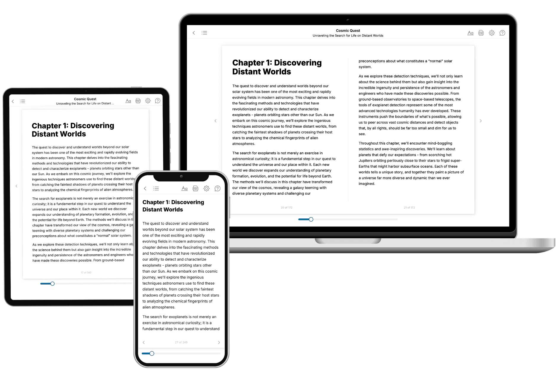
Your book is ready in 10 minutes. Read it online, download as EPUB or PDF, or send to Kindle.

Frequently asked questions
- What is TailoredRead?
TailoredRead creates complete, full-length nonfiction ebooks, written specifically for you. Tell us what you want to learn and why, and TailoredRead writes you a complete ebook on it, with charts, tables, and diagrams throughout. Read it online, on Kindle, or on any e-reader, or download it as a PDF or EPUB. Every book is written fresh the day you order it.
- How is the book tailored to me?
Before anything is written, you answer a few short questions about your goals, your background, and the topics you care about. Your book is then planned around your answers: written at your level, focused on your goals, and covering the topics you chose, instead of the one-size-fits-all book a store sells everyone.
- Will it read like generic AI content?
No. Your book isn't a chat transcript: it's planned chapter by chapter before a single word is written, told in a clear book voice, and explains ideas visually with charts, tables, and diagrams. After writing, every book goes through an editorial pass that polishes chapters so the book reads consistent from start to finish. Judge for yourself: browse thousands of sample books on our Explore Books page.
- How accurate is the content?
Our AI first checks whether your subject needs live research. Niche and fast-moving subjects are researched before writing, and the book plan and chapters are grounded in what it finds. After writing, every book goes through a quality pass that checks its facts and consistency, and researched books list their sources in a Notes section at the back.
- How long is the book?
You choose the length: Comprehensive (250-300 pages), Detailed (150-200 pages), Essential (70-100 pages), or Short (30-50 pages).
- Does the book include visuals?
Yes. Every book includes charts, tables, and diagrams: timelines, checklists, step-by-step guides, decision trees, comparison tables, matrices, and bar charts. A typical book includes dozens of these visual elements.
- Can I read it on Kindle? What formats can I download?
Yes. Every book can be read in our online reader or downloaded as a PDF or EPUB. The EPUB can be opened with Kindle, Apple Books, Kobo, and any e-reader. The Commercial and Author licenses also include an editable Word (DOCX) and Markdown download.
- How much does it cost?
About the price of a regular ebook. The exact price depends on your book's length and complexity, from $2 for the shortest books to $35 for the most comprehensive, and it's shown when you review your book, before you commit to anything. Each book is a one-time purchase, yours to keep, no subscription. Licenses that let you share, teach, or publish your book range from $35.99 to $49.99.
- Can I preview the book before purchasing?
Before you pay, you'll see what your book will cover: the title, a detailed description, the estimated length, and the complete chapter-by-chapter table of contents, so you know what you're getting before you decide. The full text itself is written after you purchase.
- How long does it take to create a book?
About 10 minutes. You can start reading the opening chapters while the rest is still being written.