The Cardiovascular Coloring Guide
Master Heart and Blood Vessel Anatomy Through Interactive Visual Learning
Build a comprehensive understanding of the cardiovascular system that sticks with you for life.
284 pages16 chaptersSep 29, 2025Anatomy
About this book
This interactive coloring guide transforms complex anatomical concepts into engaging visual experiences that make learning both effective and enjoyable. You'll explore every aspect of the heart and circulatory system through carefully designed illustrations that reveal the intricate relationships between structure and function. Each coloring exercise is strategically crafted to reinforce key concepts while building your anatomical vocabulary naturally. From the heart's four chambers to the smallest capillaries, you'll develop a clear mental map of how blood flows through your body. The combination of visual learning and hands-on coloring creates powerful memory connections that traditional textbooks simply can't match. You'll discover how the heart's electrical system coordinates each beat, understand why different blood vessels have distinct structures, and see how the entire cardiovascular network works as an integrated system. Whether you're studying for exams, teaching others, or simply curious about how your body works, this guide provides the visual foundation you need to truly grasp cardiovascular anatomy. By the time you complete this coloring journey, you'll have developed both the knowledge and confidence to understand and explain the cardiovascular system with clarity and precision.
Table of contents
- IntroductionThe Power of Visual Learning, Why Coloring Enhances Understanding, How to Use This Book, Effective Coloring TechniquesRead below
- Chapter 1: Cardiovascular System OverviewComponents of the Cardiovascular System, Primary Functions and Purposes, Understanding Circulation Patterns, Integration with Other Body Systems
- Chapter 2: Heart Anatomy FundamentalsHeart Location and Orientation, Heart Size and Shape, The Pericardium and Protective Layers, Heart Surfaces and Borders
- Chapter 3: Heart Chambers and StructureThe Four Heart Chambers, Atrial Anatomy and Features, Ventricular Anatomy and Features, Spatial Relationships Between Chambers
- Chapter 4: Heart Valves and Flow ControlTypes of Heart Valves, Atrioventricular Valves, Semilunar Valves, Valve Function and Blood Flow Direction
- Chapter 5: Heart Wall Layers and TissueEpicardium - The Outer Layer, Myocardium - The Muscle Layer, Endocardium - The Inner Layer, Tissue Properties and Functions
- Chapter 6: Coronary CirculationCoronary Arteries and Branches, Coronary Veins and Drainage, Blood Supply Patterns to Heart Muscle, Coronary Dominance Variations
- Chapter 7: Electrical Conduction SystemOverview of Electrical Conduction, Sinoatrial Node - The Pacemaker, Atrioventricular Node and Bundle, Bundle Branches and Purkinje Fibers
- Chapter 8: The Cardiac CyclePhases of the Cardiac Cycle, Systole - Contraction Events, Diastole - Relaxation Events, Pressure and Volume Changes
- Chapter 9: Blood Vessel ClassificationMajor Categories of Blood Vessels, Structural Differences Between Vessel Types, Functional Specialization of Each Type, Size Distribution Throughout the Body
- Chapter 10: Arterial System AnatomyArterial Wall Structure, Elastic Arteries, Muscular Arteries, Arterioles and Resistance Vessels
- Chapter 11: Major Arterial PathwaysAortic Regions and Segments, Head and Neck Arterial Supply, Upper Limb Arterial Pathways, Lower Limb Arterial Pathways
- Chapter 12: Venous System AnatomyVenous Wall Structure, Venous Valves and Function, Venules and Collection Vessels, Mechanisms of Venous Return
- Chapter 13: Major Venous PathwaysSuperior and Inferior Vena Cava, Head and Neck Venous Drainage, Upper Limb Venous Pathways, Lower Limb Venous Pathways
- Chapter 14: Capillary Networks and MicrocirculationCapillary Structure and Types, Capillary Bed Organization, Exchange Mechanisms at Capillaries, Specialized Capillary Networks
- Chapter 15: Pulmonary CirculationPulmonary Circuit Overview, Pulmonary Arteries and Branches, Pulmonary Veins and Return, Vessels Involved in Gas Exchange
- Chapter 16: Systemic Circulation PatternsSystemic Circuit Overview, Parallel Circulation Circuits, Series Circulation Circuits, Circulation Regulation Mechanisms
- ConclusionReview of Key Concepts, Integrating Your Understanding
Introduction
The cardiovascular system represents one of the most intricate and vital networks in the human body, yet its complexity often makes it challenging to understand through traditional learning methods. This coloring guide transforms that challenge into an engaging journey of discovery, where each stroke of your colored pencil builds deeper comprehension of how your heart and blood vessels work together. Unlike passive reading or memorization, the act of coloring creates active neural pathways that strengthen memory formation and enhance spatial understanding. When you color the chambers of the heart or trace the path of blood through arteries and veins, you're not just creating a beautiful illustration—you're building lasting knowledge that will serve you throughout your studies and beyond.
Visual learning has emerged as one of the most effective approaches for mastering anatomical concepts, particularly for complex systems like the cardiovascular network. Research consistently demonstrates that students who engage with visual materials retain information significantly longer than those who rely solely on text-based learning[1]. The combination of visual processing and motor activity involved in coloring creates what educators call multimodal learning, where multiple senses work together to reinforce understanding. This approach proves especially valuable when studying the cardiovascular system, where spatial relationships between structures directly impact their function. By coloring these relationships, you develop an intuitive grasp of how blood flows from the heart through arteries, exchanges nutrients at capillaries, and returns through veins.
This guide serves learners at various stages of their anatomical education, from students encountering cardiovascular anatomy for the first time to educators seeking effective teaching tools and healthcare enthusiasts deepening their knowledge. Each illustration has been carefully designed to reveal the logical organization underlying the cardiovascular system's apparent complexity. You'll discover that what initially seems like an overwhelming network of vessels and chambers actually follows predictable patterns based on functional requirements. The heart's four-chambered design, the branching patterns of arteries, and the convergence of venous return all reflect elegant solutions to the challenge of circulating blood throughout the body. Through systematic coloring exercises, these patterns become not just visible but memorable, creating a foundation for understanding that extends far beyond individual facts or structures.
The Power of Visual Learning
Visual learning taps into the brain's natural preference for processing images over text, a phenomenon that neuroscientists call the picture superiority effect. When you encounter anatomical information visually, your brain processes it through multiple pathways simultaneously, creating richer and more durable memories than text alone can provide. The visual cortex, which occupies approximately 30% of the brain's processing power, works in concert with spatial reasoning centers to build three-dimensional mental models of anatomical structures[2]. This explains why medical students who use anatomical atlases and visual aids consistently outperform those who rely primarily on textbooks. In the context of cardiovascular anatomy, visual learning becomes even more critical because the system's function depends entirely on spatial relationships—how chambers connect, where valves are positioned, and how blood vessels branch and converge.
The effectiveness of visual learning becomes particularly evident when studying complex three-dimensional structures like the heart. Traditional textbook descriptions might explain that the left ventricle has thicker walls than the right ventricle, but a visual representation immediately shows you the dramatic difference in muscle mass and helps you understand why this difference exists. The left ventricle must generate enough pressure to pump blood throughout the entire body, while the right ventricle only needs to send blood to the nearby lungs. When you color these structures, you're not just memorizing facts—you're building an understanding of the relationship between form and function that will help you predict and explain cardiovascular phenomena throughout your studies.
Visual learning also excels at revealing patterns that might remain hidden in text-based descriptions. Consider how arteries branch throughout the body: they follow consistent patterns based on metabolic demands, with highly active organs receiving more extensive vascular networks. When you color the coronary arteries that supply the heart muscle, you'll notice how they wrap around the heart's surface, ensuring that every region of this constantly working muscle receives adequate blood supply. Similarly, coloring the arterial supply to the brain reveals the remarkable redundancy built into cerebral circulation, with multiple pathways ensuring continuous blood flow to this vital organ. These patterns become intuitive when experienced visually, creating a framework for understanding vascular anatomy throughout the body.
The spatial nature of anatomical knowledge makes visual learning particularly powerful for understanding cardiovascular function. Blood flow follows specific pathways determined by anatomical structure, and these pathways must be visualized to be truly understood. When you color the path of blood from the right ventricle through the pulmonary arteries to the lungs and back through pulmonary veins to the left atrium, you're tracing the actual route that oxygen-poor blood takes to become oxygenated. This visual journey creates a mental map that helps you understand not just where structures are located, but how they work together as an integrated system. The act of following these pathways with your coloring tools reinforces the sequential nature of circulation and helps you grasp the timing and coordination required for effective cardiovascular function.
Research in educational psychology has identified several specific advantages of visual learning for anatomical subjects:
- Enhanced spatial reasoning: Visual materials improve your ability to understand three-dimensional relationships between structures
- Pattern recognition: Repeated visual exposure helps you identify consistent organizational principles across different body regions
- Memory consolidation: Visual memories tend to be more stable and easier to recall than purely verbal information
- Transfer of learning: Visual understanding gained in one context more readily applies to new situations and problems
The cardiovascular system particularly benefits from visual learning approaches because its organization reflects functional requirements that become obvious when seen but remain abstract when only described. The branching pattern of arteries mirrors the branching of rivers, with larger vessels dividing into progressively smaller ones to distribute blood efficiently throughout tissues. The convergence pattern of veins reverses this process, collecting blood from capillary networks and channeling it back toward the heart through increasingly larger vessels. These patterns make intuitive sense when visualized, but can seem arbitrary when encountered only through text descriptions. By engaging with these patterns visually through coloring, you develop an understanding that extends beyond memorization to genuine comprehension of cardiovascular design principles.
Visual learning also supports the development of what anatomists call anatomical literacy—the ability to read and interpret anatomical images fluently. This skill proves invaluable whether you're studying for exams, teaching others, or simply trying to understand medical information about your own health. When you can visualize how blood flows through coronary arteries, you better understand discussions about cardiovascular health and disease. When you can picture the relationship between heart chambers and valves, explanations of heart murmurs or valve disorders become more meaningful. The visual foundation you build through coloring exercises creates a framework for understanding cardiovascular concepts throughout your life, making you a more informed participant in discussions about health and medicine.
The power of visual learning extends beyond individual comprehension to communication and teaching. When you truly understand cardiovascular anatomy visually, you can explain it more effectively to others, whether you're helping classmates, teaching students, or discussing health topics with family members. Visual understanding translates naturally into clear explanations because you can draw upon mental images to guide your descriptions. This ability to visualize and communicate anatomical concepts represents one of the most valuable outcomes of visual learning approaches. The time you invest in coloring cardiovascular structures pays dividends not only in your own understanding but in your ability to share that knowledge with others who might benefit from your insights.
Why Coloring Enhances Understanding
Coloring engages multiple cognitive processes simultaneously, creating what learning scientists call elaborative processing—a deep form of learning that builds lasting understanding rather than temporary memorization. When you color anatomical structures, you must pay attention to boundaries, relationships, and details that might be overlooked during passive reading. This focused attention activates working memory systems that encode information more thoroughly than simple visual observation alone[3]. The motor activity involved in coloring also engages kinesthetic learning pathways, adding a physical dimension to your understanding that strengthens memory formation. This combination of visual, motor, and cognitive engagement creates multiple retrieval pathways for the same information, making it easier to recall when needed.
The act of choosing colors for different structures forces you to make decisions about anatomical relationships and functional differences. When coloring the heart, you might choose red for oxygen-rich blood in arteries and blue for oxygen-poor blood in veins, immediately reinforcing the fundamental difference between these vessel types. Similarly, you might use different shades to distinguish between the thick muscular walls of the left ventricle and the thinner walls of the right ventricle, visually representing the functional differences between these chambers. These color choices aren't arbitrary—they reflect your growing understanding of cardiovascular function and help cement important concepts in your memory. The decision-making process involved in coloring transforms passive learning into active problem-solving, engaging higher-order thinking skills that promote deeper understanding.
Coloring also slows down the learning process in beneficial ways, encouraging the kind of careful observation that leads to genuine understanding. In our fast-paced world, we often rush through educational materials without taking time to truly absorb their content. Coloring naturally creates a more deliberate pace, giving your brain time to process information thoroughly and make connections between related concepts. When you spend time carefully coloring the intricate network of coronary arteries, you're not just learning their names and locations—you're developing an appreciation for the complexity and elegance of cardiovascular design. This slower, more thoughtful approach to learning often reveals insights and connections that would be missed during hurried reading or cramming sessions.
The repetitive nature of coloring movements also contributes to learning effectiveness through what psychologists call procedural reinforcement. As you repeatedly trace the outlines of heart chambers or follow the paths of blood vessels, you're creating motor memories that complement your visual and cognitive understanding. These motor memories can actually trigger recall of associated information, so the physical act of drawing or tracing cardiovascular structures can help you remember their names, functions, and relationships. This explains why many students find that they can better recall anatomical information during exams if they imagine themselves coloring or drawing the relevant structures. The motor component of coloring creates an additional memory pathway that enhances overall retention and recall.
Coloring exercises also promote what educators call active construction of knowledge, where learners build understanding through direct engagement rather than passive reception of information. When you color a cross-section of an artery, you must understand which layers you're coloring and how they differ from each other. This requires you to actively think about arterial structure rather than simply reading about it. The process of translating written descriptions into colored illustrations forces you to grapple with anatomical concepts in a hands-on way that reveals gaps in understanding and promotes deeper learning. If you can't decide how to color a particular structure, it often indicates that you need to review that concept more thoroughly—the coloring process itself becomes a form of self-assessment.
The benefits of coloring extend beyond individual learning sessions to long-term retention and application. Studies of medical students have shown that those who use coloring-based learning materials retain anatomical information significantly longer than those who rely solely on traditional study methods[4]. The multi-sensory nature of coloring creates what memory researchers call elaborated encoding, where information is stored through multiple pathways that reinforce each other over time. This means that cardiovascular concepts you learn through coloring are more likely to be available when you need them months or years later, whether for advanced coursework, professional applications, or personal health decisions. The investment in coloring-based learning pays dividends far beyond immediate academic requirements.
Coloring also addresses different learning preferences and cognitive styles, making anatomical education more inclusive and effective for diverse learners. Some people learn best through visual channels, others through kinesthetic activity, and still others through careful analysis and decision-making. Coloring incorporates all of these approaches, ensuring that learners with different strengths can engage effectively with cardiovascular content. This multi-modal approach is particularly valuable in anatomy education, where students often struggle with traditional text-heavy materials that don't match their learning preferences. By providing visual, motor, and analytical components, coloring exercises create multiple pathways to understanding that accommodate different cognitive styles and learning needs.
The creative aspect of coloring also enhances motivation and engagement, factors that significantly impact learning effectiveness. When learning feels enjoyable and personally meaningful, students invest more effort and attention, leading to better outcomes. Coloring transforms what might otherwise be tedious memorization into an engaging creative activity that maintains interest over extended study sessions. This increased engagement translates directly into improved learning, as motivated students process information more deeply and persist longer when facing challenging concepts. The satisfaction of creating beautiful, accurate anatomical illustrations provides intrinsic motivation that sustains learning effort over time, making it easier to master the extensive content required for cardiovascular understanding.
Finally, coloring creates tangible products that serve as personalized study materials and reference tools. Unlike highlighted textbooks or written notes, colored anatomical illustrations become unique learning resources that reflect your individual understanding and effort. These personalized materials often prove more effective for review than generic study aids because they incorporate your own learning process and decision-making. When you review cardiovascular anatomy using illustrations you've colored yourself, you're not just seeing the structures—you're remembering the process of learning about them, which provides additional context and meaning that enhances recall and understanding.
How to Use This Book
This coloring guide follows a carefully designed progression that builds your understanding systematically from basic concepts to complex integrated systems. The organization reflects the way anatomical knowledge develops most effectively, beginning with foundational concepts and gradually adding layers of detail and complexity. Each chapter focuses on specific aspects of cardiovascular anatomy while maintaining connections to previously covered material, ensuring that you develop both detailed knowledge and broad understanding of how all components work together. The sequential approach means that concepts introduced in early chapters provide the foundation for more advanced topics later, so following the intended order will maximize your learning effectiveness and minimize confusion.
Each chapter begins with an overview that establishes the context and importance of the structures you'll be coloring, followed by detailed sections that explore specific anatomical features and their functional significance. The coloring exercises are integrated throughout each chapter rather than separated from explanatory text, ensuring that you understand the relevance and meaning of each structure as you color it. This integration prevents the common problem of mindless coloring without comprehension, keeping you actively engaged with the educational content throughout each session. The explanations provided with each illustration include not only anatomical details but also functional insights that help you understand why structures are organized as they are.
To maximize your learning effectiveness, approach each coloring exercise with specific strategies that enhance comprehension and retention:
- Read first, then color: Always read the accompanying text before beginning to color, so you understand what you're illustrating and why it matters
- Color systematically: Work through structures in a logical order rather than randomly, following blood flow patterns or anatomical relationships
- Use consistent color schemes: Develop personal color conventions (such as red for arteries, blue for veins) and apply them consistently throughout the book
- Take breaks for reflection: Pause periodically to review what you've colored and consider how it relates to previously learned material
- Create your own labels: Add your own labels or notes to illustrations to personalize your learning and reinforce key concepts
The book includes several types of illustrations designed to support different aspects of learning. Overview diagrams provide broad perspectives on cardiovascular organization, helping you understand how individual structures fit into larger systems. Detailed anatomical illustrations focus on specific structures, revealing the intricate features that determine function. Cross-sectional views show internal organization and layered structures that aren't visible from surface perspectives. Flow diagrams trace the paths of blood circulation, helping you understand the dynamic aspects of cardiovascular function. Each type of illustration serves specific educational purposes, and understanding these purposes will help you approach each coloring exercise with appropriate expectations and strategies.
The progression through cardiovascular anatomy follows a logical sequence that mirrors how medical professionals learn and think about these systems. You'll begin with the heart as the central pump, understanding its structure and function before exploring the vessels that carry blood throughout the body. Within each major topic, the organization moves from general principles to specific details, ensuring that you always have a framework for understanding new information. This approach prevents the overwhelming feeling that often accompanies anatomical study by providing clear organizational principles that make sense of apparent complexity. Each new concept builds logically on previous learning, creating a coherent understanding rather than a collection of isolated facts.
Special attention has been given to the relationships between structure and function throughout the book. Rather than simply memorizing anatomical features, you'll learn to recognize how each structure's design reflects its specific role in cardiovascular function. This functional approach makes anatomical details more meaningful and memorable because you understand their purpose and importance. When you color the thick walls of the left ventricle, you'll understand that this thickness reflects the high pressures this chamber must generate. When you color the thin walls of capillaries, you'll appreciate how this design facilitates the exchange of nutrients and waste products. This emphasis on structure-function relationships transforms anatomical study from memorization into genuine understanding.
The book also includes review sections and integration exercises that help you synthesize information from multiple chapters and see the cardiovascular system as a unified whole. These sections are particularly important because cardiovascular function depends on the coordinated activity of all components working together. While it's necessary to study individual structures in detail, the ultimate goal is understanding how they integrate to maintain circulation throughout the body. The review exercises help you make these connections explicit, ensuring that your detailed anatomical knowledge contributes to broader physiological understanding.
To get the most from this book, establish a regular study routine that allows for both focused coloring sessions and broader review periods. Anatomical learning benefits from distributed practice—shorter, regular sessions are more effective than infrequent marathon study periods. Plan to spend 30-45 minutes on focused coloring and reading, followed by brief review of previously covered material. This approach allows new information to consolidate while keeping earlier learning fresh and accessible. The physical nature of coloring makes it an excellent study activity when you're too tired for intensive reading or problem-solving but still want to make productive use of study time.
Finally, remember that this book is designed to be a working tool rather than a pristine reference. Don't hesitate to write notes in margins, add your own labels, or modify illustrations to reflect your understanding. The most effective learning often involves personalizing educational materials to match your individual needs and learning style. Your colored and annotated version of this book will become a unique study resource that reflects your learning journey and provides personalized review materials for future reference. The goal is not to create perfect illustrations but to build lasting understanding of cardiovascular anatomy and function.
Effective Coloring Techniques
The choice of coloring materials significantly impacts both the quality of your illustrations and the effectiveness of your learning experience. Colored pencils offer the most control and precision for anatomical coloring, allowing you to create subtle gradations and fine details that enhance understanding of structural relationships. High-quality colored pencils with soft leads work best because they provide rich color saturation while remaining easy to control for detailed work. Avoid waxy pencils that can create uneven coverage or make it difficult to add layers of color. Fine-tip markers can be useful for bold outlines and large areas, but use them carefully to avoid bleeding through pages or creating harsh lines that obscure anatomical details. Gel pens work well for adding labels and fine details after the main coloring is complete.
Developing a consistent color coding system enhances both the visual appeal of your illustrations and their educational value. Establish conventions that reflect anatomical and physiological principles, making your color choices meaningful rather than arbitrary. A widely used system includes:
- Red: Oxygen-rich blood, arteries, and arterial structures
- Blue: Oxygen-poor blood, veins, and venous structures
- Purple: Mixed blood or transitional areas
- Yellow: Nervous tissue and electrical conduction pathways
- Green: Lymphatic vessels and related structures
- Brown: Fibrous tissues, valves, and supportive structures
Effective shading and layering techniques can transform flat illustrations into three-dimensional representations that enhance spatial understanding. Start with light base colors and gradually build intensity through multiple layers, allowing you to create depth and dimension that reflects anatomical reality. Use darker shades to indicate areas that would naturally be in shadow or represent deeper structures, and lighter shades for surfaces that would catch light or represent superficial features. This approach helps you visualize the three-dimensional nature of cardiovascular structures, which is essential for understanding their spatial relationships and functional interactions. The layering process also slows down your coloring, providing more time for reflection and comprehension.
Pay special attention to boundaries and transitions between different structures, as these areas often contain important anatomical information. Use slightly different shades or subtle color changes to indicate where one structure ends and another begins, helping you understand the precise relationships between adjacent components. For example, when coloring the layers of the heart wall, use gradual color transitions to show how the epicardium, myocardium, and endocardium relate to each other while maintaining their distinct identities. These boundary techniques help you develop the kind of precise anatomical understanding that distinguishes superficial knowledge from deep comprehension.
Directional coloring techniques can effectively represent the flow of blood through cardiovascular structures, adding a dynamic element to your illustrations that reinforces functional understanding. When coloring blood vessels, use directional strokes that follow the direction of blood flow, creating visual momentum that helps you remember circulation patterns. For arteries, color from the heart outward toward the tissues; for veins, color from the tissues back toward the heart. This technique transforms static anatomical illustrations into dynamic representations of cardiovascular function, helping you understand the system as an active, flowing network rather than a collection of static structures.
Cross-hatching and texture techniques can differentiate between various tissue types and structural components, adding educational value to your coloring while creating visually interesting illustrations. Use different stroke patterns to represent different types of tissues:
- Smooth, even strokes: For muscle tissue and uniform structures
- Cross-hatching: For fibrous tissues and connective structures
- Stippling: For areas of high cellular density or complex organization
- Circular motions: For rounded structures and smooth surfaces
Highlighting and emphasis techniques draw attention to the most important features of each illustration, helping you focus on key learning objectives while maintaining awareness of supporting details. Use brighter colors or bolder outlines for structures that are central to the current learning objective, and more subdued colors for supporting elements. This selective emphasis helps prevent information overload while ensuring that critical concepts receive appropriate attention. For example, when studying heart valves, use bright colors for the valves themselves while coloring surrounding structures in more muted tones. This technique guides your attention to the most important elements while maintaining the context necessary for complete understanding.
Annotation and labeling techniques transform your colored illustrations into comprehensive study tools that combine visual and verbal learning. Add your own labels using fine-tip pens or pencils, including both anatomical names and functional descriptions that help you remember why each structure is important. Create small arrows or lines that connect labels to specific features, ensuring clarity and precision in your annotations. Consider adding brief functional notes that explain what each structure does or how it relates to overall cardiovascular function. These personalized annotations make your illustrations more valuable for review and help you develop the kind of integrated understanding that combines anatomical knowledge with physiological insight.
Finally, develop quality control habits that ensure your coloring enhances rather than obscures anatomical details. Work in good lighting that allows you to see subtle differences in color and detail. Take breaks to step back and evaluate your work, ensuring that your coloring choices support rather than interfere with learning objectives. Don't be afraid to start over if your initial color choices prove confusing or counterproductive—the goal is effective learning, not perfect artwork. Remember that the process of coloring is more important than the final product, so focus on techniques that enhance your understanding rather than those that simply create attractive illustrations. Your colored anatomical illustrations should serve as effective learning tools that support your educational goals and provide valuable resources for future review and reference.
Notes
Introduction
- 1. R. E. Mayer, Multimedia Learning, Cambridge University Press, 2009
- 2. S. M. Kosslyn, Image and Brain, MIT Press, 1994
- 3. F. I. M. Craik and R. S. Lockhart, "Levels of processing," Journal of Verbal Learning and Verbal Behavior, 1972
- 4. S. Nayak and K. Ramnarayan, "Learning anatomy," Medical Teacher, 2006
This is the end of the free sample. The full book has 16 chapters.
Want this book written for you?
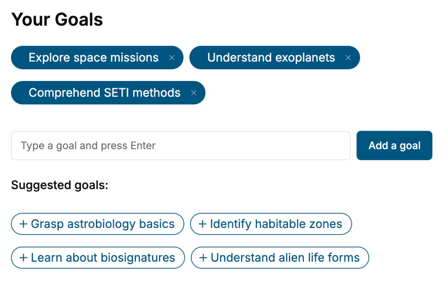
Tell us your goals and background, and get your own version of this book, tailored by AI to you.


Benefits of AI-tailored books
- Read one book, not ten:
- all the Anatomy knowledge you need, gathered into a single book.
- Save days of learning:
- tell us your goals, choose your topics, and the whole book is built around them.
- Learn effortlessly:
- Anatomy book written for your specific background and expertise.
- Understand at a glance:
- includes charts, tables, timelines, checklists, decision trees, and more.
- Stay current:
- written the day you order it, not years ago.
- Trust what you read:
- every book is checked for accuracy, with real research when your subject needs it.
Create your unique book in 3 steps
- 1. Select your focus
Select the focus of your Anatomy book and share your background
- 2. Personalize your book
Specify your goals and choose sub-topics to include
- 3. Get your tailored book
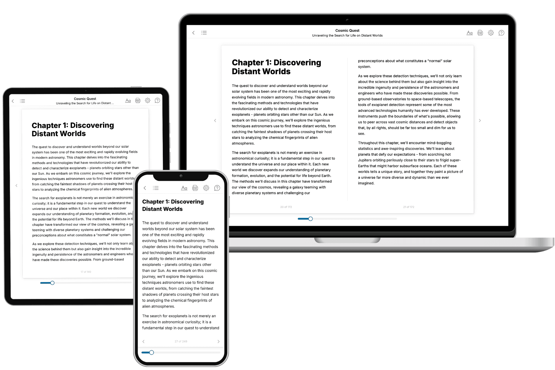
Your book is ready in 10 minutes. Read it online, download as EPUB or PDF, or send to Kindle.

Frequently asked questions
- What is TailoredRead?
TailoredRead creates complete, full-length nonfiction ebooks, written specifically for you. Tell us what you want to learn and why, and TailoredRead writes you a complete ebook on it, with charts, tables, and diagrams throughout. Read it online, on Kindle, or on any e-reader, or download it as a PDF or EPUB. Every book is written fresh the day you order it.
- How is the book tailored to me?
Before anything is written, you answer a few short questions about your goals, your background, and the topics you care about. Your book is then planned around your answers: written at your level, focused on your goals, and covering the topics you chose, instead of the one-size-fits-all book a store sells everyone.
- Will it read like generic AI content?
No. Your book isn't a chat transcript: it's planned chapter by chapter before a single word is written, told in a clear book voice, and explains ideas visually with charts, tables, and diagrams. After writing, every book goes through an editorial pass that polishes chapters so the book reads consistent from start to finish. Judge for yourself: browse thousands of sample books on our Explore Books page.
- How accurate is the content?
Our AI first checks whether your subject needs live research. Niche and fast-moving subjects are researched before writing, and the book plan and chapters are grounded in what it finds. After writing, every book goes through a quality pass that checks its facts and consistency, and researched books list their sources in a Notes section at the back.
- How long is the book?
You choose the length: Comprehensive (250-300 pages), Detailed (150-200 pages), Essential (70-100 pages), or Short (30-50 pages).
- Does the book include visuals?
Yes. Every book includes charts, tables, and diagrams: timelines, checklists, step-by-step guides, decision trees, comparison tables, matrices, and bar charts. A typical book includes dozens of these visual elements.
- Can I read it on Kindle? What formats can I download?
Yes. Every book can be read in our online reader or downloaded as a PDF or EPUB. The EPUB can be opened with Kindle, Apple Books, Kobo, and any e-reader. The Commercial and Author licenses also include an editable Word (DOCX) and Markdown download.
- How much does it cost?
About the price of a regular ebook. The exact price depends on your book's length and complexity, from $2 for the shortest books to $35 for the most comprehensive, and it's shown when you review your book, before you commit to anything. Each book is a one-time purchase, yours to keep, no subscription. Licenses that let you share, teach, or publish your book range from $35.99 to $49.99.
- Can I preview the book before purchasing?
Before you pay, you'll see what your book will cover: the title, a detailed description, the estimated length, and the complete chapter-by-chapter table of contents, so you know what you're getting before you decide. The full text itself is written after you purchase.
- How long does it take to create a book?
About 10 minutes. You can start reading the opening chapters while the rest is still being written.