The Circulatory System Coloring Guide
Learn How Your Heart, Blood, and Vessels Keep You Alive Through Interactive Anatomy
Most people think the circulatory system is simply about the heart pumping blood—but the reality is far more intricate and fascinating.
222 pages10 chaptersFeb 2, 2026Anatomy
About this book
Your cardiovascular system is a sophisticated network of organs and vessels working in perfect coordination to keep every cell in your body alive. This coloring guide transforms abstract anatomical concepts into tangible, visual learning experiences. By actively coloring and labeling detailed illustrations, you'll develop a genuine understanding of how your heart chambers work, how blood flows through arteries and veins, and where the critical exchange of oxygen and nutrients happens. Each chapter combines scientifically accurate diagrams with clear explanations, allowing you to see the connections between structure and function. Whether you're a student, healthcare professional, or simply curious about how your body works, this interactive approach makes complex circulatory concepts accessible and memorable. Discover the remarkable engineering of your cardiovascular system and gain insights that will enhance your appreciation for your body's most vital functions.
Table of contents
- IntroductionWhy Understanding Your Circulatory System Matters, How to Use This Coloring Guide, The Power of Visual Learning and Coloring, Your Journey Through Cardiovascular AnatomyRead below
- Chapter 1: The Heart: Structure and ChambersLocation and Size of the Heart, The Four Chambers: Atria and Ventricles, Heart Walls and the Pericardium, Coloring Activity: Label and Color the Heart Chambers
- Chapter 2: Heart Valves and Blood Flow DirectionHow Valves Prevent Backflow, The Four Heart Valves Explained, Understanding Heart Sounds and Valve Function, Coloring Activity: Valve Pathways and Blood Direction
- Chapter 3: Arteries: Carrying Blood Away from the HeartArtery Wall Layers and Elasticity, Major Arteries of the Body, Pulse and Blood Pressure in Arteries, Coloring Activity: Tracing Major Arterial Pathways
- Chapter 4: Veins: Returning Blood to the HeartVein Wall Layers and Valves, Major Veins of the Body, How Blood Returns Against Gravity, Coloring Activity: Mapping Venous Return Pathways
- Chapter 5: Capillaries: Where Exchange HappensMicroscopic Structure of Capillaries, Oxygen and Nutrient Exchange at the Cellular Level, Capillary Beds and Tissue Perfusion, Coloring Activity: Capillary Networks and Exchange
- Chapter 6: Pulmonary Circulation: Oxygenating Your BloodThe Path to and from the Lungs, Gas Exchange in the Pulmonary Capillaries, Pulmonary Arteries and Veins, Coloring Activity: Tracing Pulmonary Circulation
- Chapter 7: Systemic Circulation: Delivering Life to Every CellThe Complete Systemic Circuit, The Aorta and Its Major Branches, Blood Supply to Major Organs, Coloring Activity: Complete Systemic Circulation Map
- Chapter 8: Blood Pressure and Vessel ResistanceHow Blood Pressure Is Generated, Systolic and Diastolic Pressure Explained, Vessel Diameter and Resistance, Coloring Activity: Blood Pressure Zones and Vessel Types
- Chapter 9: The Cardiac Cycle: One Heartbeat at a TimeSystole and Diastole: Contraction and Relaxation, Atrial and Ventricular Contractions, Electrical Signals Controlling the Heartbeat, Coloring Activity: Stages of the Cardiac Cycle
- Chapter 10: Special Circulations and Unique PathwaysCoronary Circulation: Feeding the Heart Itself, Cerebral Circulation: Protecting the Brain, Fetal Circulation and Birth Changes, Coloring Activity: Special Circulatory Pathways
- ConclusionIntegrating Your Circulatory Knowledge, How Understanding Circulation Supports Your Health
- Glossary of Circulatory Terms
- Further Reading and Resources
Introduction
Every second of your life, your circulatory system performs an extraordinary feat of engineering. Your heart beats approximately 100,000 times each day, pumping roughly 2,000 gallons of blood through a network of vessels that, if laid end to end, would stretch over 60,000 miles. This intricate system delivers oxygen and nutrients to trillions of cells while simultaneously removing waste products that would otherwise poison your tissues. Yet despite its constant, life-sustaining work, most people have only a vague understanding of how this remarkable system actually functions. This coloring guide bridges that gap by transforming abstract anatomical concepts into tangible, visual experiences that engage both your analytical and creative thinking.
Traditional anatomy textbooks often present the circulatory system as a series of disconnected facts and labeled diagrams that you're expected to memorize. This approach misses a fundamental truth about how humans learn best: we understand complex systems more deeply when we actively engage with them through multiple sensory pathways. When you color an illustration of the heart's chambers, trace the pathway of blood through the pulmonary circulation, or label the major arteries branching from the aorta, you're not just passively receiving information. You're creating neural connections that link visual patterns, spatial relationships, and functional concepts in ways that pure reading cannot achieve. Research in educational psychology consistently demonstrates that this kind of active, multimodal learning produces stronger retention and deeper comprehension than passive study methods alone[1].
This guide is designed specifically for learners who already have a foundational understanding of basic anatomy and are ready to develop a more sophisticated, integrated knowledge of cardiovascular function. You won't find oversimplified explanations or cartoon-like illustrations here. Instead, you'll encounter scientifically accurate diagrams paired with clear, accessible explanations that respect your intelligence while making complex concepts understandable. Each chapter builds systematically on previous knowledge, guiding you from the heart's structural chambers through the intricate pathways of circulation that reach every corner of your body. By the time you complete this journey, you'll possess not just memorized facts but a genuine understanding of how structure determines function in the cardiovascular system—knowledge that will serve you whether you're pursuing healthcare education, teaching others, or simply seeking to understand the remarkable machinery that keeps you alive.
Why Understanding Your Circulatory System Matters
Cardiovascular disease remains the leading cause of death globally, claiming approximately 17.9 million lives each year according to the World Health Organization[2]. This staggering statistic isn't meant to frighten you but to illustrate a crucial point: understanding how your circulatory system works empowers you to make informed decisions about your health. When you comprehend how blood pressure is generated, how vessel diameter affects resistance, or why coronary arteries are particularly vulnerable to blockage, you gain context for medical advice that might otherwise seem arbitrary. Recommendations about exercise, diet, stress management, and medication make more sense when you understand the underlying anatomy and physiology they're designed to protect.
For students pursuing careers in healthcare, mastery of circulatory anatomy is absolutely foundational. Nurses need to understand venous pathways when establishing IV access or recognizing signs of deep vein thrombosis. Physicians must visualize coronary circulation when interpreting cardiac catheterization results or explaining bypass surgery to patients. Physical therapists rely on knowledge of blood flow dynamics when designing rehabilitation programs for cardiac patients. Emergency medical technicians assess pulse points and blood pressure as critical vital signs, interpretations that depend entirely on understanding arterial pathways and cardiac function. Even if you're not entering a medical profession, this knowledge enhances your ability to communicate effectively with healthcare providers, understand your own medical test results, and advocate for appropriate care.
Beyond practical health applications, studying the circulatory system offers profound insights into biological design principles that appear throughout nature. The heart's four-chambered structure represents an elegant solution to the challenge of maintaining separate pulmonary and systemic circuits while maximizing efficiency. The progressive branching of arteries into smaller vessels demonstrates how biological systems optimize distribution networks. The microscopic architecture of capillaries reveals how form follows function at the cellular level, with walls just one cell thick to facilitate rapid exchange of gases and nutrients. These aren't just anatomical curiosities—they're examples of evolutionary problem-solving that engineers and designers study when developing artificial organs, drug delivery systems, and microfluidic devices.
Understanding circulation also transforms your relationship with your own body. When you exercise and feel your heart rate increase, you'll recognize this as your cardiovascular system responding to increased oxygen demand from working muscles. When you stand up quickly and feel momentarily dizzy, you'll understand this as a temporary lag in venous return and blood pressure adjustment. When you see the blue veins visible beneath your skin, you'll know these are vessels returning deoxygenated blood to your heart, not vessels carrying "blue blood" as common misconceptions suggest. This knowledge converts your body from a mysterious black box into a comprehensible system whose signals and responses make logical sense.
The circulatory system also provides a perfect case study in how body systems integrate and depend on each other. Your cardiovascular system doesn't function in isolation—it works in constant coordination with your respiratory system to oxygenate blood, your digestive system to absorb nutrients into the bloodstream, your urinary system to regulate blood volume and pressure, and your nervous system to adjust heart rate and vessel diameter in response to changing demands. By understanding these connections, you develop a more holistic view of human physiology that recognizes the body as an integrated whole rather than a collection of separate parts. This systems-thinking approach is increasingly recognized as essential for both medical practice and personal health management.
Finally, there's an intrinsic value in understanding the machinery that sustains your life. Your circulatory system has been working without conscious effort since before you were born, adapting to every activity, emotion, and environmental change you've experienced. It deserves your attention and appreciation. When you study the precise coordination required for blood to flow through four heart chambers in perfect sequence, when you trace the pathway that carries oxygen from your lungs to your toes in less than a minute, when you examine the microscopic capillaries where life-sustaining exchange occurs—you're engaging with one of nature's most remarkable achievements. This knowledge enriches your understanding of what it means to be alive and functioning in a human body.
Whether your motivation is professional preparation, health literacy, scientific curiosity, or personal enrichment, the time you invest in understanding your circulatory system will yield returns throughout your life. The knowledge you gain here isn't temporary information you'll forget after an exam—it's foundational understanding that will inform countless future learning experiences and real-world situations. Each time you encounter cardiovascular topics in news articles, medical appointments, or further education, you'll have a framework for integrating new information. This guide provides that framework through a combination of accurate anatomical detail, clear functional explanations, and active learning strategies that transform passive reading into engaged understanding.
How to Use This Coloring Guide
This book is designed as an interactive learning tool, not a passive reading experience. Each chapter combines detailed anatomical illustrations with explanatory text and structured coloring activities that reinforce your understanding through active engagement. To get the most from this guide, you'll need a set of colored pencils or fine-tipped markers in a variety of colors. Colored pencils are generally preferable because they allow for more precise work in detailed areas and can be layered to create different shades. You'll want at least twelve colors, including multiple shades of red and blue for distinguishing oxygenated and deoxygenated blood, as well as contrasting colors for different structures like chambers, vessels, and valves.
The recommended approach is to work through the chapters sequentially rather than jumping around randomly. The book is structured to build knowledge progressively, with each chapter introducing concepts that depend on understanding from previous sections. For example, you'll grasp pulmonary and systemic circulation much more easily after you've studied heart chambers, valves, and the basic differences between arteries and veins. That said, if you're using this guide to supplement a course with a different sequence, you can adapt the order to match your curriculum—just be prepared to reference earlier chapters when you encounter unfamiliar terms or concepts. Each chapter includes cross-references to related sections to help you navigate these connections.
Within each chapter, you'll find a consistent structure designed to optimize learning. The chapter begins with explanatory text that introduces key concepts and anatomical structures. Read this material carefully before you begin coloring, as understanding the function and relationships of structures will guide your color choices and help you recognize what you're looking at in the illustrations. After the explanatory sections, you'll encounter dedicated coloring activities with specific instructions. These activities aren't just artistic exercises—they're carefully designed to reinforce spatial relationships, functional pathways, and structural distinctions that are central to understanding how the circulatory system works.
When you reach a coloring activity, follow these steps for maximum learning benefit:
- Read all instructions completely before you begin coloring to understand what you're being asked to identify and distinguish
- Choose your color scheme thoughtfully, using the suggested colors when provided or selecting contrasting colors that make different structures clearly distinguishable
- Color slowly and carefully, paying attention to boundaries between structures and the spatial relationships between different components
- Label structures as you color them, writing the anatomical terms neatly to reinforce terminology
- Trace pathways with your finger or a pencil to follow the flow of blood through the system, verbalizing the sequence as you go
- Review the completed illustration by covering the labels and testing yourself on structure identification
A critical strategy for using this guide effectively is to establish a consistent color-coding system that you maintain throughout the book. This consistency will help you recognize patterns and make connections across different chapters. Here's a recommended color scheme that aligns with standard anatomical conventions:
- Bright red for oxygenated blood and the vessels carrying it (arteries in systemic circulation, veins in pulmonary circulation)
- Dark blue or purple for deoxygenated blood and its vessels (veins in systemic circulation, arteries in pulmonary circulation)
- Light pink or peach for heart muscle (myocardium) and vessel walls
- Yellow or gold for fatty tissue, the pericardium, or connective tissue
- Green for lymphatic vessels when they appear in illustrations
- Different shades for different chambers—perhaps light blue for right atrium, darker blue for right ventricle, light red for left atrium, and darker red for left ventricle
Don't rush through the coloring activities. Research on learning retention shows that the physical act of coloring, when done mindfully and deliberately, creates stronger memory traces than simply looking at pre-colored diagrams[3]. The time you spend carefully coloring the walls of the left ventricle or tracing the pathway of the aorta and its branches is time spent encoding that information into long-term memory. Think of coloring not as a childish activity but as a form of active meditation on anatomical structure—a way of slowing down and really seeing the details that you might skim over when reading text alone.
As you work through each chapter, take advantage of the margins to make your own notes. When you encounter a concept that connects to something you learned in a previous chapter, jot down that connection. If a particular structure reminds you of something from your own experience—perhaps you've felt your pulse at the radial artery, or you remember a relative who had coronary bypass surgery—write that association in the margin. These personal connections and cross-references transform the book from a generic educational resource into your own customized learning tool. When you review later, these notes will trigger recall more effectively than the printed text alone.
Finally, use the coloring activities as self-assessment tools. After completing a chapter, return to the illustrations a day or two later and test yourself. Can you identify all the labeled structures without looking at the key? Can you trace the pathway of blood through the heart chambers in the correct sequence? Can you explain why arteries have thicker walls than veins? This spaced repetition—reviewing material after a delay rather than immediately—is one of the most effective strategies for converting short-term learning into long-term retention. The coloring guide format makes this kind of self-testing easy and visually engaging, turning review sessions into active problem-solving rather than passive re-reading.
The Power of Visual Learning and Coloring
The human brain processes visual information with remarkable speed and efficiency. Studies estimate that approximately 50% of your brain's resources are devoted to visual processing, and you can comprehend a visual scene in as little as 13 milliseconds[4]. This visual dominance in human cognition explains why anatomical illustrations are so much more effective than text descriptions alone for understanding spatial relationships and structural organization. When you read that the left ventricle has thicker walls than the right ventricle, you're processing abstract language. When you see an illustration showing this difference and color the left ventricular wall with more layers, you're creating a visual memory that your brain can retrieve much more readily.
Coloring takes visual learning a step further by adding kinesthetic and tactile dimensions to the experience. When you physically color a structure, you're engaging motor cortex regions of your brain in addition to visual processing areas. This multi-sensory engagement creates what neuroscientists call "elaborative encoding"—the formation of memory traces through multiple neural pathways simultaneously. The more pathways involved in encoding information, the more retrieval routes you'll have available when you need to recall that information later. This is why students who actively draw or color anatomical structures typically outperform those who only study pre-made diagrams, even when the final visual products are identical.
The act of choosing colors for different structures also engages your decision-making and categorization abilities in ways that support learning. When you decide to color all arteries red and all veins blue, you're making a categorical distinction that reinforces the functional difference between these vessel types. When you choose different shades for the four heart chambers, you're creating a visual system that helps you remember their separate identities and functions. These color-coding decisions aren't arbitrary—they're cognitive acts that require you to think about the relationships and distinctions between structures, which deepens your understanding of how the system is organized.
Coloring also naturally slows down the learning process in beneficial ways. In our age of rapid information consumption, there's a tendency to skim through material quickly, creating an illusion of understanding without genuine comprehension. When you color an anatomical illustration, you can't rush. You must pay attention to boundaries, stay within lines, and notice details about shape and position. This enforced slowness gives your brain time to process information more deeply. You might notice, for example, that the pulmonary arteries branch in a particular pattern, or that the heart valves are positioned at specific angles—details you would likely miss if you were just glancing at a pre-colored diagram.
Research on the "generation effect" in memory provides additional support for coloring as a learning strategy. This well-established psychological principle states that information you generate yourself is remembered better than information you passively receive[5]. When you color an illustration based on instructions and your understanding of the text, you're generating a visual representation rather than simply viewing one created by someone else. Even though the outline is provided, your color choices, shading decisions, and labeling create a unique product that your brain recognizes as your own creation. This sense of ownership enhances memory consolidation and makes the information feel more personally relevant.
The meditative quality of coloring also offers cognitive benefits that extend beyond simple memorization. When you're focused on carefully coloring within the lines of a capillary network or shading the layers of an artery wall, you enter a state of concentrated attention that psychologists call "flow." In this state, your mind is fully engaged with the task at hand, free from the distractions and mental chatter that often interfere with learning. This focused attention allows for deeper processing of information and can make study sessions more productive and less mentally fatiguing. Many students report that coloring anatomy diagrams is actually relaxing despite being educational—a rare combination in academic work.
Visual learning through coloring is particularly effective for understanding the circulatory system because this system is fundamentally about spatial relationships and pathways. Blood doesn't just exist in abstract categories—it flows through specific routes, from chamber to chamber, through branching vessels, into microscopic capillaries, and back again. These pathways have three-dimensional geometry that's difficult to grasp from text alone. When you color the pathway of blood from the right atrium through the pulmonary circulation and back to the left atrium, you're tracing a spatial journey that helps you understand not just what happens but where it happens. This spatial understanding is essential for making sense of clinical scenarios, such as understanding how a blockage in a specific coronary artery affects particular regions of heart muscle.
Finally, the visual products you create through coloring serve as personalized study materials for future review. A textbook page looks the same every time you open it, but a page you've colored yourself carries visual cues that trigger your memory of the learning session when you created it. You might remember that you colored the aorta while sitting in a particular location, or that you had a specific insight about valve function while working on that illustration. These contextual memories, called "encoding specificity" in cognitive psychology, provide additional retrieval cues that make it easier to access the anatomical information when you need it. Your colored illustrations become not just study aids but memory anchors that connect factual knowledge to personal experience.
Your Journey Through Cardiovascular Anatomy
This guide takes you on a systematic journey through the circulatory system, beginning with the heart's structure and progressing outward through the vessels that carry blood throughout your body. The organization follows both anatomical logic and functional relationships, ensuring that you build foundational knowledge before tackling more complex concepts. Chapter 1 starts with the heart itself, examining its location, size, and four-chambered structure. You'll learn to distinguish between atria and ventricles, understand the significance of the muscular walls that separate chambers, and explore the protective pericardium that surrounds the heart. This structural foundation is essential because everything else in the circulatory system ultimately connects to these chambers.
Chapter 2 focuses on the heart valves—the one-way gates that ensure blood flows in the correct direction through the cardiac chambers. You'll discover how the tricuspid, pulmonary, mitral, and aortic valves open and close in precise sequence, preventing backflow and maintaining the efficiency of the cardiac pump. Understanding valve function is crucial for making sense of the cardiac cycle and for recognizing what goes wrong in common valve disorders. The coloring activities in this chapter will help you visualize the spatial arrangement of valves and trace the pathway of blood as it moves through each valve in turn.
Chapters 3, 4, and 5 examine the three types of blood vessels—arteries, veins, and capillaries—each with distinct structures suited to their specific functions. You'll learn why arteries have thick, elastic walls capable of withstanding high pressure, while veins have thinner walls and internal valves to assist blood return against gravity. The capillary chapter reveals the elegant simplicity of these microscopic vessels, whose walls are just one cell thick to facilitate rapid exchange of oxygen, nutrients, and waste products. These three chapters provide the anatomical vocabulary you'll need to understand the circulation pathways that follow.
Chapters 6 and 7 trace the two major circulation loops: pulmonary circulation (between heart and lungs) and systemic circulation (between heart and the rest of the body). In the pulmonary circulation chapter, you'll follow blood from the right ventricle through the pulmonary arteries to the lung capillaries, where gas exchange occurs, and back through pulmonary veins to the left atrium. The systemic circulation chapter takes you on a more extensive journey, following oxygenated blood from the left ventricle through the aorta and its many branches to capillary beds throughout the body, then back through the venous system to the right atrium. These chapters integrate everything you've learned about heart chambers, valves, and vessel types into complete functional circuits.
Chapter 8 explores blood pressure and vascular resistance—the forces that drive blood through the circulatory system. You'll understand how the heart's contractions generate pressure, how vessel diameter affects resistance to flow, and why blood pressure decreases progressively as blood moves from arteries through capillaries to veins. This chapter provides the physiological context for understanding why certain cardiovascular conditions develop and how treatments work. The concepts here connect structure to function in ways that make the entire system more comprehensible.
Chapter 9 examines the cardiac cycle in detail, breaking down what happens during a single heartbeat into distinct phases. You'll learn the difference between systole (contraction) and diastole (relaxation), understand how atrial and ventricular contractions are coordinated, and discover the electrical conduction system that controls heart rhythm. This chapter brings together concepts from earlier chapters—chambers, valves, and pressure—to show how they interact in the rhythmic cycle that sustains your life. The coloring activities here will help you visualize the sequence of events and understand the timing relationships between different cardiac events.
Chapter 10 explores special circulations that have unique characteristics: coronary circulation that supplies the heart muscle itself, cerebral circulation that protects and nourishes the brain, and fetal circulation that functions before birth when the lungs aren't yet operational. These specialized pathways demonstrate how the basic circulatory plan adapts to meet specific physiological challenges. Understanding these variations deepens your appreciation for the system's flexibility and reveals important clinical considerations—for example, why coronary artery disease is so dangerous, or why certain congenital heart defects occur when fetal circulation fails to convert properly at birth.
Throughout this journey, you'll encounter recurring themes that tie the chapters together. One central theme is the relationship between structure and function—how the anatomy of each component is precisely suited to its role in the system. Another is the concept of the circulatory system as a closed loop, where blood continuously cycles through the same pathways rather than being consumed and replaced. A third theme is integration, recognizing how the cardiovascular system coordinates with respiratory, digestive, urinary, and nervous systems to maintain homeostasis. These themes will help you see the circulatory system not as a collection of isolated facts but as a coherent, integrated whole.
As you progress through the chapters, you'll notice that your understanding becomes increasingly sophisticated. Early chapters present individual structures and basic concepts. Middle chapters show how these components connect into functional pathways. Later chapters reveal the dynamic processes and regulatory mechanisms that keep the system running smoothly. This progression mirrors how experts in any field develop their knowledge—starting with vocabulary and basic concepts, then building connections and relationships, and finally achieving an integrated understanding where individual facts cohere into meaningful patterns. By the end of this guide, you'll have developed that expert-level integrated understanding of cardiovascular anatomy and physiology.
The coloring activities throughout the book are carefully sequenced to support this progressive learning. Early activities focus on identification and labeling, helping you learn anatomical vocabulary and recognize structures. Middle activities emphasize tracing pathways and understanding spatial relationships. Later activities challenge you to integrate information across multiple systems and predict functional outcomes. This progression ensures that you're always working at an appropriate level of challenge—not so easy that you're bored, but not so difficult that you're overwhelmed. Each completed activity builds your confidence and prepares you for the next level of complexity.
Remember that this journey through cardiovascular anatomy is not a race. The goal isn't to finish the book as quickly as possible but to develop genuine, lasting understanding. Take your time with each chapter. Reread sections that seem confusing. Complete the coloring activities carefully and thoughtfully. Make notes about connections you notice. Ask questions and seek out additional resources when you want to explore a topic more deeply. The knowledge you're building here is foundational for understanding human biology, whether you're pursuing professional healthcare education or simply seeking to understand your own body better. Every minute you invest in careful, thoughtful study will pay dividends in comprehension and retention that will serve you for years to come.
Notes
Introduction
- 1. R. E. Mayer, Multimedia Learning (2nd ed.), Cambridge University Press, 2009
- 2. World Health Organization, Cardiovascular diseases (CVDs) fact sheet, 2021
- 3. W. R. Klemm, Memory Power 101, Skyhorse Publishing, 2013
- 4. M. C. Potter and others, "Detecting meaning in RSVP at 13 ms per picture," Attention, Perception, & Psychophysics, 2014
- 5. N. J. Slamecka and P. Graf, "The generation effect," Journal of Experimental Psychology: Human Learning and Memory, 1978
This is the end of the free sample. The full book has 10 chapters.
Want this book written for you?
Tell us your goals and background, and get your own version of this book, tailored by AI to you.


Benefits of AI-tailored books
- Read one book, not ten:
- all the Anatomy knowledge you need, gathered into a single book.
- Save days of learning:
- tell us your goals, choose your topics, and the whole book is built around them.
- Learn effortlessly:
- Anatomy book written for your specific background and expertise.
- Understand at a glance:
- includes charts, tables, timelines, checklists, decision trees, and more.
- Stay current:
- written the day you order it, not years ago.
- Trust what you read:
- every book is checked for accuracy, with real research when your subject needs it.
Create your unique book in 3 steps
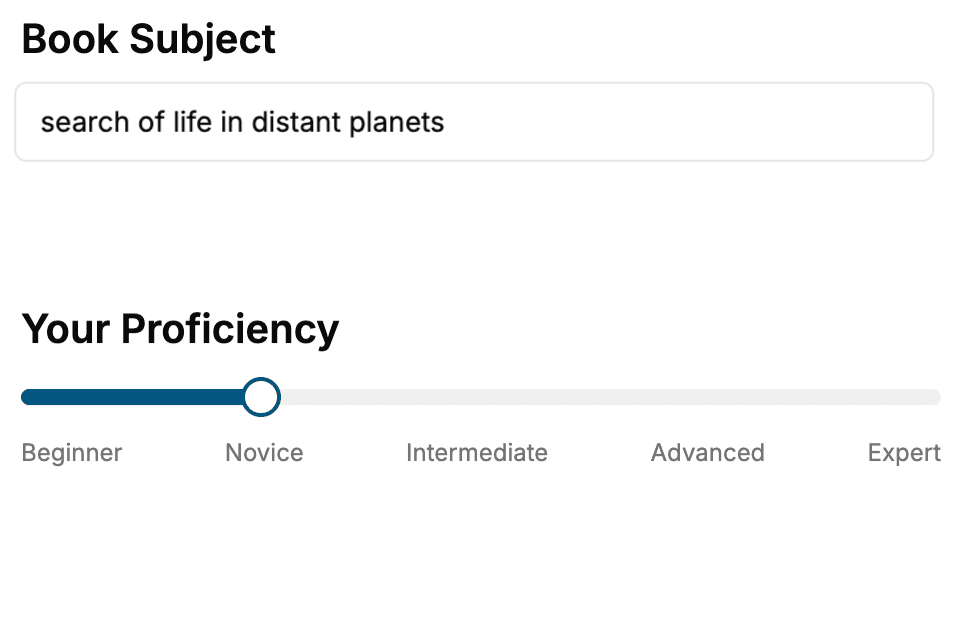
- 1. Select your focus
Select the focus of your Anatomy book and share your background
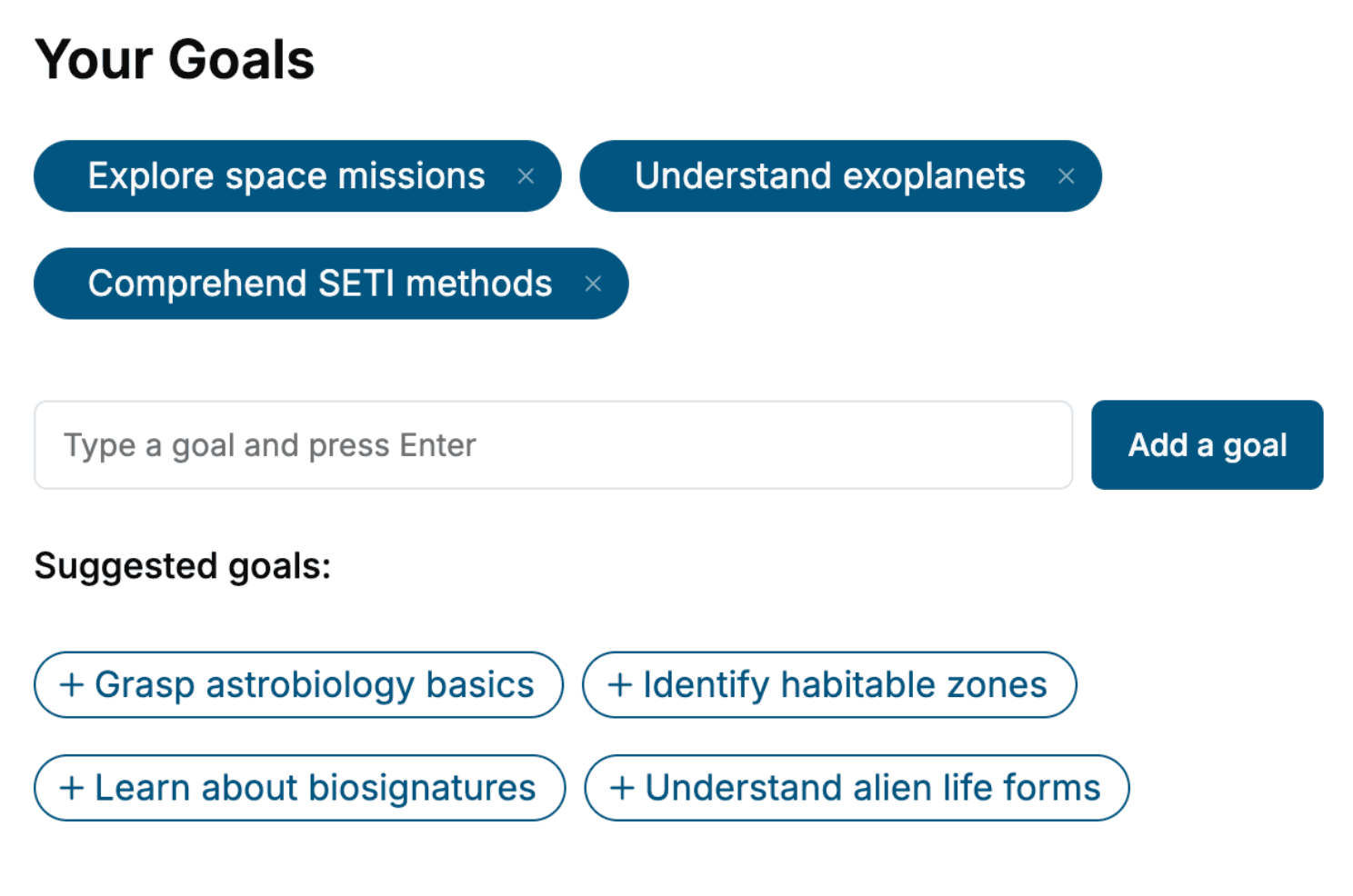
- 2. Personalize your book
Specify your goals and choose sub-topics to include
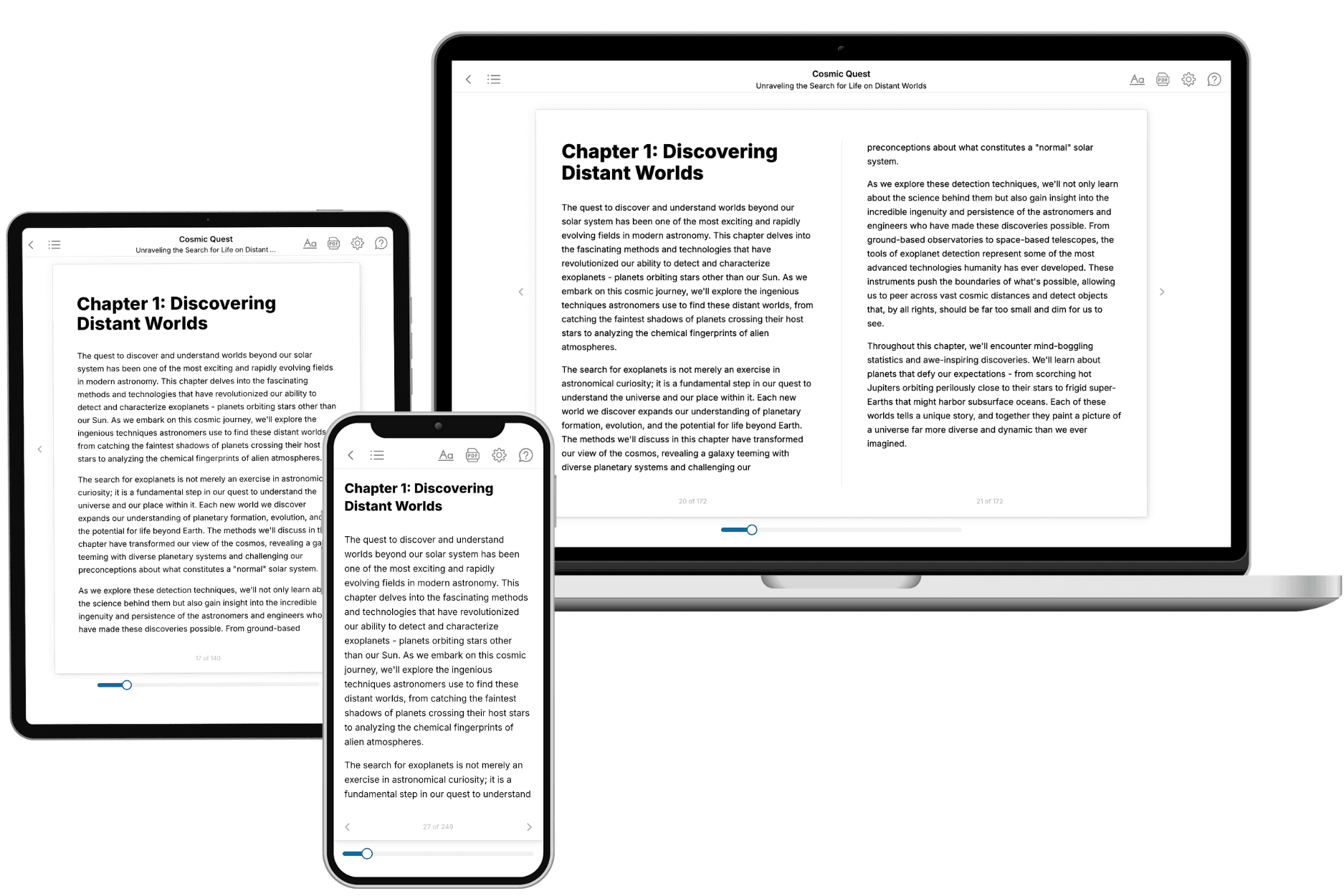
- 3. Get your tailored book
Your book is ready in 10 minutes. Read it online, download as EPUB or PDF, or send to Kindle.
Frequently asked questions
- What is TailoredRead?
TailoredRead creates complete, full-length nonfiction ebooks, written specifically for you. Tell us what you want to learn and why, and TailoredRead writes you a complete ebook on it, with charts, tables, and diagrams throughout. Read it online, on Kindle, or on any e-reader, or download it as a PDF or EPUB. Every book is written fresh the day you order it.
- How is the book tailored to me?
Before anything is written, you answer a few short questions about your goals, your background, and the topics you care about. Your book is then planned around your answers: written at your level, focused on your goals, and covering the topics you chose, instead of the one-size-fits-all book a store sells everyone.
- Will it read like generic AI content?
No. Your book isn't a chat transcript: it's planned chapter by chapter before a single word is written, told in a clear book voice, and explains ideas visually with charts, tables, and diagrams. After writing, every book goes through an editorial pass that polishes chapters so the book reads consistent from start to finish. Judge for yourself: browse thousands of sample books on our Explore Books page.
- How accurate is the content?
Our AI first checks whether your subject needs live research. Niche and fast-moving subjects are researched before writing, and the book plan and chapters are grounded in what it finds. After writing, every book goes through a quality pass that checks its facts and consistency, and researched books list their sources in a Notes section at the back.
- How long is the book?
You choose the length: Comprehensive (250-300 pages), Detailed (150-200 pages), Essential (70-100 pages), or Short (30-50 pages).
- Does the book include visuals?
Yes. Every book includes charts, tables, and diagrams: timelines, checklists, step-by-step guides, decision trees, comparison tables, matrices, and bar charts. A typical book includes dozens of these visual elements.
- Can I read it on Kindle? What formats can I download?
Yes. Every book can be read in our online reader or downloaded as a PDF or EPUB. The EPUB can be opened with Kindle, Apple Books, Kobo, and any e-reader. The Commercial and Author licenses also include an editable Word (DOCX) and Markdown download.
- How much does it cost?
About the price of a regular ebook. The exact price depends on your book's length and complexity, from $2 for the shortest books to $35 for the most comprehensive, and it's shown when you review your book, before you commit to anything. Each book is a one-time purchase, yours to keep, no subscription. Licenses that let you share, teach, or publish your book range from $35.99 to $49.99.
- Can I preview the book before purchasing?
Before you pay, you'll see what your book will cover: the title, a detailed description, the estimated length, and the complete chapter-by-chapter table of contents, so you know what you're getting before you decide. The full text itself is written after you purchase.
- How long does it take to create a book?
About 10 minutes. You can start reading the opening chapters while the rest is still being written.