Clinical Anatomy Essentials
From Embryology to Clinical Practice—A Comprehensive Guide to Human Structure and Function
How well do your students understand why anatomical variations occur, or why certain nerve injuries produce predictable patterns?
222 pages12 chaptersFeb 27, 2026Human AnatomyCreated by Seth
About this book
This comprehensive reference bridges the gap between classical anatomy and modern clinical practice by systematically connecting embryological development to adult anatomy, imaging findings, and surgical applications. Rather than presenting anatomy as static structures, this book reveals the dynamic developmental processes that shape human form and function. You'll explore how germ layer derivatives organize into complex systems, how embryological folding mechanisms create three-dimensional relationships, and how understanding these principles transforms your ability to interpret imaging, predict clinical presentations, and teach anatomy with deeper insight. Designed for anatomy educators and advanced practitioners, this resource integrates cutting-edge neuroanatomical imaging correlation with embryological principles, providing the conceptual framework needed to master peripheral nerve anatomy, vascular systems, and clinical variation. Whether preparing students for board examinations or advancing your own expertise, this book offers the integrated perspective that distinguishes exceptional anatomists from competent ones.
Table of contents
- IntroductionWhy Embryology Matters in Clinical Practice, The Integration Framework: Connecting Development to Anatomy, How to Use This Book for Maximum LearningRead below
- Chapter 1: Embryological FoundationsGerm Layer Derivatives and Tissue Origins, Neural Crest Derivatives: The Architects of Peripheral Systems, Somite Differentiation and Segmental Organization, Notochord Development and Axial Skeleton Formation
- Chapter 2: Embryological Folding MechanismsThree-Dimensional Organization from Folding, Neural Tube Formation and Segmentation, Body Wall Formation and Cavity Development, Clinical Implications of Embryological Folding Defects
- Chapter 3: Spinal Cord Segmental OrganizationSegmental Anatomy and Functional Levels, Motor Pathways and Segmental Control, Sensory Organization and Dermatome Mapping, Autonomic Segmentation and Visceral Control
- Chapter 4: Cranial Nerve Nuclei MappingBrainstem Organization and Nuclear Columns, Motor Nuclei Mapping and Functional Organization, Sensory Nuclei Organization and Pathways, Clinical Brainstem Syndromes and Nuclear Lesions
- Chapter 5: Peripheral Nervous System ArchitectureNerve Formation and Developmental Pathways, Plexus Organization and Functional Grouping, Terminal Nerve Distribution and End-Organ Innervation
- Chapter 6: Brachial Plexus Anatomy and VariationsPlexus Formation and Embryological Origins, Anatomical Relationships and Spatial Organization, Terminal Branches and Functional Distribution, Anatomical Variations and Clinical Significance
- Chapter 7: Peripheral Nerve Injury PatternsInjury Classification and Severity Grading, Predictable Injury Patterns Based on Anatomy, Recovery Mechanisms and Regeneration Pathways, Clinical Assessment and Prognostic Indicators
- Chapter 8: Cutaneous Nerve Distribution MapsDermatome Organization and Segmental Distribution, Peripheral Nerve Territories and Overlap Patterns, Clinical Sensory Testing and Anatomical Correlation
- Chapter 9: Autonomic Nervous System AnatomySympathetic Organization and Segmental Origins, Parasympathetic Pathways and Cranial Nerve Distribution, Visceral Innervation Patterns and Functional Integration, Autonomic Clinical Correlations and Dysfunction Patterns
- Chapter 10: Vascular System ArchitectureArterial Development and Embryological Origins, Venous Organization and Drainage Patterns, Vascular Variations and Anatomical Anomalies
- Chapter 11: Lymphatic Drainage PatternsLymphatic Development and Embryological Origins, Regional Drainage Patterns and Node Organization, Clinical Lymphatic Assessment and Pathological Spread
- Chapter 12: Neuroanatomical Imaging CorrelationImaging Modalities and Technical Principles, Cross-Sectional Anatomy and Three-Dimensional Reconstruction, Normal Variants and Anatomical Anomalies on Imaging, Pathological Correlation and Clinical Interpretation
- ConclusionIntegrating Knowledge for Clinical Excellence, Future Directions in Anatomical Science
Introduction
Anatomy has never been a static discipline. From the earliest dissections in Renaissance Europe to today's high-resolution MRI tractography, the study of human structure has always evolved alongside the tools and questions of its era. Yet one tension has persisted across centuries: the gap between knowing what a structure looks like and understanding why it exists in that form. This book is built around the conviction that closing that gap requires embryology. When you understand how the body is built, you understand why it looks the way it does, why it varies the way it does, and why it fails the way it does.
The audience for this reference is specific and intentional. If you are an anatomy professor, a clinical anatomist, or an advanced practitioner who teaches or applies structural knowledge at a sophisticated level, this book was designed with your needs in mind. It assumes a strong foundational knowledge of gross anatomy and histology. It does not spend time on introductory definitions. Instead, it moves directly into the integrative territory where embryological principles meet clinical imaging, surgical planning, and the interpretation of anatomical variation. That integration is the core value this book offers.
Each chapter in this book follows a consistent logic: development first, then structure, then clinical application. This sequence is not arbitrary. It reflects the actual causal chain that produces the human body. A nerve does not simply appear in a particular location; it migrates there, guided by molecular signals, constrained by surrounding mesenchyme, and organized by segmental identity established weeks before birth. Understanding that sequence transforms how you teach nerve injury patterns, how you interpret imaging anomalies, and how you explain anatomical variation to students who otherwise memorize without comprehension. The chapters ahead are organized to build that understanding systematically, from embryological foundations through neuroanatomical imaging correlation.
Before moving into the content itself, this introduction addresses three foundational questions. First, why does embryology matter in clinical practice, beyond its traditional role as a preclinical subject? Second, what is the integration framework that connects developmental biology to adult anatomy throughout this book? Third, how should you use this reference to extract maximum value, whether you are preparing a lecture series, reviewing for professional development, or building a curriculum around embryological principles? These three subchapters set the conceptual stage for everything that follows.
Why Embryology Matters in Clinical Practice
There is a common assumption in medical education that embryology is a preclinical subject, something to be learned in the first year and then largely set aside as clinical training takes over. This assumption is costly. Embryology is not background knowledge. It is explanatory knowledge, and the difference matters enormously when you are trying to understand why a patient presents with an unexpected finding, why a surgical approach carries a particular risk, or why a nerve injury produces a deficit that does not match a simple anatomical map.
Consider the clinical problem of anatomical variation. Variations in vascular anatomy, nerve branching patterns, and organ position are encountered regularly in surgical and radiological practice. A surgeon who encounters an aberrant right subclavian artery arising as the last branch of the aortic arch may be surprised if they have not connected that finding to the embryological regression of the right fourth aortic arch. A radiologist who sees a persistent left superior vena cava on a chest CT needs to recognize it as a remnant of the left anterior cardinal vein, a structure that normally regresses during the fifth to eighth weeks of development.[1] In both cases, embryological knowledge converts a surprising finding into a predictable one.
The predictive power of embryology extends directly into nerve injury interpretation. The brachial plexus, for example, is not organized arbitrarily. Its roots, trunks, divisions, cords, and terminal branches reflect the segmental identity of the somites from which the limb bud musculature derives. When an upper trunk injury produces weakness in shoulder abduction and elbow flexion but spares hand intrinsics, that pattern is not a coincidence. It is the direct consequence of C5 and C6 segmental identity, which was established during somite differentiation in the fourth week of development. Teaching this connection gives students a framework for predicting deficits rather than memorizing them.
Embryology also explains why certain congenital anomalies cluster together. Conditions that affect neural crest cell migration, for instance, tend to produce deficits across multiple systems simultaneously, because neural crest derivatives include peripheral ganglia, craniofacial cartilage, cardiac outflow tract structures, and melanocytes. DiGeorge syndrome, which results from a deletion at chromosome 22q11.2, produces cardiac defects, parathyroid hypoplasia, and thymic aplasia together because all three structures depend on neural crest cells migrating through the third and fourth pharyngeal arches.[2] Without embryological context, these associations appear coincidental. With it, they are mechanistically coherent.
In the context of imaging interpretation, embryological knowledge becomes a tool for distinguishing normal variants from pathology. The thyroglossal duct cyst, for example, appears along the midline of the neck anywhere from the base of the tongue to the thyroid cartilage, because it follows the path of thyroid gland descent from the foramen cecum. A clinician who knows this developmental pathway can immediately recognize the anatomical logic of the cyst's location and understand why surgical excision must include the central portion of the hyoid bone to prevent recurrence. Without that developmental context, the anatomy of the procedure seems arbitrary.
Beyond individual clinical scenarios, embryological understanding shapes how anatomy is taught at a conceptual level. Students who learn anatomy as a collection of named structures face a significant memorization burden with limited retention. Students who learn anatomy as the product of developmental processes have a generative framework. They can reason about structures they have not explicitly memorized, because they understand the rules that govern how the body is organized. This is the pedagogical argument for embryology, and it is one of the strongest justifications for the approach taken throughout this book.
Finally, embryology matters because it is increasingly visible in clinical imaging. High-resolution fetal MRI, three-dimensional ultrasound, and advanced postnatal neuroimaging now reveal developmental structures and processes that were previously accessible only through histological sections or cadaveric dissection. Clinicians and educators who understand embryological principles are better equipped to interpret these images, to recognize developmental anomalies, and to communicate findings accurately to colleagues and patients. The integration of embryology with imaging is not a future aspiration. It is a present clinical reality.
Key insight: Embryology transforms anatomical variation from a source of surprise into a source of prediction. Every structure that deviates from the textbook norm does so for a developmental reason. Finding that reason is the work of integrated anatomical thinking.
The Integration Framework: Connecting Development to Anatomy
This book uses a consistent integration framework across all chapters. Understanding that framework before you begin reading will help you extract more value from each section and see the connections between chapters that might otherwise appear to cover separate topics. The framework has three layers: developmental origin, structural organization, and clinical application. Every major anatomical system covered in this book is approached through all three layers, in that sequence.
The first layer, developmental origin, asks where a structure comes from. This means identifying the germ layer, the specific embryological tissue, and the developmental process responsible for producing the structure. For the peripheral nervous system, this means tracing neurons and Schwann cells back to neural crest cells, which delaminate from the dorsal neural tube during neurulation. For the vascular system, it means identifying which aortic arch arteries persist, which regress, and which are remodeled into the great vessels of the adult thorax. This layer is not simply historical background. It is the causal foundation for everything that follows.
The second layer, structural organization, asks how the structure is arranged in the adult body and why that arrangement takes the form it does. This is where classical gross anatomy intersects with embryological explanation. The organization of the brachial plexus into roots, trunks, divisions, cords, and terminal branches reflects the segmental identity of the contributing spinal cord levels and the spatial constraints imposed by the developing limb bud and surrounding mesenchyme. The arrangement of cranial nerve nuclei in longitudinal columns within the brainstem reflects the segmental organization of the rhombencephalon during early neural tube development. In each case, the adult structure is legible once the developmental logic is understood.
The third layer, clinical application, asks how developmental origin and structural organization inform diagnosis, imaging interpretation, and surgical planning. This layer is where the framework pays its most direct dividends for practicing clinicians and educators. Consider the following examples of how the three layers connect in practice:
- Peripheral nerve injury: Neural crest origin explains why Schwann cells can support axonal regeneration (they retain developmental plasticity); structural organization explains the predictable deficit pattern; clinical application identifies the prognostic implications of injury level relative to the cell body.
- Vascular anomalies: Aortic arch development explains why a double aortic arch forms (failure of one arch to regress); structural organization describes the vascular ring it creates; clinical application identifies the tracheal and esophageal compression that results.
- Dermatome mapping: Somite differentiation explains why dermatomes follow segmental patterns; structural organization describes the overlap between adjacent dermatomes; clinical application guides sensory testing and localizes spinal cord lesions.
This three-layer framework also governs how anatomical variation is treated throughout the book. Variations are not presented as exceptions to be memorized separately from normal anatomy. They are presented as predictable outcomes of developmental processes that can deviate within defined parameters. A prefixed brachial plexus, in which C4 contributes significantly to the upper trunk, is not a random anomaly. It reflects a shift in the segmental contribution to the limb bud, a shift that occurs along a continuum and that has predictable consequences for the distribution of motor and sensory function in the upper limb.[3]
The framework also integrates imaging throughout, rather than treating it as a separate topic confined to a single chapter. Cross-sectional anatomy is referenced in the context of each system, because the ability to interpret imaging requires the same three-layer understanding that governs the rest of the book. A radiologist reading a lumbar spine MRI needs to know the developmental origin of the intervertebral disc (notochord and annular fibrosus from sclerotome), the structural organization of the disc relative to the spinal cord and nerve roots, and the clinical implications of disc herniation at specific levels. Chapter 12 provides a dedicated treatment of neuroanatomical imaging correlation, but imaging references appear throughout the book wherever they reinforce the integration framework.
One important feature of this framework is that it is explicitly bidirectional. You can move from development to clinical application, as described above, but you can also move in the reverse direction. When you encounter an unexpected clinical finding, an unusual imaging variant, or a surgical complication that does not fit the standard anatomical description, the framework gives you a method for working backward to the developmental explanation. This reverse reasoning is one of the most valuable skills an advanced anatomist can develop, and it is one that this book is specifically designed to support.
The integration framework also acknowledges the limits of current knowledge. Embryological explanations for some anatomical variations remain incompletely understood, and the book is explicit about those gaps. Where the developmental basis for a variation is well established and supported by experimental or clinical evidence, that evidence is cited. Where the explanation is hypothetical or contested, that uncertainty is noted. This intellectual honesty is part of what makes the framework useful as a teaching tool, because it models the kind of critical thinking that distinguishes expert anatomists from those who simply reproduce received knowledge.
Framework summary: Every system in this book is analyzed through three questions: Where does it come from developmentally? How is it organized structurally in the adult? And how does that developmental origin and structural organization inform clinical practice? Keeping these three questions active as you read will maximize the conceptual value of each chapter.
How to Use This Book for Maximum Learning
This book is designed to function as both a systematic reference and a conceptual resource. Those two functions call for different reading strategies, and understanding which mode you are in at any given time will help you use the book more effectively. The chapters are organized to support sequential reading from beginning to end, but they are also written to stand independently for readers who need to access specific systems or topics without reading the entire text in order.
If you are approaching this book sequentially, the recommended path is to read Chapters 1 and 2 before moving into the system-specific chapters. These two chapters, covering embryological foundations and folding mechanisms, establish the conceptual vocabulary and developmental logic that all subsequent chapters build on. Readers who skip directly to, say, the brachial plexus chapter or the autonomic nervous system chapter will find the content accessible, but they will miss the deeper explanatory connections that make the material genuinely integrative rather than simply descriptive.
If you are using this book as a reference for specific topics, the following navigation strategy is recommended:
- Identify the system or structure of interest using the table of contents or the index.
- Read the developmental origin section of the relevant chapter first, even if you are primarily interested in the clinical application content. The developmental context is short but essential for understanding why the clinical patterns take the form they do.
- Cross-reference related chapters using the internal references provided throughout the text. The peripheral nervous system, brachial plexus, and cutaneous nerve distribution chapters, for example, are closely interconnected and reward reading in combination.
- Consult the imaging correlation chapter (Chapter 12) alongside any system chapter where imaging interpretation is a priority for your teaching or practice.
For anatomy educators specifically, this book offers several features designed to support curriculum development and lecture preparation. Each chapter's three-layer structure maps directly onto a lecture format: developmental origin as the conceptual hook, structural organization as the core content, and clinical application as the takeaway that motivates student engagement. The clinical examples and case scenarios embedded throughout the chapters can be adapted directly into teaching cases, problem-based learning scenarios, or examination questions. The footnotes and citations provide a starting point for deeper literature review on any topic covered.
The book also supports a teaching approach centered on predictive anatomy, the practice of asking students to reason about structures and patterns they have not explicitly memorized, using developmental and organizational principles as their tools. For example, rather than asking students to list the muscles innervated by the radial nerve, a predictive anatomy question might ask: given that the radial nerve carries fibers from C5 through T1 with a predominance of C7, and given that it runs in the posterior compartment of the arm and forearm, which functional deficits would you expect from a mid-humeral shaft fracture? This kind of question requires integrated understanding, not memorization, and this book provides the conceptual foundation needed to teach and answer it.
For readers engaged in professional development rather than formal teaching, the book functions as a structured review that fills the gaps left by traditional anatomy education. Most anatomy training, even at the graduate level, treats embryology and gross anatomy as separate subjects with limited cross-referencing. This book systematically repairs that separation. Reading it alongside clinical cases, imaging studies, or surgical atlases will produce the most durable learning, because the conceptual framework becomes anchored to concrete examples from your own practice.
A note on the use of clinical examples throughout the text: the examples provided are drawn from well-documented anatomical and clinical literature and are intended to illustrate principles, not to serve as clinical guidelines. Anatomy is the foundation of clinical reasoning, but clinical decision-making requires integration of anatomy with physiology, pathology, patient history, and current evidence-based practice. The clinical correlations in this book are educational tools. They are designed to deepen anatomical understanding, not to substitute for clinical judgment or professional training.
The chapters on neuroanatomical imaging (Chapter 12) deserve special mention for readers whose primary interest is imaging interpretation. That chapter is written to be read in conjunction with actual imaging studies, not as a standalone text. The descriptions of cross-sectional anatomy and three-dimensional reconstruction are most valuable when you can simultaneously view the structures being described on a real or digitally reproduced image. If you have access to an anatomy imaging atlas or a digital imaging platform, using it alongside Chapter 12 will significantly enhance the learning value of that material.
Finally, consider how you engage with the anatomical variation content distributed throughout the book. Variations are not presented as curiosities or footnotes. They are presented as evidence for the developmental principles being discussed. When you encounter a variation, the productive question is not simply "how common is this?" but rather "what does this variation tell me about the developmental process that produced it, and what clinical implications follow from that developmental logic?" Approaching variations with that question in mind will reinforce the integration framework and build the kind of flexible anatomical reasoning that distinguishes expert practitioners from those who rely on memorized norms.
Practical tip for educators: Each chapter's clinical application section can be converted into a case-based learning exercise by presenting the clinical scenario first and asking students to reason backward to the anatomical and developmental explanation. This reversal of the chapter's sequence is a powerful pedagogical tool for developing integrative thinking.
The chapters ahead cover a wide range of systems and topics, from the molecular signals that guide neural crest migration to the imaging characteristics of brainstem lesions. What connects them is not simply the subject matter of anatomy, but the consistent application of a developmental perspective that makes anatomical knowledge explanatory rather than merely descriptive. That perspective is the core contribution of this book, and it is the lens through which every chapter is best read.
This is the end of the free sample. The full book has 12 chapters.
Want this book written for you?
Tell us your goals and background, and get your own version of this book, tailored by AI to you.


Benefits of AI-tailored books
- Read one book, not ten:
- all the Human Anatomy knowledge you need, gathered into a single book.
- Save days of learning:
- tell us your goals, choose your topics, and the whole book is built around them.
- Learn effortlessly:
- Human Anatomy book written for your specific background and expertise.
- Understand at a glance:
- includes charts, tables, timelines, checklists, decision trees, and more.
- Stay current:
- written the day you order it, not years ago.
- Trust what you read:
- every book is checked for accuracy, with real research when your subject needs it.
Create your unique book in 3 steps
- 1. Select your focus
Select the focus of your Human Anatomy book and share your background
- 2. Personalize your book
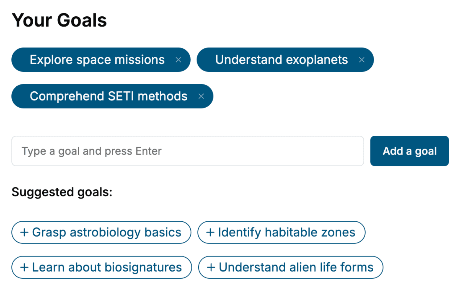
Specify your goals and choose sub-topics to include
- 3. Get your tailored book
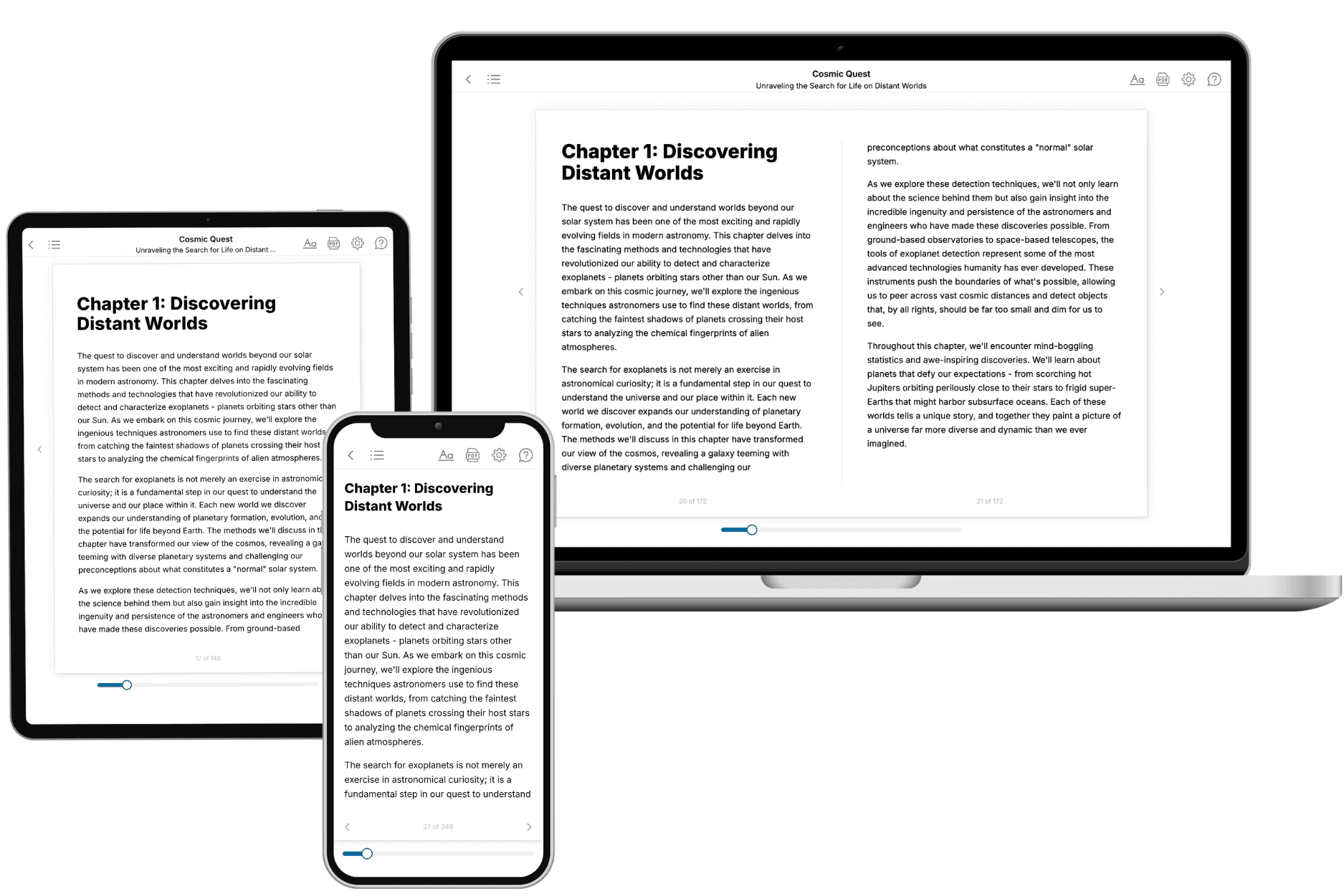
Your book is ready in 10 minutes. Read it online, download as EPUB or PDF, or send to Kindle.

Frequently asked questions
- What is TailoredRead?
TailoredRead creates complete, full-length nonfiction ebooks, written specifically for you. Tell us what you want to learn and why, and TailoredRead writes you a complete ebook on it, with charts, tables, and diagrams throughout. Read it online, on Kindle, or on any e-reader, or download it as a PDF or EPUB. Every book is written fresh the day you order it.
- How is the book tailored to me?
Before anything is written, you answer a few short questions about your goals, your background, and the topics you care about. Your book is then planned around your answers: written at your level, focused on your goals, and covering the topics you chose, instead of the one-size-fits-all book a store sells everyone.
- Will it read like generic AI content?
No. Your book isn't a chat transcript: it's planned chapter by chapter before a single word is written, told in a clear book voice, and explains ideas visually with charts, tables, and diagrams. After writing, every book goes through an editorial pass that polishes chapters so the book reads consistent from start to finish. Judge for yourself: browse thousands of sample books on our Explore Books page.
- How accurate is the content?
Our AI first checks whether your subject needs live research. Niche and fast-moving subjects are researched before writing, and the book plan and chapters are grounded in what it finds. After writing, every book goes through a quality pass that checks its facts and consistency, and researched books list their sources in a Notes section at the back.
- How long is the book?
You choose the length: Comprehensive (250-300 pages), Detailed (150-200 pages), Essential (70-100 pages), or Short (30-50 pages).
- Does the book include visuals?
Yes. Every book includes charts, tables, and diagrams: timelines, checklists, step-by-step guides, decision trees, comparison tables, matrices, and bar charts. A typical book includes dozens of these visual elements.
- Can I read it on Kindle? What formats can I download?
Yes. Every book can be read in our online reader or downloaded as a PDF or EPUB. The EPUB can be opened with Kindle, Apple Books, Kobo, and any e-reader. The Commercial and Author licenses also include an editable Word (DOCX) and Markdown download.
- How much does it cost?
About the price of a regular ebook. The exact price depends on your book's length and complexity, from $2 for the shortest books to $35 for the most comprehensive, and it's shown when you review your book, before you commit to anything. Each book is a one-time purchase, yours to keep, no subscription. Licenses that let you share, teach, or publish your book range from $35.99 to $49.99.
- Can I preview the book before purchasing?
Before you pay, you'll see what your book will cover: the title, a detailed description, the estimated length, and the complete chapter-by-chapter table of contents, so you know what you're getting before you decide. The full text itself is written after you purchase.
- How long does it take to create a book?
About 10 minutes. You can start reading the opening chapters while the rest is still being written.