Clinical Neuroanatomy Essentials
Mastering Brain Structure, Function, and Pathology Through Real-World Cases
Master the intricate relationship between brain structure and clinical presentation through a comprehensive exploration of neuroanatomy grounded in real-world cases.
269 pages13 chaptersFeb 27, 2026NeuroanatomyCreated by Seth
About this book
This advanced reference integrates detailed anatomical knowledge with clinical correlation, enabling you to rapidly localize lesions, interpret complex neuroimaging, and understand the mechanisms underlying neurological syndromes. From brainstem nuclei and cranial nerve pathways to spinal cord organization and white matter tracts, each system is presented with anatomical precision and clinical relevance. You'll explore how vascular territories predict stroke patterns, how specific tract lesions produce characteristic deficits, and how to recognize classic syndromes like Wallenberg's and internuclear ophthalmoplegia. The book emphasizes three-dimensional anatomical relationships, somatotopic organization, and the integrated circuits connecting cortex, basal ganglia, cerebellum, and brainstem. Whether you're teaching advanced students, refining your diagnostic skills, or preparing comprehensive examinations, this resource provides the depth and clinical integration necessary to excel in neuroanatomical practice and education.
Table of contents
- IntroductionWhy Clinical Integration Matters in Neuroanatomy, How to Use This Book for Maximum Learning, Core Anatomical Principles and TerminologyRead below
- Chapter 1: Neuroanatomical Foundations and OrganizationCentral Nervous System Organization and Divisions, Embryological Basis of Neural Structure, Vascular Principles and Blood-Brain Barrier
- Chapter 2: Cerebral Anatomy and Cortical LocalizationLobar Organization and Functional Areas, Cortical Mapping and Somatotopic Representation, White Matter Systems and Interhemispheric Connections
- Chapter 3: Vascular Territories and Stroke PatternsCerebral Circulation: Major Arteries and Territories, Stroke Syndromes and Vascular Correlations, Interpreting Vascular Imaging and Perfusion Studies
- Chapter 4: Brainstem Anatomy and Clinical SyndromesBrainstem Organization: Midbrain, Pons, and Medulla, Motor and Sensory Pathways Through the Brainstem, Classic Brainstem Syndromes and Lesion Patterns
- Chapter 5: Cranial Nerves: Anatomy and Clinical ExaminationCranial Nerve Nuclei and Brainstem Organization, Individual Cranial Nerves: Anatomy and Function, Systematic Cranial Nerve Examination and Interpretation
- Chapter 6: Motor Pathways and Pyramidal SystemPyramidal Tract Organization and Decussation, Pyramidal Tract Lesions and Clinical Patterns, Upper Motor Neuron Signs and Localization
- Chapter 7: Sensory Pathways and Spinal Cord OrganizationSpinothalamic Tract: Pain and Temperature Pathways, Dorsal Column Pathways: Proprioception and Vibration, Spinal Cord Lesion Patterns and Dissociated Sensory Loss
- Chapter 8: Spinal Nerve Roots and Peripheral DistributionsSpinal Nerve Root Anatomy and Emergence, Dermatomes, Myotomes, and Reflex Arcs, Peripheral Nerve Distributions and Clinical Correlation
- Chapter 9: Extrapyramidal Motor System and Basal GangliaBasal Ganglia Circuits and Motor Control, Extrapyramidal Motor Signs and Dysfunction Patterns, Movement Disorders and Anatomical Correlations
- Chapter 10: Cerebellar Anatomy and FunctionCerebellar Circuits and Afferent-Efferent Pathways, Cerebellar Zones and Functional Organization, Cerebellar Dysfunction and Clinical Manifestations
- Chapter 11: Vestibular System and Eye Movement ControlVestibular Anatomy and Brainstem Connections, Eye Movement Pathways and Nuclear Organization, Vertigo, Nystagmus, and Central-Peripheral Differentiation
- Chapter 12: Neuroimaging Essentials and Anatomical CorrelationNeuroimaging Modalities and Technical Principles, Anatomical Landmarks and Orientation on Imaging, Recognizing Pathology and Anatomical Abnormalities
- Chapter 13: Integrated Clinical Cases and LocalizationSystematic Approach to Clinical Case Analysis, Complex Cases Requiring Multi-System Integration, Diagnostic Reasoning and Anatomical Hypothesis Testing
- ConclusionSynthesizing Neuroanatomical Knowledge, Continuing Your Neuroanatomical Mastery
- Glossary of Neuroanatomical Terms
- Further Reading and Resources
Introduction
Neuroanatomy sits at the intersection of structure and meaning. Every sulcus, nucleus, and fiber tract exists not as an isolated anatomical curiosity but as a functional unit whose disruption produces predictable, recognizable clinical consequences. For those who teach and practice at the advanced level, this relationship between structure and deficit is the central intellectual challenge of the discipline. It demands more than memorization. It requires a three-dimensional, dynamic understanding of how neural systems are organized, how they interact, and how lesions at specific locations produce the precise patterns of dysfunction seen at the bedside.
The gap between knowing neuroanatomy and applying it clinically is wider than most textbooks acknowledge. A student can correctly label every structure on a brainstem cross-section and still struggle to explain why a patient with a lateral medullary infarct loses pain sensation on the left side of the face but the right side of the body. Bridging that gap requires a fundamentally different approach to learning and teaching the subject. It requires anchoring every anatomical fact to a clinical consequence, and every clinical observation to an anatomical substrate. That is the philosophy driving every chapter of this book.
This resource is designed for neuroanatomy educators, clinical neuroscientists, and advanced practitioners who already possess a strong foundational knowledge base and are seeking to deepen their command of structure-function relationships. The goal is not to introduce neuroanatomy from scratch but to elevate existing expertise to the level where localization becomes rapid, imaging interpretation becomes precise, and teaching becomes genuinely illuminating. Whether you are preparing a lecture on brainstem syndromes, refining your approach to a complex clinical case, or building a curriculum around integrated neuroanatomical reasoning, this book provides the depth and clinical grounding to support that work.
Each chapter is organized around a core anatomical system, presented with enough structural detail to satisfy rigorous academic standards while remaining anchored to clinical relevance throughout. Real-world cases, specific syndrome descriptions, and imaging correlations appear throughout the text, not as supplementary additions but as integral components of the anatomical narrative. The result is a reference that functions equally well as a teaching guide, a clinical refresher, and a framework for advanced self-directed study.
Why Clinical Integration Matters in Neuroanatomy
Pure anatomical knowledge, divorced from clinical context, has a short half-life in the memory of even the most dedicated learner. Research in cognitive science consistently shows that information anchored to meaningful, real-world scenarios is retained far more effectively than abstract facts presented in isolation.[1] In neuroanatomy, this principle has direct practical implications. When a learner understands that the medial longitudinal fasciculus (MLF) connects the abducens nucleus on one side to the contralateral oculomotor nucleus, that fact becomes memorable because it immediately explains internuclear ophthalmoplegia. The anatomy and the clinical syndrome reinforce each other.
Clinical integration also transforms the way anatomical relationships are understood spatially. Consider the internal capsule. Its posterior limb carries corticospinal fibers, and its genu carries corticobulbar fibers. A small lacunar infarct in the posterior limb produces contralateral hemiplegia with relative sparing of the face. A slightly more anterior lesion at the genu produces prominent facial and bulbar weakness. These distinctions are not just academic. They are the difference between localizing a lesion to the posterior limb versus the genu on imaging, and they carry direct implications for prognosis and management. Anatomy taught with this level of clinical specificity becomes genuinely useful.
For educators, clinical integration solves one of the most persistent challenges in teaching neuroanatomy: maintaining student engagement with material that can feel abstract and overwhelming. When a lecture on the spinothalamic tract opens with a patient presenting with dissociated sensory loss after a spinal cord injury, the anatomical details that follow have immediate relevance. Students are not memorizing pathways in the abstract. They are building the tools to understand a real clinical problem. This pedagogical approach consistently produces deeper learning and better retention than structure-first, function-later teaching sequences.[2]
Clinical integration also sharpens the precision of anatomical thinking. When you know that a lesion of the right facial nerve nucleus produces ipsilateral upper and lower facial weakness (unlike a cortical lesion, which spares the forehead), you are forced to understand why. The answer lies in the bilateral cortical representation of the upper face and the unilateral representation of the lower face. That understanding requires knowing the anatomy of the corticobulbar pathway, the organization of the facial nucleus, and the clinical examination findings that distinguish central from peripheral facial palsy. Each piece of knowledge reinforces the others, building a robust, interconnected understanding rather than a fragile list of isolated facts.
The clinical cases and syndrome descriptions throughout this book are not decorative. They are the primary vehicle through which anatomical knowledge is organized and made durable. Each case is chosen to illuminate a specific anatomical principle, and each syndrome is presented in enough clinical detail to make the underlying anatomy vivid and memorable. The goal is to develop the kind of integrated, flexible neuroanatomical knowledge that allows rapid, confident localization at the bedside and precise interpretation of neuroimaging findings.
For those who teach, this approach also provides a ready-made framework for curriculum design. The cases and clinical correlations in each chapter can be adapted directly into lectures, small-group discussions, or problem-based learning sessions. The anatomical principles are presented at a level of depth that supports advanced teaching, while the clinical anchors make the material accessible and engaging for learners at multiple stages of training. Clinical integration is not a pedagogical luxury. In neuroanatomy, it is the most effective path to genuine mastery.
A practical example: When teaching the Wallenberg syndrome (lateral medullary infarction), begin with the clinical presentation: ipsilateral facial pain and temperature loss, contralateral body pain and temperature loss, ipsilateral Horner syndrome, dysphagia, hoarseness, and ipsilateral limb ataxia. Then work backward through the anatomy of the lateral medulla to identify each affected structure: the spinal trigeminal nucleus and tract, the spinothalamic tract, the descending sympathetic fibers, the nucleus ambiguus, and the inferior cerebellar peduncle. The syndrome becomes a map of the lateral medulla, and the lateral medulla becomes the explanation for the syndrome.
This bidirectional reasoning, moving from syndrome to anatomy and from anatomy to syndrome, is the core cognitive skill that clinical integration develops. It is the skill that separates a neuroanatomist who can label structures from one who can localize lesions. Every chapter in this book is designed to build and reinforce that skill through repeated, varied practice with real clinical material.
How to Use This Book for Maximum Learning
This book is organized to support multiple modes of engagement, depending on your goals and the context in which you are using it. It can be read sequentially as a comprehensive review of clinical neuroanatomy, used as a reference for specific systems or syndromes, or mined for teaching material to support lectures and case-based learning sessions. Understanding how the content is structured will help you extract maximum value from each chapter.
Each chapter follows a consistent internal structure. It opens with the core anatomy of the system under discussion, presented with enough detail to support advanced clinical reasoning. This is followed by a discussion of the major pathways, circuits, or organizational principles relevant to that system. The chapter then moves into clinical correlation, where specific syndromes, lesion patterns, and imaging findings are discussed in detail. Cases are integrated throughout rather than segregated into separate sections, so the anatomical and clinical content remain in constant dialogue.
For those using this book primarily as a teaching resource, the following approach is recommended:
- Identify the anatomical system or clinical syndrome you plan to teach.
- Read the relevant chapter in full to build a comprehensive understanding of the material.
- Select one or two of the clinical cases or syndrome descriptions as the anchor for your teaching session.
- Use the anatomical detail in the chapter to build the explanatory framework around that clinical anchor.
- Supplement with the imaging correlations provided to give learners a visual representation of the anatomy and pathology.
For those using this book primarily for clinical skill development, a different approach works better. Rather than reading chapters sequentially, consider starting with a clinical problem you have encountered recently and locating the relevant chapter. Read the clinical correlation sections first to orient yourself to the syndrome, then work through the anatomical detail to understand the structural basis of the findings. This reverse-engineering approach mirrors the clinical reasoning process and reinforces the habit of moving between clinical observation and anatomical explanation.
The final chapter on integrated clinical cases is particularly valuable for both educators and clinicians. It presents complex, multi-system cases that require simultaneous reasoning across several anatomical domains. These cases are designed to challenge even experienced practitioners and to model the kind of sophisticated localization reasoning that distinguishes expert neuroanatomical thinking. Working through these cases systematically, ideally before reading the provided analysis, is one of the most effective ways to consolidate and test the knowledge built in earlier chapters.
Neuroimaging is woven throughout the book rather than confined to a single chapter. Each system is discussed with reference to how its anatomy appears on standard imaging modalities, and the dedicated neuroimaging chapter provides a framework for systematic image interpretation. For those who regularly interpret neuroimaging in clinical or educational settings, the imaging correlations in each chapter can be used to build a mental library of anatomical landmarks and pathological patterns that supports rapid, confident image reading.
Footnotes and references are provided throughout to support deeper exploration of specific topics. These are not exhaustive literature reviews but targeted pointers to foundational texts, landmark studies, and authoritative references that can extend your understanding of particular areas. The further reading section at the end of the book provides a curated list of resources organized by topic for those who wish to pursue specific areas in greater depth.
One practical suggestion: keep a running list of clinical cases or teaching scenarios that resonate with you as you read. The most effective neuroanatomical educators build their teaching around a personal repertoire of cases that they know deeply and can present with confidence. This book provides the raw material for building that repertoire. The cases and syndromes described here are starting points. Over time, you will supplement them with cases from your own clinical experience, creating a personalized library of anatomical-clinical correlations that becomes one of your most valuable teaching assets.
Recommended reading sequence for educators: Begin with the Introduction and Chapter 1 to establish the organizational framework. Then proceed to the chapters most relevant to your current teaching responsibilities. Return to Chapter 13 (Integrated Clinical Cases) after completing at least four or five system-specific chapters, as the integrated cases will be most valuable once you have a broad enough knowledge base to engage with multi-system reasoning.
Core Anatomical Principles and Terminology
Before moving into system-specific content, it is worth establishing a shared set of anatomical principles and terminological conventions that apply throughout the book. These are not elementary definitions for beginners. They are the conceptual foundations that underpin advanced neuroanatomical reasoning, and revisiting them explicitly helps ensure that the more complex material in later chapters rests on a solid and consistent base.
Directional terminology in neuroanatomy follows conventions that differ from general anatomy and can be a source of confusion, particularly in the brainstem and spinal cord. The following terms are used consistently throughout this book:
- Rostral: toward the front of the brain (anterior in the cerebrum; superior in the brainstem and spinal cord)
- Caudal: toward the tail (posterior in the cerebrum; inferior in the brainstem and spinal cord)
- Dorsal: toward the back surface (posterior in the cerebrum; posterior in the spinal cord)
- Ventral: toward the belly surface (anterior in the cerebrum; anterior in the spinal cord)
- Ipsilateral: on the same side as the lesion or reference point
- Contralateral: on the opposite side from the lesion or reference point
The concept of decussation is central to neuroanatomical localization and deserves explicit attention. Most major motor and sensory pathways cross the midline at some point in their course, meaning that lesions above the decussation produce contralateral deficits, while lesions below it produce ipsilateral deficits. The corticospinal tract decussates at the pyramidal decussation in the caudal medulla. The dorsal column-medial lemniscal pathway decussates in the caudal medulla at the level of the nucleus gracilis and nucleus cuneatus. The spinothalamic tract decussates within one to two spinal cord segments of the level at which its fibers enter the cord. Knowing where each pathway crosses is essential for accurate lesion localization.
Somatotopic organization refers to the systematic spatial arrangement of neural representations of body parts within a given structure. This principle applies throughout the nervous system, from the primary motor and somatosensory cortices to the spinal cord, the thalamus, and even individual brainstem nuclei. In the spinal cord, for example, the corticospinal tract is organized so that fibers serving the sacral segments are most lateral, with lumbar, thoracic, and cervical fibers arranged progressively more medially. This somatotopic arrangement explains why an expanding lateral cord lesion, such as an extramedullary tumor, may initially produce sacral-sparing sensory loss, while an intramedullary lesion at the same level may preferentially affect the central fibers serving the arms and trunk.[3]
The distinction between upper motor neuron (UMN) and lower motor neuron (LMN) signs is one of the most clinically important principles in neuroanatomy. Upper motor neurons originate in the motor cortex and descend through the corticospinal and corticobulbar tracts to synapse on lower motor neurons in the anterior horn of the spinal cord or in cranial nerve motor nuclei. Lesions of UMNs produce a characteristic pattern of weakness with increased tone, hyperreflexia, and an extensor plantar response (Babinski sign). Lesions of LMNs produce weakness with decreased tone, hyporeflexia or areflexia, and muscle atrophy. This distinction immediately narrows the anatomical location of a lesion and guides the subsequent diagnostic workup.
Vascular territory anatomy is a principle that recurs throughout the book and deserves early emphasis. The brain's arterial supply is organized into well-defined territories, and ischemic strokes respect these boundaries. Understanding which structures lie within a given vascular territory allows you to predict the clinical syndrome that will result from occlusion of that vessel, and conversely, to identify the likely occluded vessel from a patient's clinical presentation. For example, the posterior inferior cerebellar artery (PICA) supplies the lateral medulla and the inferior cerebellum. Occlusion of PICA produces the lateral medullary (Wallenberg) syndrome, with its characteristic combination of ipsilateral facial and contralateral body sensory loss, ipsilateral Horner syndrome, and cerebellar ataxia. Knowing the vascular territory makes the syndrome predictable; knowing the syndrome makes the vascular territory identifiable.
Functional column organization in the brainstem is a concept that greatly simplifies the otherwise bewildering arrangement of cranial nerve nuclei. The brainstem nuclei are organized into longitudinal columns based on their functional type, derived from the embryological alar and basal plates. These columns include:
- General somatic afferent (GSA): receives general sensation from skin and deep structures (e.g., trigeminal sensory nuclei)
- Special somatic afferent (SSA): receives special senses of hearing and balance (e.g., cochlear and vestibular nuclei)
- General visceral afferent (GVA): receives visceral sensation (e.g., nucleus tractus solitarius, caudal portion)
- Special visceral afferent (SVA): receives taste (e.g., nucleus tractus solitarius, rostral portion)
- General somatic efferent (GSE): innervates skeletal muscles derived from somites (e.g., oculomotor, trochlear, abducens, hypoglossal nuclei)
- Special visceral efferent (SVE): innervates muscles derived from pharyngeal arches (e.g., trigeminal motor, facial, nucleus ambiguus)
- General visceral efferent (GVE): parasympathetic outflow (e.g., Edinger-Westphal nucleus, dorsal motor nucleus of vagus)
Understanding this columnar organization transforms the apparent complexity of brainstem nuclei into a logical, predictable arrangement. Once you know that GSE nuclei are located near the midline and SVE nuclei are located more laterally, you can predict the approximate location of any cranial nerve motor nucleus and understand why lesions at specific brainstem levels affect particular combinations of cranial nerves.
Finally, the concept of crossed findings deserves explicit mention as a guiding principle for brainstem localization. A lesion that produces ipsilateral cranial nerve signs combined with contralateral long tract signs (motor or sensory) is almost certainly located in the brainstem, at the level of the affected cranial nerve nucleus. This is because the cranial nerve nucleus is affected before the long tracts have crossed, producing ipsilateral cranial nerve dysfunction, while the long tracts are affected before they decussate, producing contralateral limb signs. This pattern, sometimes called an alternating hemiplegia or crossed syndrome, is one of the most reliable localizing signs in clinical neurology and will be revisited in detail in the brainstem chapter.[4]
These core principles, directional terminology, decussation patterns, somatotopic organization, UMN versus LMN distinctions, vascular territory anatomy, functional column organization, and crossed findings, form the conceptual toolkit that makes advanced neuroanatomical reasoning possible. They are not background knowledge to be assumed and forgotten. They are active analytical tools that should be applied consciously and consistently throughout every clinical encounter and every teaching session. The chapters that follow are built on these foundations, and returning to them whenever a clinical or anatomical puzzle arises will consistently point toward the correct answer.
Notes
Introduction
- 1. J. D. Bransford and others, How People Learn, National Academy Press, 2000
- 2. V. L. Patel and others, "Expertise and tacit knowledge in medicine," R.J. Sternberg & J.A. Horvath, Tacit Knowledge in Professional Practice. Lawrence Erlbaum Associates, 1999
- 3. Standring and S. (Ed.), Gray's Anatomy, Elsevier, 2020
- 4. H. Blumenfeld, Neuroanatomy Through Clinical Cases (2nd ed.), Sinauer Associates, 2010
This is the end of the free sample. The full book has 13 chapters.
Want this book written for you?
Tell us your goals and background, and get your own version of this book, tailored by AI to you.


Benefits of AI-tailored books
- Read one book, not ten:
- all the Neuroanatomy knowledge you need, gathered into a single book.
- Save days of learning:
- tell us your goals, choose your topics, and the whole book is built around them.
- Learn effortlessly:
- Neuroanatomy book written for your specific background and expertise.
- Understand at a glance:
- includes charts, tables, timelines, checklists, decision trees, and more.
- Stay current:
- written the day you order it, not years ago.
- Trust what you read:
- every book is checked for accuracy, with real research when your subject needs it.
Create your unique book in 3 steps
- 1. Select your focus
Select the focus of your Neuroanatomy book and share your background
- 2. Personalize your book
Specify your goals and choose sub-topics to include
- 3. Get your tailored book
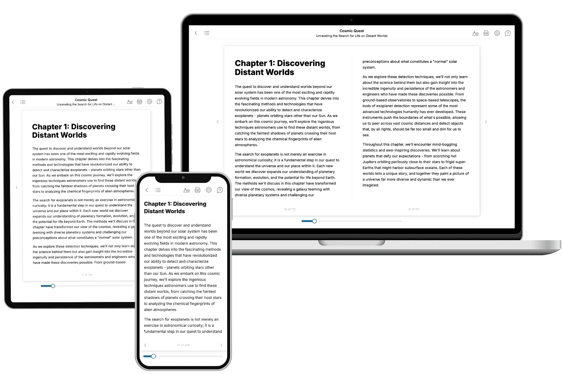
Your book is ready in 10 minutes. Read it online, download as EPUB or PDF, or send to Kindle.

Frequently asked questions
- What is TailoredRead?
TailoredRead creates complete, full-length nonfiction ebooks, written specifically for you. Tell us what you want to learn and why, and TailoredRead writes you a complete ebook on it, with charts, tables, and diagrams throughout. Read it online, on Kindle, or on any e-reader, or download it as a PDF or EPUB. Every book is written fresh the day you order it.
- How is the book tailored to me?
Before anything is written, you answer a few short questions about your goals, your background, and the topics you care about. Your book is then planned around your answers: written at your level, focused on your goals, and covering the topics you chose, instead of the one-size-fits-all book a store sells everyone.
- Will it read like generic AI content?
No. Your book isn't a chat transcript: it's planned chapter by chapter before a single word is written, told in a clear book voice, and explains ideas visually with charts, tables, and diagrams. After writing, every book goes through an editorial pass that polishes chapters so the book reads consistent from start to finish. Judge for yourself: browse thousands of sample books on our Explore Books page.
- How accurate is the content?
Our AI first checks whether your subject needs live research. Niche and fast-moving subjects are researched before writing, and the book plan and chapters are grounded in what it finds. After writing, every book goes through a quality pass that checks its facts and consistency, and researched books list their sources in a Notes section at the back.
- How long is the book?
You choose the length: Comprehensive (250-300 pages), Detailed (150-200 pages), Essential (70-100 pages), or Short (30-50 pages).
- Does the book include visuals?
Yes. Every book includes charts, tables, and diagrams: timelines, checklists, step-by-step guides, decision trees, comparison tables, matrices, and bar charts. A typical book includes dozens of these visual elements.
- Can I read it on Kindle? What formats can I download?
Yes. Every book can be read in our online reader or downloaded as a PDF or EPUB. The EPUB can be opened with Kindle, Apple Books, Kobo, and any e-reader. The Commercial and Author licenses also include an editable Word (DOCX) and Markdown download.
- How much does it cost?
About the price of a regular ebook. The exact price depends on your book's length and complexity, from $2 for the shortest books to $35 for the most comprehensive, and it's shown when you review your book, before you commit to anything. Each book is a one-time purchase, yours to keep, no subscription. Licenses that let you share, teach, or publish your book range from $35.99 to $49.99.
- Can I preview the book before purchasing?
Before you pay, you'll see what your book will cover: the title, a detailed description, the estimated length, and the complete chapter-by-chapter table of contents, so you know what you're getting before you decide. The full text itself is written after you purchase.
- How long does it take to create a book?
About 10 minutes. You can start reading the opening chapters while the rest is still being written.