The Complete Thyroid Anatomy Coloring Guide
Master Thyroid Structure Through Interactive Visual Learning
Picture yourself confidently identifying every aspect of thyroid anatomy with crystal-clear precision.
284 pages15 chaptersSep 15, 2025Anatomy
About this book
You'll move beyond memorizing isolated facts to truly understanding how the thyroid's intricate structure supports its vital functions in your body. This comprehensive coloring guide transforms the complex world of thyroid anatomy into an engaging, hands-on learning experience. You'll discover how the thyroid's unique butterfly shape, its relationship with surrounding neck structures, and its microscopic organization all work together as an integrated system. Each carefully designed illustration guides you through progressive levels of anatomical detail, from basic thyroid positioning to advanced histological features. Through systematic coloring exercises, you'll build lasting visual memories of thyroid anatomy that traditional textbooks simply cannot provide. The interactive approach helps you understand not just what structures look like, but why they're positioned and shaped as they are. You'll explore the thyroid's blood supply, nerve connections, and cellular architecture while developing the visual literacy essential for anyone studying human anatomy. Whether you're preparing for exams, enhancing your anatomical knowledge, or simply fascinated by how your body works, this guide provides the perfect blend of scientific accuracy and engaging visual learning that makes complex anatomy both accessible and memorable.
Table of contents
- IntroductionThe Science Behind Coloring for Learning, How to Use This Guide Effectively, Essential Coloring Techniques for Anatomy, Your Journey Through Thyroid AnatomyRead below
- Chapter 1: Thyroid FundamentalsThyroid Location and Orientation, Understanding the Butterfly Shape, Normal Size and Weight Variations, Surface Anatomy Landmarks
- Chapter 2: Gross Thyroid AnatomyRight and Left Thyroid Lobes, The Connecting Isthmus, Pyramidal Lobe Variations, Anterior and Posterior Surfaces
- Chapter 3: Thyroid's Neck RelationshipsRelationship to the Trachea, Connections to Laryngeal Structures, Proximity to the Esophagus, Surrounding Muscle and Fascial Layers
- Chapter 4: Thyroid Capsule and CoveringsThe True Thyroid Capsule, False Capsule and Pretracheal Fascia, Capsular Extensions and Septa, Ligamentous Attachments
- Chapter 5: Thyroid Arterial SupplySuperior Thyroid Artery, Inferior Thyroid Artery, Thyroidea Ima Artery Variations, Arterial Anastomoses and Collaterals
- Chapter 6: Thyroid Venous DrainageSuperior Thyroid Veins, Middle Thyroid Veins, Inferior Thyroid Veins, Thyroid Venous Plexus
- Chapter 7: Thyroid Lymphatic SystemIntrathyroidal Lymphatic Vessels, Regional Lymph Node Groups, Primary and Secondary Drainage Pathways, Anatomical Variations in Lymphatic Drainage
- Chapter 8: Thyroid InnervationSympathetic Nerve Supply, Parasympathetic Nerve Supply, Recurrent Laryngeal Nerve Relationship, Anatomical Nerve Variations
- Chapter 9: Microscopic Thyroid StructureThyroid Follicle Organization, Follicular Cell Characteristics, Colloid Composition and Function, Parafollicular C-Cells
- Chapter 10: Cellular ArchitectureFollicular Epithelial Organization, Basement Membrane Structure, Intercellular Junctions and Connections, Cellular Polarity and Orientation
- Chapter 11: Thyroid Connective Tissue FrameworkInterfollicular Stromal Elements, Collagen Fiber Networks, Reticular Fiber Framework, Extracellular Matrix Components
- Chapter 12: Thyroid Developmental AnatomyEmbryonic Thyroid Origins, Thyroglossal Duct Development, Thyroid Descent and Migration, Common Developmental Variations
- Chapter 13: Anatomical Variations and AnomaliesShape and Size Variations, Ectopic Thyroid Tissue, Vascular Supply Anomalies, Positional and Structural Variants
- Chapter 14: Comparative Thyroid AnatomyAge-Related Anatomical Changes, Gender-Based Anatomical Differences, Population and Ethnic Variations, Structure-Function Correlations
- Chapter 15: Integrated Thyroid Anatomy ReviewThyroid as Part of Endocrine System, Synthesis of Anatomical Knowledge, Anatomical Basis for Understanding Function, Self-Assessment and Knowledge Consolidation
- Glossary
- Further Reading
Introduction
Welcome to an innovative approach to mastering thyroid anatomy that transforms complex anatomical structures into an engaging, memorable learning experience. The thyroid gland, with its distinctive butterfly shape nestled in your neck, represents one of the most fascinating and functionally important organs in the human body. This comprehensive coloring guide bridges the gap between traditional textbook learning and hands-on visual engagement, creating a pathway to deep anatomical understanding that goes far beyond simple memorization. Through carefully designed illustrations and systematic coloring exercises, you'll develop the kind of three-dimensional anatomical visualization that forms the foundation of true expertise in human anatomy.
The journey through thyroid anatomy requires more than passive reading or rote memorization of anatomical terms. Your brain processes visual information differently than text, creating stronger neural pathways when you actively engage with anatomical structures through coloring and illustration. This guide harnesses the power of active learning, where each stroke of your colored pencil reinforces anatomical relationships and builds lasting visual memories. You'll discover how the thyroid's position relative to the trachea, its intricate blood supply, and its microscopic organization all contribute to its remarkable functional capabilities. The interactive nature of coloring allows you to explore these relationships at your own pace while building confidence in your anatomical knowledge.
Every illustration in this guide has been meticulously designed to progress from fundamental concepts to advanced anatomical details, ensuring that you build a solid foundation before tackling more complex structures. You'll begin with basic thyroid positioning and orientation, then systematically explore gross anatomy, vascular relationships, microscopic structure, and developmental patterns. This progressive approach mirrors how anatomical knowledge naturally builds upon itself, with each new concept reinforcing and expanding your understanding of previous material. The coloring process itself becomes a form of active meditation on anatomical structure, allowing you to internalize the spatial relationships and functional connections that make the thyroid such a remarkable organ.
The Science Behind Coloring for Learning
Research in cognitive neuroscience has consistently demonstrated that visual learning engages multiple brain regions simultaneously, creating more robust and lasting memories than traditional text-based study methods. When you color anatomical structures, your brain activates visual processing centers, motor control areas, and spatial reasoning networks all at once. This multi-modal engagement creates what neuroscientists call elaborative encoding, where information becomes embedded in multiple neural pathways rather than stored in isolated memory traces. Studies have shown that students who engage in visual-motor learning activities, such as anatomical coloring, demonstrate significantly improved retention rates compared to those using traditional study methods alone[1].
The act of coloring requires sustained attention and deliberate decision-making about color choices, spatial relationships, and structural boundaries. This focused engagement activates your brain's executive attention networks, which are crucial for deep learning and memory consolidation. Unlike passive reading, where your mind might wander while your eyes scan text, coloring demands continuous cognitive engagement with the material. You must constantly evaluate anatomical relationships, make decisions about color coding, and maintain awareness of structural boundaries and connections. This sustained attention creates what educational psychologists term desirable difficulties – challenges that initially require effort but ultimately lead to stronger, more durable learning outcomes.
Visual-spatial learning through coloring also taps into your brain's natural pattern recognition abilities, which evolved to help humans navigate complex three-dimensional environments. When you color the thyroid's butterfly shape, trace its vascular supply, or differentiate between follicular and parafollicular cells, you're engaging the same neural systems that help you understand spatial relationships in the physical world. This connection between anatomical learning and spatial cognition explains why many students find that coloring helps them visualize anatomical structures during exams, even when they're working from memory. The colored images become mental templates that you can recall and manipulate in your mind's eye.
The repetitive nature of coloring also supports memory consolidation through what neuroscientists call spaced repetition and interleaving. As you work through different anatomical views and cross-sections of the thyroid, you encounter the same structures from multiple perspectives, reinforcing your understanding through varied contexts. This approach mirrors how anatomical knowledge is actually used in practice, where you need to recognize structures from different angles, in different states, and in relationship to surrounding tissues. The coloring process naturally creates these varied exposures, building flexible, adaptable knowledge rather than rigid memorization of isolated facts.
Motor learning research has also revealed that the physical act of coloring creates proprioceptive memories – memories stored in your muscles and motor cortex. These kinesthetic memories complement visual and verbal memories, creating what learning theorists call multimodal memory traces. When you later encounter thyroid anatomy in other contexts, your brain can draw upon visual memories of the colored illustrations, motor memories of the coloring process, and conceptual memories of anatomical relationships. This redundancy in memory storage significantly improves both recall accuracy and the speed of information retrieval, making coloring an exceptionally efficient learning strategy for complex anatomical material.
Furthermore, the creative aspect of choosing colors and making aesthetic decisions about your illustrations engages brain regions associated with motivation and reward processing. This emotional engagement with the learning material creates what educational researchers call intrinsic motivation – the internal drive to continue learning because the process itself is rewarding. Unlike external motivators such as grades or requirements, intrinsic motivation leads to deeper engagement, better retention, and continued interest in the subject matter. The satisfaction of completing a detailed anatomical illustration provides immediate positive feedback that reinforces your learning efforts and encourages continued study.
The meditative quality of focused coloring also reduces stress and anxiety, which are known to interfere with memory formation and recall. When you're relaxed and focused, your brain produces optimal levels of neurotransmitters like acetylcholine and dopamine, which facilitate learning and memory consolidation. The rhythmic, repetitive motions of coloring can induce a state similar to mindfulness meditation, where your attention is fully present with the task at hand. This focused attention state, combined with reduced stress levels, creates ideal conditions for deep learning and long-term memory formation. Many students report that they remember anatomical details more clearly after coloring sessions than after traditional study periods.
How to Use This Guide Effectively
Maximizing your learning from this coloring guide requires a systematic approach that balances structure with flexibility, allowing you to adapt the material to your individual learning style and goals. Begin each chapter by reading through the introductory text to understand the anatomical concepts you'll be exploring, then examine the illustrations before you start coloring to get a sense of the overall structural relationships. This preview process activates your brain's predictive processing systems, creating a framework for organizing new information as you encounter it. Take time to identify key structures, note their relative positions, and consider how they might relate to anatomical knowledge you already possess. This preliminary survey creates cognitive scaffolding that supports deeper learning as you progress through the coloring exercises.
Develop a consistent color-coding system that you'll use throughout the guide to represent different types of anatomical structures. For example, you might choose to color all arteries red, veins blue, nerves yellow, and different tissue types in distinct colors that you'll remember and apply consistently. This systematic approach serves multiple learning purposes:
- Pattern Recognition: Consistent colors help you quickly identify structure types across different illustrations
- Memory Association: Color associations create additional retrieval cues for anatomical information
- Visual Organization: Systematic coloring makes complex illustrations easier to interpret and remember
- Cross-Reference Learning: Consistent colors allow you to compare structures across different anatomical views
Work through the chapters in sequence rather than jumping randomly between topics, as each chapter builds upon knowledge from previous sections. The thyroid's anatomy is interconnected, with vascular supply relating to gross structure, innervation patterns connecting to developmental origins, and microscopic organization reflecting functional requirements. This interconnected nature means that understanding early concepts thoroughly will make later chapters more accessible and meaningful. However, don't hesitate to return to earlier chapters when working on advanced topics – this backward review reinforces learning and helps you see connections that might not have been apparent during your initial study. The spiral nature of anatomical learning means that concepts become clearer and more meaningful with each review cycle.
Engage actively with each illustration by asking yourself questions as you color: Why is this structure positioned here?
How does this relate to what I learned in the previous chapter?
What would happen if this structure were damaged or absent?
This questioning approach transforms passive coloring into active inquiry, engaging your critical thinking skills and promoting deeper understanding. Write your questions and observations in the margins of your guide – these personal notes become valuable review materials and help you track your evolving understanding of thyroid anatomy. The process of formulating questions also helps you identify areas where you need additional study or clarification.
Use the coloring process as an opportunity for spaced repetition by returning to completed illustrations after several days or weeks. During these review sessions, try to identify structures from memory before checking the labels, then use different colors or shading techniques to highlight aspects you want to reinforce. This review process helps transfer information from short-term to long-term memory while allowing you to assess your progress and identify areas that need additional attention. You might also try covering labels and testing yourself on structure identification, using the colored illustrations as visual cues to trigger anatomical knowledge stored in your memory.
Supplement your coloring activities with additional learning strategies that complement visual learning. Create concept maps connecting different anatomical structures, write brief summaries of key concepts in your own words, or explain anatomical relationships to study partners or family members. These complementary activities engage different learning modalities and help consolidate the visual knowledge you're building through coloring. Consider photographing your completed illustrations to create digital flashcards or study materials that you can review on mobile devices during spare moments throughout your day.
Set realistic goals and maintain consistent study habits rather than attempting marathon coloring sessions that might lead to fatigue or superficial engagement with the material. Research suggests that distributed practice – shorter, regular study sessions spread over time – produces better learning outcomes than massed practice sessions. Aim for 30-45 minute coloring sessions that allow for focused attention without mental fatigue. During each session, try to complete one subsection thoroughly rather than rushing through multiple sections superficially. This approach ensures that you're building solid understanding at each step rather than accumulating fragmented knowledge that might be difficult to integrate later.
Track your progress through the guide by maintaining a learning journal where you record insights, questions, and connections you discover during your coloring sessions. Note which concepts you find challenging and which seem to come naturally, as this self-awareness will help you allocate study time effectively and identify your learning strengths. Document aha moments
when anatomical relationships suddenly become clear, as these insights often indicate that you're successfully integrating new knowledge with existing understanding. Your learning journal becomes a personalized study resource that reflects your unique journey through thyroid anatomy and can serve as a valuable review tool as you prepare for exams or practical applications.
Essential Coloring Techniques for Anatomy
Mastering specific coloring techniques will significantly enhance both the educational value and visual appeal of your anatomical illustrations. The choice of coloring materials affects not only the final appearance of your work but also the learning process itself. Colored pencils offer the greatest control and precision for anatomical coloring, allowing you to create subtle gradations, fine details, and layered effects that reflect the complexity of biological structures. High-quality colored pencils with soft, blendable cores work best for anatomical illustration, as they allow for smooth color application and easy blending between adjacent structures. Avoid markers or crayons for detailed anatomical work, as they lack the precision needed for small structures and can bleed through paper, making your illustrations difficult to use for review purposes.
Develop your pressure control to create different intensities and textures that reflect the varying characteristics of anatomical structures. Light pressure creates subtle, translucent colors perfect for representing delicate structures like capillary networks or thin fascial layers. Medium pressure produces solid, even colors suitable for major organs and tissue masses. Heavy pressure creates deep, saturated colors that work well for emphasizing important structures or creating contrast between different anatomical elements. Practice varying your pressure within single structures to show depth, curvature, and three-dimensional form. For example, when coloring the thyroid lobes, use lighter pressure on areas that would catch light and heavier pressure in shadowed regions to create a sense of volume and dimensionality.
Master the technique of directional shading to enhance the three-dimensional appearance of anatomical structures. Follow the natural contours and fiber directions of tissues when applying color – for muscle tissue, color in the direction of fiber orientation; for blood vessels, follow the vessel's path; for curved surfaces like the thyroid lobes, use curved strokes that follow the organ's natural shape. This directional approach not only creates more realistic illustrations but also reinforces your understanding of tissue organization and structural orientation. The physical act of following anatomical directions with your coloring strokes creates kinesthetic memories that complement visual learning and help you remember structural characteristics more effectively.
Utilize layering techniques to represent the complex, overlapping nature of anatomical structures. Begin with light base colors for deeper structures, then gradually add layers for more superficial elements, using progressively darker or more saturated colors for structures closest to the surface. This layering approach mirrors the actual three-dimensional organization of anatomical structures and helps you understand spatial relationships between different tissue types. For example, when coloring cross-sections of the thyroid, start with light colors for the deepest follicular structures, add medium tones for interfollicular tissue, and use darker colors for capsular and surface elements. This technique creates depth and helps you visualize how structures would appear in actual anatomical specimens.
Employ strategic use of white space and highlighting to represent important anatomical features that might otherwise be overlooked. Leave small areas uncolored to represent light reflection on moist surfaces, highlight the lumens of blood vessels or follicular spaces, or emphasize the boundaries between different tissue types. Use a white colored pencil or gel pen to add highlights after completing your base coloring, creating effects that make your illustrations more dynamic and easier to interpret. These highlighting techniques are particularly important for representing the thyroid's highly vascularized nature and the complex internal organization of follicular structures.
Create effective contrast between adjacent structures to clearly delineate anatomical boundaries and relationships. Use complementary colors (colors opposite each other on the color wheel) for structures that need to be easily distinguished from one another. For example, if you color arteries in warm reds, use cool blues for adjacent veins to create clear visual separation. Vary the intensity of colors to create hierarchical emphasis – use bright, saturated colors for the most important structures in each illustration and more muted tones for supporting elements. This contrast strategy helps prevent your illustrations from becoming muddy or confusing while ensuring that key anatomical relationships remain clearly visible.
Develop consistent techniques for representing different tissue types that you can apply throughout the guide. Create distinctive patterns or textures for different anatomical elements:
- Smooth, even coloring for solid organs and muscle tissue
- Stippling or dotting for glandular tissue and cellular details
- Cross-hatching for fibrous connective tissue
- Parallel lines for fascial planes and membrane structures
- Circular or spiral patterns for follicular organizations
Pay special attention to edge quality and line work to enhance the clarity and professional appearance of your anatomical illustrations. Use sharp, clean lines for distinct anatomical boundaries like organ capsules or vessel walls, and softer, more diffuse edges for gradual transitions between tissue types. Vary your line weight to create visual hierarchy – use thicker, darker lines for major structural boundaries and thinner, lighter lines for subtle anatomical details. Consider using a fine-tipped pen or sharp pencil to reinforce important structural outlines after completing your coloring, ensuring that key anatomical relationships remain clearly defined even in complex, multi-colored illustrations. This attention to line quality transforms simple coloring exercises into sophisticated anatomical illustrations that serve as valuable long-term study resources.
Your Journey Through Thyroid Anatomy
Your exploration of thyroid anatomy through this comprehensive coloring guide follows a carefully structured progression that mirrors how anatomical knowledge naturally builds from fundamental concepts to sophisticated understanding. You'll begin with the thyroid's basic position and orientation in the neck, establishing a solid foundation of spatial relationships that will support all subsequent learning. This foundational knowledge includes understanding how the thyroid's distinctive butterfly shape relates to surrounding neck structures, why it's positioned at the level of the fifth cervical to first thoracic vertebrae, and how its anterior location makes it accessible for examination and procedures. These fundamental concepts provide the spatial framework necessary for understanding more complex anatomical relationships you'll encounter in later chapters.
The journey progresses systematically through gross anatomical features, where you'll explore the thyroid's bilateral lobes, connecting isthmus, and occasional pyramidal lobe variations. Each gross anatomical feature serves specific functional purposes that become apparent through detailed illustration and coloring exercises. You'll discover how the thyroid's shape optimizes its relationship with the trachea and larynx, why the isthmus typically crosses the second and third tracheal rings, and how anatomical variations in thyroid shape relate to developmental patterns and functional requirements. This gross anatomical foundation prepares you for understanding the complex vascular and neural relationships that support thyroid function and integrate it with broader physiological systems.
Your study of thyroid vasculature reveals one of the most richly perfused organs in the human body, with blood flow patterns that reflect the thyroid's high metabolic activity and endocrine function. Through detailed coloring of arterial supply patterns, you'll understand how the superior thyroid arteries arise from the external carotid system while inferior thyroid arteries branch from the thyrocervical trunk, creating redundant blood supply that ensures consistent thyroid perfusion. The venous drainage system presents equally complex patterns, with superior, middle, and inferior thyroid veins creating multiple pathways for blood return. These vascular relationships become particularly meaningful when you understand how they support the thyroid's role in hormone production and distribution throughout the body.
The lymphatic system chapter introduces you to the thyroid's extensive lymphatic drainage, which plays crucial roles in immune surveillance and fluid balance within the gland. You'll explore how intrathyroidal lymphatic vessels connect to regional lymph node groups, creating drainage pathways that extend from local cervical nodes to distant mediastinal collections. This lymphatic organization reflects the thyroid's embryological origins and its functional integration with the body's immune system. Understanding lymphatic drainage patterns also provides insight into how thyroid disorders might spread and why certain lymph node groups become important in various thyroid conditions.
Neural control of the thyroid involves both autonomic innervation and important relationships with nearby cranial nerves, particularly the recurrent laryngeal nerves. Your exploration of thyroid innervation reveals how sympathetic fibers from the cervical sympathetic chain modulate thyroid blood flow and cellular activity, while the proximity of recurrent laryngeal nerves creates important anatomical relationships that influence both thyroid and laryngeal function. These neural relationships demonstrate how anatomical structures that might seem unrelated actually function as integrated systems, with the thyroid's position and innervation reflecting its developmental history and functional requirements.
The transition from gross to microscopic anatomy represents a shift in perspective that reveals how the thyroid's macroscopic organization supports its cellular functions. You'll discover how the thyroid's follicular organization creates an efficient system for hormone synthesis and storage, with each follicle functioning as a microscopic hormone factory. The relationship between follicular cells, colloid storage, and parafollicular C-cells demonstrates how different cell types work together within the same organ to produce different hormones with distinct physiological roles. This microscopic perspective helps you understand why the thyroid's gross anatomical features are organized as they are and how structure supports function at every level of organization.
Developmental anatomy provides crucial insights into why the thyroid is structured and positioned as it is in the adult body. Your exploration of embryological development explains the thyroid's midline origins, its descent through the neck during fetal development, and how developmental processes create both normal anatomical variations and potential anomalies. Understanding developmental patterns helps you appreciate why ectopic thyroid tissue might be found along the thyroglossal duct pathway, why pyramidal lobes occur in some individuals, and how developmental timing influences final thyroid anatomy. This developmental perspective transforms anatomical facts into logical consequences of biological processes, making anatomical knowledge more meaningful and memorable.
The guide concludes with integrated reviews that help you synthesize your detailed anatomical knowledge into comprehensive understanding of the thyroid as a complete anatomical and functional system. You'll explore how gross anatomy, microscopic structure, vascular supply, innervation, and developmental patterns all work together to create an organ perfectly adapted for its endocrine functions. This integration phase helps you move beyond memorization of isolated facts to develop the kind of three-dimensional, functionally-oriented anatomical understanding that serves as the foundation for advanced study in physiology, pathology, and other health sciences. Your completed coloring guide becomes a personalized anatomical atlas that reflects your journey from basic concepts to sophisticated understanding of one of the body's most important endocrine organs.
Notes
Introduction
- 1. W. R. Klemm, "Computer slide shows," College Teaching, 2007
This is the end of the free sample. The full book has 15 chapters.
Want this book written for you?
Tell us your goals and background, and get your own version of this book, tailored by AI to you.


Benefits of AI-tailored books
- Read one book, not ten:
- all the Anatomy knowledge you need, gathered into a single book.
- Save days of learning:
- tell us your goals, choose your topics, and the whole book is built around them.
- Learn effortlessly:
- Anatomy book written for your specific background and expertise.
- Understand at a glance:
- includes charts, tables, timelines, checklists, decision trees, and more.
- Stay current:
- written the day you order it, not years ago.
- Trust what you read:
- every book is checked for accuracy, with real research when your subject needs it.
Create your unique book in 3 steps
- 1. Select your focus
Select the focus of your Anatomy book and share your background
- 2. Personalize your book
Specify your goals and choose sub-topics to include
- 3. Get your tailored book
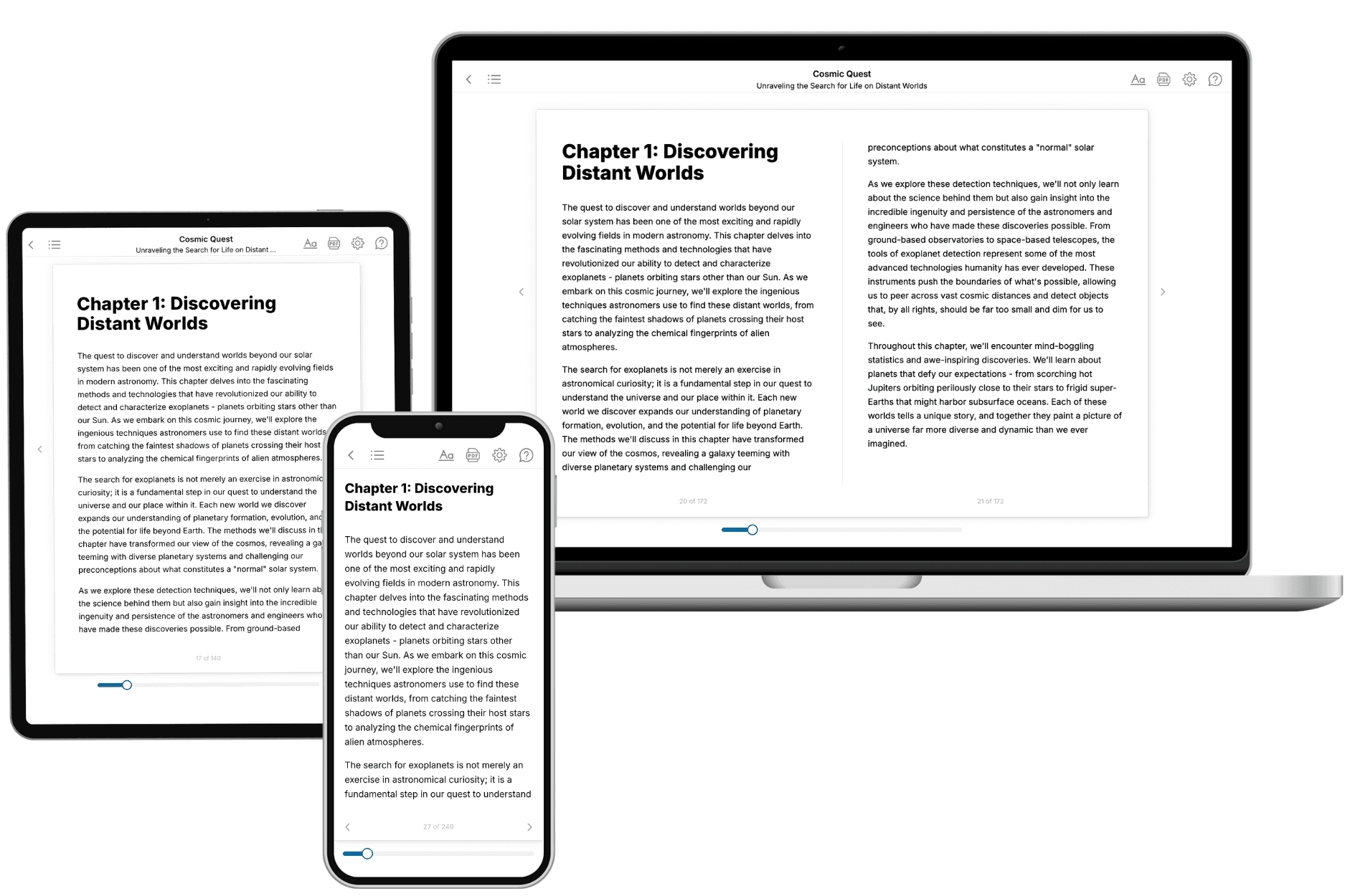
Your book is ready in 10 minutes. Read it online, download as EPUB or PDF, or send to Kindle.

Frequently asked questions
- What is TailoredRead?
TailoredRead creates complete, full-length nonfiction ebooks, written specifically for you. Tell us what you want to learn and why, and TailoredRead writes you a complete ebook on it, with charts, tables, and diagrams throughout. Read it online, on Kindle, or on any e-reader, or download it as a PDF or EPUB. Every book is written fresh the day you order it.
- How is the book tailored to me?
Before anything is written, you answer a few short questions about your goals, your background, and the topics you care about. Your book is then planned around your answers: written at your level, focused on your goals, and covering the topics you chose, instead of the one-size-fits-all book a store sells everyone.
- Will it read like generic AI content?
No. Your book isn't a chat transcript: it's planned chapter by chapter before a single word is written, told in a clear book voice, and explains ideas visually with charts, tables, and diagrams. After writing, every book goes through an editorial pass that polishes chapters so the book reads consistent from start to finish. Judge for yourself: browse thousands of sample books on our Explore Books page.
- How accurate is the content?
Our AI first checks whether your subject needs live research. Niche and fast-moving subjects are researched before writing, and the book plan and chapters are grounded in what it finds. After writing, every book goes through a quality pass that checks its facts and consistency, and researched books list their sources in a Notes section at the back.
- How long is the book?
You choose the length: Comprehensive (250-300 pages), Detailed (150-200 pages), Essential (70-100 pages), or Short (30-50 pages).
- Does the book include visuals?
Yes. Every book includes charts, tables, and diagrams: timelines, checklists, step-by-step guides, decision trees, comparison tables, matrices, and bar charts. A typical book includes dozens of these visual elements.
- Can I read it on Kindle? What formats can I download?
Yes. Every book can be read in our online reader or downloaded as a PDF or EPUB. The EPUB can be opened with Kindle, Apple Books, Kobo, and any e-reader. The Commercial and Author licenses also include an editable Word (DOCX) and Markdown download.
- How much does it cost?
About the price of a regular ebook. The exact price depends on your book's length and complexity, from $2 for the shortest books to $35 for the most comprehensive, and it's shown when you review your book, before you commit to anything. Each book is a one-time purchase, yours to keep, no subscription. Licenses that let you share, teach, or publish your book range from $35.99 to $49.99.
- Can I preview the book before purchasing?
Before you pay, you'll see what your book will cover: the title, a detailed description, the estimated length, and the complete chapter-by-chapter table of contents, so you know what you're getting before you decide. The full text itself is written after you purchase.
- How long does it take to create a book?
About 10 minutes. You can start reading the opening chapters while the rest is still being written.