The Digestive System Coloring Book
Learn Anatomy Through Interactive Illustrations and Detailed Exploration
Understanding how your digestive system works is fundamental to appreciating your body's remarkable ability to convert food into energy and nutrients.
222 pages10 chaptersMay 12, 2026Anatomy
About this book
Yet many people struggle to visualize the complex anatomy and interconnected processes involved in digestion. This coloring book bridges that gap by combining detailed, scientifically accurate illustrations with interactive coloring activities that transform passive reading into active learning. Each chapter focuses on a specific region or function of the digestive system, from the mouth through the colon, with carefully designed illustrations that highlight anatomical structures, their relationships, and their roles in the digestive process. By engaging both your visual and kinesthetic learning styles, you'll develop a deeper, more intuitive understanding of how your digestive system functions. Whether you're a student, healthcare professional, educator, or simply curious about your body, this book provides an accessible yet comprehensive exploration of one of your body's most vital systems.
Table of contents
- IntroductionWhy Study Digestive System Anatomy?, How to Use This Coloring Book, Overview of the Digestive Journey, Coloring Tips and TechniquesRead below
- Chapter 1: The Mouth and Oral CavityTeeth Structure and Types, The Tongue and Taste Buds, Salivary Glands and Saliva Production
- Chapter 2: The Esophagus and SwallowingEsophageal Anatomy and Layers, Peristalsis: The Wave-Like Motion, The Swallowing Mechanism
- Chapter 3: The StomachRegions and Chambers of the Stomach, Stomach Lining and Gastric Glands, Mechanical and Chemical Digestion
- Chapter 4: The Small IntestineDuodenum, Jejunum, and Ileum, Villi and Microvilli: Absorption Structures, Nutrient Absorption Process
- Chapter 5: The Large Intestine and ColonColon Structure and Regions, Water Reabsorption and Electrolytes, The Rectum and Anal Sphincters
- Chapter 6: The LiverLiver Lobes and Hepatic Lobules, Bile Production and Function, Hepatic Blood Supply and Circulation
- Chapter 7: The Pancreas and GallbladderPancreatic Structure and Regions, Pancreatic Enzymes and Hormones, Gallbladder Storage and Bile Release
- Chapter 8: Digestive Enzymes and Chemical BreakdownMajor Digestive Enzymes and Their Roles, Carbohydrate Breakdown, Protein and Fat Digestion
- Chapter 9: The Peritoneum and Supporting StructuresPeritoneal Membranes and Layers, Mesentery and Omentum, Organ Positioning and Support
- Chapter 10: Blood Supply and Nervous ControlArterial Supply to Digestive Organs, Venous Drainage and Portal Circulation, Nervous System Control of Digestion
- ConclusionThe Digestive System as an Integrated Whole, Continuing Your Anatomical Learning
- Glossary of Digestive Terms
- Further Reading and Resources
Introduction
Every time you eat a meal, your body launches one of its most sophisticated operations. From the moment food touches your lips to the moment waste is eliminated, a remarkable series of organs, muscles, enzymes, and nerves work in precise coordination to extract everything your body needs to survive and thrive. This process, digestion, is so automatic that most people never stop to think about the extraordinary anatomy making it possible. Yet understanding that anatomy changes everything about how you see your own body.
This coloring book was designed to make that understanding both accessible and lasting. Research consistently shows that active learning strategies, including drawing, coloring, and labeling, produce significantly stronger retention than passive reading alone.[1] When you pick up a colored pencil and shade in the layers of the stomach wall or trace the path of bile from the liver to the duodenum, you are not just filling in a picture. You are building a mental map that sticks. The combination of visual recognition and physical engagement creates memory pathways that reading alone simply cannot match.
The digestive system is also one of the most clinically relevant systems in the human body. Conditions like irritable bowel syndrome, Crohn's disease, gastroesophageal reflux, and liver disease affect hundreds of millions of people worldwide.[2] Whether you are a student preparing for an anatomy exam, a healthcare professional refreshing your foundational knowledge, an educator looking for engaging teaching tools, or simply someone curious about how your body works, a solid grasp of digestive anatomy gives you a framework for understanding health and disease in a deeply practical way. This book meets you wherever you are on that journey.
Each chapter in this book focuses on a specific region or functional component of the digestive system, moving from the mouth all the way through to the colon, and then exploring the accessory organs and supporting structures that make the whole system work. The illustrations are scientifically accurate and carefully designed to highlight the structures most important for understanding function. Labels, coloring guides, and brief explanatory text accompany every illustration so that the visual and conceptual learning reinforce each other at every step. By the time you reach the final chapter, you will have built a comprehensive, spatially grounded understanding of one of your body's most vital systems.
Why Study Digestive System Anatomy?
The digestive system does far more than process food. It is the gateway through which your body acquires every nutrient, vitamin, mineral, and calorie it needs to function. Without a properly working digestive tract, even the healthiest diet becomes useless. Nutrients that cannot be absorbed cannot fuel your cells, build your tissues, or support your immune system. This is why digestive anatomy is not just an academic subject. It is the foundation of understanding human health at its most fundamental level.
For students in medicine, nursing, dietetics, pharmacy, or any allied health field, digestive anatomy is a core competency. Clinical scenarios involving abdominal pain, malabsorption, gastrointestinal bleeding, or surgical planning all require a precise mental picture of where organs sit, how they connect, and what they do. A student who can visualize the relationship between the common bile duct, the pancreatic duct, and the duodenum will understand conditions like pancreatitis and choledocholithiasis far more intuitively than one who has only memorized definitions.
For healthcare professionals already working in clinical settings, revisiting digestive anatomy through a visual, interactive format can sharpen skills that routine practice sometimes dulls. Nurses assessing bowel sounds, radiographers interpreting abdominal scans, and dietitians counseling patients with malabsorption disorders all benefit from a refreshed, spatially accurate understanding of the system they work with every day. This book provides that refresher in a format that is engaging rather than tedious.
For educators, the coloring book format offers a ready-made teaching tool. The illustrations in this book can serve as the basis for classroom activities, lab exercises, or self-study assignments. Students who color and label anatomical structures during a lesson demonstrate better recall on subsequent assessments than those who only listen or read.[3] The act of choosing colors, deciding which structure to shade next, and writing in labels forces active engagement with the material in a way that passive note-taking does not.
Even outside formal education or healthcare, understanding your digestive system has real everyday value. When you know that the stomach produces acid to break down proteins, you understand why certain medications need to be taken with food. When you understand how the small intestine absorbs nutrients through its specialized lining, you appreciate why conditions that damage that lining, like celiac disease, have such wide-ranging effects on the body. Anatomical knowledge transforms abstract health advice into something concrete and meaningful.
Finally, studying digestive anatomy cultivates a broader appreciation for the body's design. The digestive system is a masterpiece of biological engineering, roughly nine meters of specialized tubing packed into the abdominal cavity, each segment precisely adapted for its role.[4] The more closely you examine it, the more impressive it becomes. This book invites you to look closely, and to find that the details are not just informative but genuinely fascinating.
How to Use This Coloring Book
This book is designed to be flexible. You do not need to work through it from cover to cover in a single sitting, and you do not need to follow the chapters in strict order, though doing so will give you the most coherent learning experience. Each chapter is self-contained enough that you can dip into a specific region of the digestive system whenever it is relevant to what you are studying or curious about. That said, the chapters do build on each other conceptually, so starting from the beginning is recommended if you are new to digestive anatomy.
Each chapter follows a consistent structure designed to support layered learning. Here is what you will find in every chapter:
- Introductory text: A brief overview of the region or topic covered, placing it in the context of the broader digestive system.
- Anatomical illustrations: Detailed, scientifically accurate drawings with numbered or lettered labels pointing to key structures.
- Coloring guide: Suggested color assignments for each labeled structure, helping you distinguish related structures visually.
- Explanatory subchapters: Written sections that explain the function and clinical relevance of the structures you have just colored.
- Review questions: Short prompts at the end of each chapter to test your recall and reinforce what you have learned.
The coloring guides suggest specific colors for specific structures, but these are recommendations, not rules. The goal is to create visual contrast between adjacent structures so that your brain can distinguish them clearly. If you prefer a different color scheme, use it. What matters is consistency within your own system. For example, if you color all smooth muscle layers in blue throughout the book, your brain will begin to recognize that color as a cue for smooth muscle wherever it appears, which strengthens pattern recognition across chapters.
You will get the most out of this book if you read the explanatory text before you color, not after. Reading first gives you a conceptual framework so that when you pick up your pencil, you are not just filling in shapes. You are reinforcing specific knowledge about what each structure does and why it matters. After coloring, re-read the text and see how much more clearly the words connect to the images in your mind. This read-color-review cycle is the core learning loop the book is built around.
If you are using this book as part of a course or study group, consider comparing your completed illustrations with those of classmates. Discussing why you chose certain colors, or explaining a structure's function to someone else, deepens understanding significantly. Teaching is one of the most powerful forms of learning, and even informal explanation to a study partner activates the same cognitive processes.
For those using this book as a professional reference or refresher, the glossary at the back provides quick definitions of all key terms used throughout the text. The further reading section points you toward peer-reviewed resources and textbooks for deeper exploration of any topic that sparks your interest. Think of this book as a starting point and a visual anchor, not a ceiling on how far your learning can go.
Finally, do not rush. Anatomy rewards patience. Spending twenty focused minutes on a single illustration, really examining the spatial relationships between structures, will serve you better than quickly coloring through an entire chapter. The goal is not to finish the book. The goal is to genuinely understand what you are looking at.
Overview of the Digestive Journey
Before diving into the detailed anatomy of each region, it helps to have a clear picture of the digestive journey as a whole. Digestion is a one-way trip. Food enters at the mouth and travels through a continuous tube, the gastrointestinal (GI) tract, until waste exits at the anus. Along the way, that food is physically broken apart, chemically dissolved, selectively absorbed, and finally compacted for elimination. Each stage of this journey happens in a specific location, performed by specific structures, using specific tools.
The journey begins in the oral cavity, where teeth mechanically grind food into smaller pieces while saliva begins chemical digestion. Salivary amylase, an enzyme in saliva, starts breaking down carbohydrates even before you swallow. The tongue shapes the chewed food into a ball called a bolus and pushes it toward the throat. This first stage is brief but essential. The smaller and more thoroughly mixed the food is when it leaves the mouth, the more efficiently the rest of the digestive system can process it.
From the mouth, the bolus travels down the esophagus, a muscular tube roughly 25 centimeters long that connects the throat to the stomach. The esophagus does not digest food. Its job is transport, accomplished through coordinated muscle contractions called peristalsis. A ring of muscle at the bottom of the esophagus, the lower esophageal sphincter, relaxes to let food into the stomach and then closes again to prevent stomach acid from flowing back up.
The stomach is where serious mechanical and chemical digestion begins. Powerful muscular walls churn food into a semi-liquid mixture called chyme, while gastric glands in the stomach lining secrete hydrochloric acid and enzymes like pepsin that begin breaking down proteins. The stomach can hold up to about 1.5 liters of food and typically empties into the small intestine over a period of two to four hours, depending on the composition of the meal.[5]
The small intestine is where the majority of digestion and virtually all nutrient absorption takes place. Stretching approximately six to seven meters in length, it is divided into three segments: the duodenum, the jejunum, and the ileum. The duodenum receives chyme from the stomach along with digestive secretions from the pancreas and bile from the liver via the gallbladder. These secretions neutralize the acid in chyme and provide the enzymes needed to break down carbohydrates, proteins, and fats into their smallest absorbable components. The inner lining of the small intestine is covered in tiny finger-like projections called villi, which are themselves covered in even tinier projections called microvilli, creating an enormous surface area for absorption.
What the small intestine does not absorb passes into the large intestine, also called the colon. The colon's primary job is to reabsorb water and electrolytes from the remaining material, compacting it into solid waste. The colon also hosts trillions of bacteria, collectively called the gut microbiome, which ferment undigested fibers and play important roles in immune function and overall health.[6] Waste is stored in the rectum and eliminated through the anus under voluntary and involuntary muscular control.
Supporting this entire journey are three major accessory organs that sit outside the GI tract but contribute essential secretions to it. The liver produces bile, a fluid that emulsifies dietary fats so they can be digested and absorbed. The gallbladder stores and concentrates bile between meals, releasing it into the duodenum when fatty food arrives. The pancreas produces a powerful cocktail of digestive enzymes as well as hormones like insulin and glucagon that regulate blood sugar. Without these three organs, digestion would be profoundly impaired even if the GI tract itself were perfectly healthy.
Coloring Tips and Techniques
You do not need to be an artist to get full value from this coloring book. The illustrations are designed to be colored by anyone, regardless of artistic skill. What matters is not how beautiful your finished pages look but how much attention you pay to each structure as you color it. That focused attention is where the learning happens. A few practical tips, however, will help you get cleaner results and make the experience more enjoyable.
Choosing your coloring tools is the first decision. Each medium has trade-offs:
- Colored pencils: The most recommended option for anatomy coloring books. They offer precise control, blend well, and do not bleed through pages. They are ideal for fine details like the layers of the intestinal wall or the branching ducts of the liver.
- Felt-tip markers: Produce vivid, saturated color but can bleed through thin paper. If you use markers, place a blank sheet behind the page you are working on to protect the illustration underneath.
- Gel pens: Good for adding fine detail or labeling, but less practical for filling large areas. They work well as a complement to colored pencils.
- Watercolor pencils: An advanced option that allows you to blend colors smoothly with a damp brush, creating a more realistic tissue appearance. Best suited for those with some experience using them.
When it comes to color selection, the most important principle is contrast. Adjacent structures should be colored in clearly different hues so that boundaries are visually obvious. A common mistake is choosing colors that are too similar, such as light blue and light purple for neighboring structures, which makes the illustration harder to read at a glance. Aim for high contrast between structures that are anatomically distinct, and use related shades (such as light red and dark red) only for structures that are functionally related and you want to visually group together.
A practical color-coding system used by many anatomy students assigns colors by tissue type rather than by individual structure. For example:
- Red or pink: Arteries and highly vascularized tissues
- Blue or purple: Veins and venous structures
- Yellow or gold: Nerves and neural tissue
- Green: Lymphatic vessels and bile ducts
- Orange: Glandular tissue and secretory structures
- Brown or tan: Connective tissue and supporting structures
Using this system consistently across all chapters means that by the time you reach the later chapters, color itself becomes a learning cue. When you see a green structure in an illustration, your brain automatically begins thinking about secretory or lymphatic function before you have even read the label. This kind of automatic association is a sign that the learning is becoming deeply embedded.
For technique, work from light to dark. Start with a light layer of color and build up intensity gradually. This gives you more control and makes it easier to correct mistakes. Color in smooth, even strokes that follow the shape of the structure you are filling in. For tubular structures like the esophagus or intestines, stroke along the length of the tube. For rounded structures like the stomach or gallbladder, use circular strokes that follow the organ's contour. These small technique choices make the finished illustration cleaner and more informative.
Pay special attention to boundaries and borders. The edges where one structure meets another are often the most anatomically significant areas in an illustration. Take your time at these boundaries, using a sharp pencil tip or a fine-tipped marker to define the edge clearly before filling in the body of the structure. This is especially important in cross-sectional illustrations, where the layered structure of the GI wall is shown in detail. Each layer, mucosa, submucosa, muscularis, and serosa, should be clearly distinguishable from the others.
Finally, label as you go. Each illustration includes numbered or lettered labels pointing to specific structures. After coloring a structure, write its name in the corresponding label line immediately, while the structure is fresh in your mind. Do not wait until you have finished coloring the entire illustration. Writing the name reinforces the connection between the visual image and the anatomical term, which is exactly the kind of dual-coding that makes information stick.[7] If you are unsure of a name, look it up in the text or the glossary before writing it down. Guessing and getting it wrong can create a false memory that is harder to correct later.
With these tools and techniques in hand, you are ready to begin. The chapters ahead will take you on a detailed, illustrated tour of one of the body's most complex and fascinating systems. Approach each page with curiosity, take your time with the details, and trust that the act of coloring itself is doing important cognitive work. The understanding you build here will serve you for years to come.
Notes
Introduction
- 1. J. D. Wammes and others, "The drawing effect," Quarterly Journal of Experimental Psychology, 2016
- 2. G. B. D. Collaborators, "The global, regional, and national burden of inflammatory bowel disease in 195 countries and territories, 1990-2017," The Lancet Gastroenterology & Hepatology, 2017
- 3. W. H. Cliff and A. W. Wright, "Directed case study method for teaching human anatomy and physiology," Advances in Physiology EducationS19-S28, 1996
- 4. G. J. Tortora and B. Derrickson, Principles of Anatomy and Physiology (15th ed.), Wiley, 2017
- 5. J. E. Hall, Guyton and Hall Textbook of Medical Physiology (13th ed.), Elsevier, 2016
- 6. R. Sender and others, "Revised estimates for the number of human and bacteria cells in the body," Cell, 2016
- 7. A. Paivio, "Dual coding theory," Canadian Journal of Psychology, 1991
This is the end of the free sample. The full book has 10 chapters.
Want this book written for you?
Tell us your goals and background, and get your own version of this book, tailored by AI to you.


Benefits of AI-tailored books
- Read one book, not ten:
- all the Anatomy knowledge you need, gathered into a single book.
- Save days of learning:
- tell us your goals, choose your topics, and the whole book is built around them.
- Learn effortlessly:
- Anatomy book written for your specific background and expertise.
- Understand at a glance:
- includes charts, tables, timelines, checklists, decision trees, and more.
- Stay current:
- written the day you order it, not years ago.
- Trust what you read:
- every book is checked for accuracy, with real research when your subject needs it.
Create your unique book in 3 steps
- 1. Select your focus
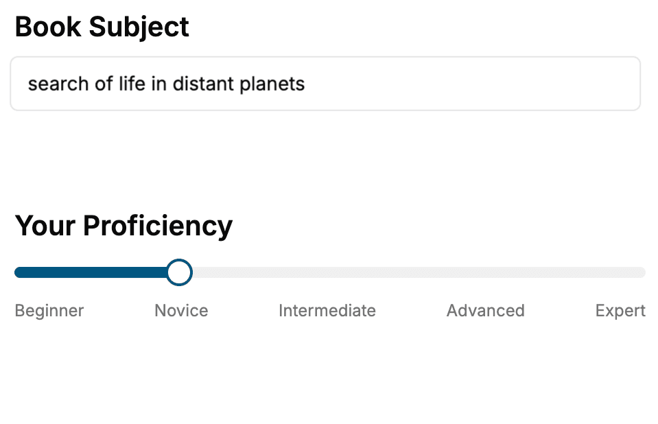
Select the focus of your Anatomy book and share your background
- 2. Personalize your book
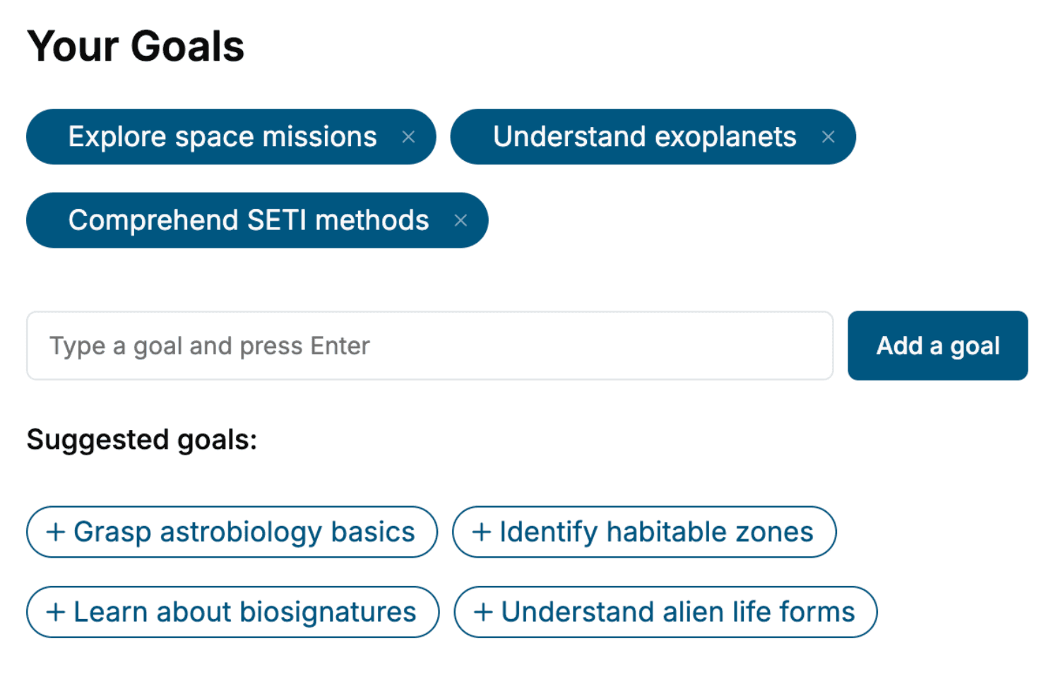
Specify your goals and choose sub-topics to include
- 3. Get your tailored book
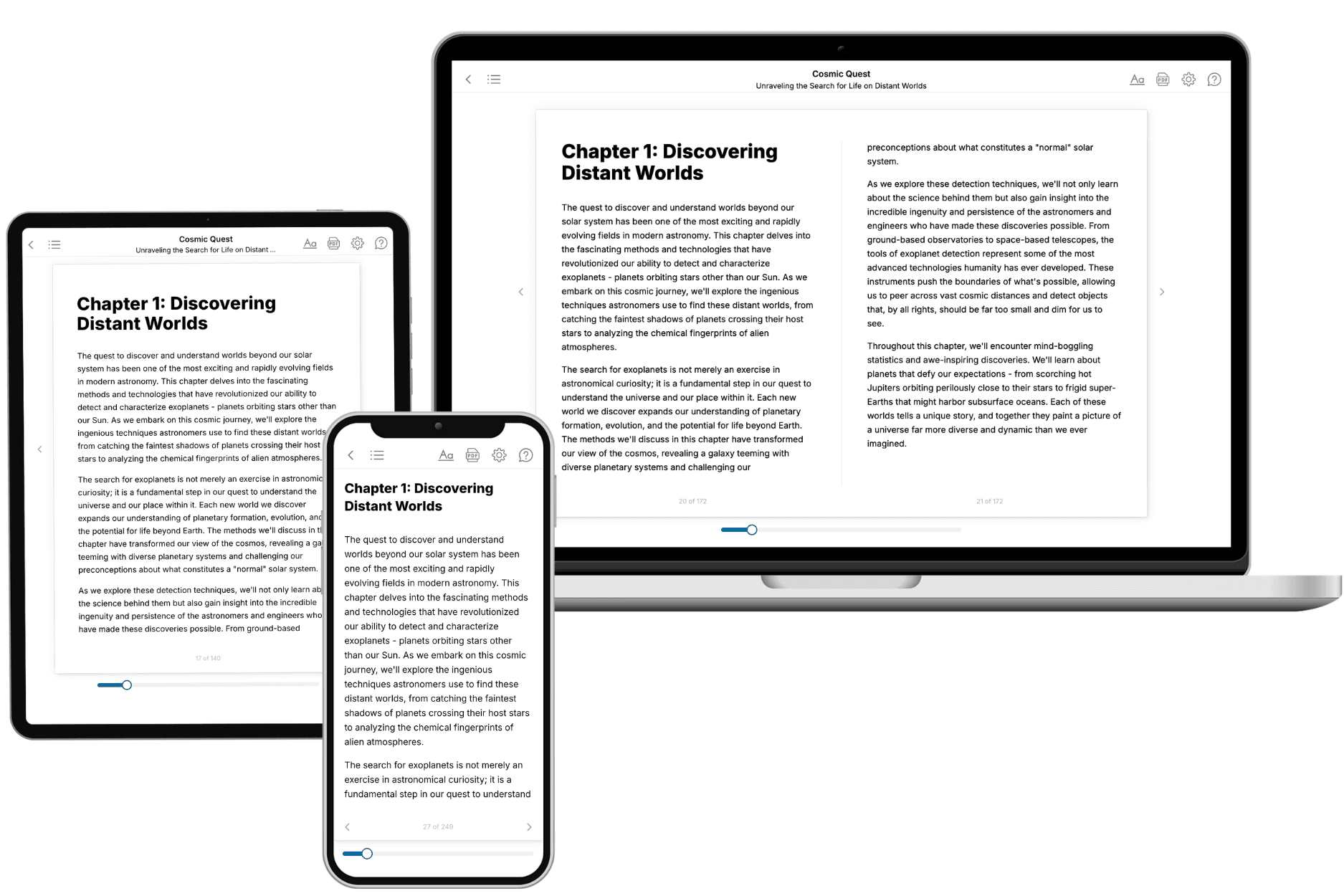
Your book is ready in 10 minutes. Read it online, download as EPUB or PDF, or send to Kindle.
Frequently asked questions
- What is TailoredRead?
TailoredRead creates complete, full-length nonfiction ebooks, written specifically for you. Tell us what you want to learn and why, and TailoredRead writes you a complete ebook on it, with charts, tables, and diagrams throughout. Read it online, on Kindle, or on any e-reader, or download it as a PDF or EPUB. Every book is written fresh the day you order it.
- How is the book tailored to me?
Before anything is written, you answer a few short questions about your goals, your background, and the topics you care about. Your book is then planned around your answers: written at your level, focused on your goals, and covering the topics you chose, instead of the one-size-fits-all book a store sells everyone.
- Will it read like generic AI content?
No. Your book isn't a chat transcript: it's planned chapter by chapter before a single word is written, told in a clear book voice, and explains ideas visually with charts, tables, and diagrams. After writing, every book goes through an editorial pass that polishes chapters so the book reads consistent from start to finish. Judge for yourself: browse thousands of sample books on our Explore Books page.
- How accurate is the content?
Our AI first checks whether your subject needs live research. Niche and fast-moving subjects are researched before writing, and the book plan and chapters are grounded in what it finds. After writing, every book goes through a quality pass that checks its facts and consistency, and researched books list their sources in a Notes section at the back.
- How long is the book?
You choose the length: Comprehensive (250-300 pages), Detailed (150-200 pages), Essential (70-100 pages), or Short (30-50 pages).
- Does the book include visuals?
Yes. Every book includes charts, tables, and diagrams: timelines, checklists, step-by-step guides, decision trees, comparison tables, matrices, and bar charts. A typical book includes dozens of these visual elements.
- Can I read it on Kindle? What formats can I download?
Yes. Every book can be read in our online reader or downloaded as a PDF or EPUB. The EPUB can be opened with Kindle, Apple Books, Kobo, and any e-reader. The Commercial and Author licenses also include an editable Word (DOCX) and Markdown download.
- How much does it cost?
About the price of a regular ebook. The exact price depends on your book's length and complexity, from $2 for the shortest books to $35 for the most comprehensive, and it's shown when you review your book, before you commit to anything. Each book is a one-time purchase, yours to keep, no subscription. Licenses that let you share, teach, or publish your book range from $35.99 to $49.99.
- Can I preview the book before purchasing?
Before you pay, you'll see what your book will cover: the title, a detailed description, the estimated length, and the complete chapter-by-chapter table of contents, so you know what you're getting before you decide. The full text itself is written after you purchase.
- How long does it take to create a book?
About 10 minutes. You can start reading the opening chapters while the rest is still being written.