The Electric Heart
Understanding the Bioelectrical Mechanisms That Power Your Heartbeat
The complexity of cardiac electrical activity often seems overwhelming, even to those with solid biology backgrounds.
300 pages15 chaptersSep 28, 2025Cardiology
About this book
Every second of every day, your heart generates and conducts electrical signals with remarkable precision, yet the intricate mechanisms behind this process remain mysterious to many. This comprehensive guide demystifies the cardiac conduction system by breaking down complex bioelectrical concepts into clear, understandable principles. You'll discover how specialized cardiac cells generate electrical impulses, how these signals travel through distinct pathways, and how the entire system coordinates to produce the rhythmic contractions that sustain life. From the molecular level of ion channels and membrane potentials to the organ-level coordination of the complete conduction pathway, you'll gain deep insights into one of biology's most elegant systems. The book explores the unique properties of cardiac action potentials, the role of calcium in excitation-contraction coupling, and how the autonomic nervous system modulates heart rhythm. Whether you're building on existing biology knowledge or preparing for advanced study, this book provides the thorough understanding you need to appreciate the remarkable bioelectrical symphony that keeps your heart beating.
Table of contents
- IntroductionThe Heart as an Electrical Organ, Bioelectricity in Living Systems, What Makes Cardiac Conduction Special, How This Book Will Guide Your LearningRead below
- Chapter 1: Cellular Foundations of Cardiac BioelectricityTypes of Cardiac Cells, Cell Membrane Structure and Function, Establishing the Resting Membrane Potential, Ion Gradients and Electrochemical Forces
- Chapter 2: Ion Channels and Membrane TransportVoltage-Gated Ion Channels, Sodium Channels and Fast Depolarization, Calcium Channels and Slow Conduction, Potassium Channels and Repolarization, Channel Kinetics and Gating Mechanisms
- Chapter 3: Cardiac Action PotentialsThe Five Phases of Cardiac Action Potentials, Ventricular Myocyte Action Potentials, Atrial Myocyte Action Potentials, Nodal Cell Action Potentials, Refractory Periods and Excitability
- Chapter 4: Pacemaker Cells and AutomaticityProperties of Pacemaker Cells, The Funny Current and Spontaneous Depolarization, The Calcium Clock Mechanism, Hierarchy of Cardiac Pacemakers
- Chapter 5: The Sinoatrial NodeAnatomical Structure of the SA Node, Electrophysiology of SA Node Cells, Impulse Generation and Rate Control, Exit Pathways from the SA Node
- Chapter 6: Atrial Conduction PathwaysAtrial Muscle Architecture, Internodal Conduction Pathways, Interatrial Conduction and Bachmann's Bundle, Sequence of Atrial Activation
- Chapter 7: The Atrioventricular NodeStructure and Location of the AV Node, AV Conduction Delay and Its Purpose, Electrophysiological Properties, Modulation of AV Conduction
- Chapter 8: The His-Purkinje SystemThe Bundle of His, Left and Right Bundle Branches, Purkinje Fiber Network, Mechanisms of Rapid Conduction, Ventricular Activation Sequence
- Chapter 9: Excitation-Contraction CouplingCalcium-Induced Calcium Release, Role of the Sarcoplasmic Reticulum, Contractile Protein Interactions, Calcium Cycling and Relaxation
- Chapter 10: Gap Junctions and Cell-to-Cell CommunicationStructure of Gap Junctions, Connexin Proteins and Channel Formation, Electrical Coupling Between Cells, Factors Affecting Conduction Velocity
- Chapter 11: Autonomic Control of Heart RhythmSympathetic Nervous System Effects, Parasympathetic Nervous System Effects, Neurotransmitter Mechanisms, Receptor Signaling Pathways
- Chapter 12: Hormonal Modulation of Cardiac ConductionCatecholamines and Beta-Adrenergic Signaling, Thyroid Hormone Effects, Other Hormonal Influences, Circadian Rhythms and Heart Rate
- Chapter 13: Understanding Conduction AbnormalitiesTypes of Conduction Blocks, Reentry and Circular Conduction, Ectopic Pacemakers and Abnormal Automaticity, Abnormal Conduction Patterns
- Chapter 14: Measuring Cardiac Electrical ActivityElectrocardiography Principles, Intracardiac Electrogram Recordings, Optical Mapping Techniques, Patch Clamp Electrophysiology
- Chapter 15: Computational Models of Cardiac ConductionSingle Cell Mathematical Models, Tissue and Organ-Level Models, Applications of Cardiac Simulations, Model Validation and Limitations
- Conclusion
- Glossary
- Further Reading
Introduction
Every moment of your life depends on one of nature's most remarkable electrical systems. Your heart generates and conducts electrical signals with extraordinary precision, creating the rhythmic contractions that pump blood throughout your body. This bioelectrical symphony operates continuously, coordinating millions of cardiac cells to work in perfect harmony. Yet despite its fundamental importance, the intricate mechanisms behind cardiac conduction remain poorly understood by many who study biology and physiology.
The complexity of cardiac bioelectricity extends far beyond simple electrical impulses traveling through heart muscle. This system involves specialized cells that generate spontaneous electrical activity, ion channels that precisely control the flow of charged particles across cell membranes, and sophisticated conduction pathways that ensure coordinated activation of the entire heart. Understanding these mechanisms requires grasping concepts that span from molecular-level interactions to organ-level coordination, making cardiac electrophysiology one of the most challenging yet fascinating areas of biological study.
This book bridges the gap between foundational biology knowledge and the advanced understanding needed to truly comprehend how your heart's electrical system functions. Rather than overwhelming you with dense technical details, it builds systematically from basic principles to complex mechanisms, providing the depth of understanding necessary to appreciate this remarkable biological system. You will discover how the heart's electrical activity differs fundamentally from other excitable tissues, why certain cells can generate spontaneous rhythms, and how the entire conduction system coordinates to produce the heartbeat that sustains life.
The journey through cardiac bioelectricity reveals insights that extend beyond academic knowledge into practical understanding of how disruptions in these electrical processes can lead to rhythm abnormalities and conduction disorders. By mastering these concepts, you gain the foundation needed for advanced study in cardiology, electrophysiology, or biomedical engineering, while developing an appreciation for one of biology's most elegant and essential systems.
The Heart as an Electrical Organ
The heart functions as both a mechanical pump and a sophisticated electrical organ, with its pumping action entirely dependent on precisely timed electrical signals. Unlike skeletal muscle, which requires external nerve stimulation to contract, the heart generates its own electrical impulses through specialized pacemaker cells. This intrinsic electrical activity allows the heart to beat independently of the nervous system, though neural and hormonal influences can modulate its rhythm. The electrical system of the heart represents a remarkable example of biological automation, where cellular mechanisms have evolved to create self-sustaining, rhythmic activity that can persist for an entire lifetime.
The heart's electrical activity originates from the movement of ions across cell membranes, creating electrical currents that spread throughout the cardiac tissue. These ionic currents are generated by specialized proteins called ion channels, which selectively allow specific ions to cross the cell membrane under particular conditions. The coordinated opening and closing of different types of ion channels creates the characteristic electrical signals known as action potentials. These action potentials serve as the fundamental units of electrical communication in the heart, carrying information from cell to cell and coordinating the timing of cardiac contractions.
What makes the heart unique among electrical organs is its ability to maintain rhythmic activity through multiple backup systems and regulatory mechanisms. The primary pacemaker, located in the sinoatrial node, normally controls heart rhythm, but secondary pacemakers throughout the conduction system can take over if the primary system fails. This redundancy ensures that electrical activity continues even when parts of the system are damaged or disrupted. Additionally, the heart's electrical system can adapt to changing physiological demands through various regulatory mechanisms that adjust heart rate, conduction velocity, and contractile strength.
The electrical and mechanical functions of the heart are intimately linked through a process called excitation-contraction coupling. When an electrical signal reaches a cardiac muscle cell, it triggers the release of calcium ions from internal storage sites. These calcium ions then interact with contractile proteins, causing the cell to shorten and generate force. This coupling between electrical excitation and mechanical contraction ensures that electrical signals are efficiently converted into the pumping action needed to circulate blood. The timing and coordination of this process across millions of cardiac cells determines the effectiveness of each heartbeat.
Understanding the heart as an electrical organ requires appreciating both its autonomous nature and its responsiveness to external influences. While the heart can beat independently, its electrical activity is constantly modulated by the autonomic nervous system, circulating hormones, and local metabolic factors. The sympathetic nervous system can increase heart rate and contractility during stress or exercise, while the parasympathetic system slows the heart during rest and recovery. This dynamic regulation allows the heart to adjust its electrical and mechanical performance to meet the body's changing circulatory demands.
The study of cardiac bioelectricity has revealed fundamental principles that apply to other excitable tissues while highlighting the unique adaptations that make cardiac conduction special. The heart's electrical system demonstrates how evolution has optimized biological processes for reliability, efficiency, and adaptability. By examining the heart as an electrical organ, we gain insights into broader principles of bioelectricity while appreciating the specific mechanisms that make cardiac function possible. This perspective forms the foundation for understanding both normal cardiac physiology and the electrical disturbances that can lead to rhythm disorders.
Modern research continues to uncover new aspects of cardiac electrical function, from the molecular details of ion channel structure to the complex patterns of electrical propagation in diseased hearts. Advanced techniques such as optical mapping, patch-clamp electrophysiology, and computational modeling have revolutionized our understanding of cardiac bioelectricity. These tools allow researchers to study electrical activity at multiple scales, from single ion channels to whole hearts, providing unprecedented insights into the mechanisms that control cardiac rhythm and conduction.
The clinical relevance of understanding the heart as an electrical organ cannot be overstated. Electrical disturbances are responsible for many forms of heart disease, from simple rhythm irregularities to life-threatening arrhythmias. Treatments for these conditions, including pacemakers, defibrillators, and antiarrhythmic drugs, all work by modifying the heart's electrical activity. As our understanding of cardiac bioelectricity continues to advance, new therapeutic approaches are being developed that target specific aspects of the electrical system, offering hope for more effective treatments of cardiac electrical disorders.
Bioelectricity in Living Systems
Bioelectricity represents one of the fundamental properties of living cells, arising from the separation of electrical charges across cell membranes. All living cells maintain an electrical potential difference between their interior and exterior, created by the unequal distribution of ions on either side of the cell membrane. This electrical potential, typically ranging from -50 to -90 millivolts in most cells, provides the energy source for many cellular processes and serves as the basis for electrical signaling in excitable tissues. The generation and maintenance of bioelectrical potentials require sophisticated cellular machinery, including ion pumps, channels, and transporters that precisely control the movement of charged particles.
The origins of bioelectricity can be traced to the selective permeability of cell membranes and the active transport of ions against their concentration gradients. Cell membranes are composed of lipid bilayers that are naturally impermeable to most ions, but specialized proteins embedded in these membranes create pathways for controlled ion movement. Ion pumps, such as the sodium-potassium ATPase, use cellular energy to maintain concentration gradients by actively transporting ions against their electrochemical gradients. These pumps create the conditions necessary for bioelectrical activity by establishing and maintaining the ionic imbalances that generate membrane potentials.
Different types of cells have evolved specialized mechanisms for using bioelectricity to perform specific functions. Nerve cells use rapid changes in membrane potential to transmit information over long distances, while muscle cells use electrical signals to trigger contraction. Sensory cells convert various forms of energy into electrical signals that can be processed by the nervous system. Each cell type has developed unique combinations of ion channels and transport mechanisms that allow them to generate characteristic electrical patterns suited to their particular functions. This diversity in bioelectrical mechanisms reflects the versatility of electrical signaling in biological systems.
The heart represents a particularly sophisticated example of bioelectrical organization, where multiple cell types work together to create coordinated electrical activity. Cardiac pacemaker cells generate spontaneous electrical impulses, while cardiac muscle cells respond to these signals with characteristic action potentials that trigger contraction. Specialized conduction cells rapidly propagate electrical signals throughout the heart, ensuring synchronized activation of the entire organ. This multi-cellular electrical system demonstrates how bioelectricity can be organized at the tissue and organ level to perform complex physiological functions.
Understanding bioelectricity requires grasping several key concepts that apply across different biological systems. The concept of equilibrium potential describes the electrical potential at which there is no net movement of a particular ion across the membrane. The Goldman-Hodgkin-Katz equation quantifies how the permeabilities and concentrations of different ions contribute to the overall membrane potential. These mathematical relationships provide the theoretical framework for understanding how changes in ion concentrations or membrane permeabilities affect cellular electrical activity. Mastering these concepts is essential for understanding more complex bioelectrical phenomena.
The measurement and study of bioelectricity have been crucial for advancing our understanding of cellular and organ function. Techniques such as intracellular recording, patch-clamp electrophysiology, and extracellular field potential recording allow researchers to monitor electrical activity at different scales and in various experimental conditions. These methods have revealed the detailed mechanisms underlying bioelectrical phenomena and have enabled the development of mathematical models that can predict electrical behavior under different conditions. The ability to measure and manipulate bioelectrical activity has also led to numerous clinical applications, from diagnostic techniques to therapeutic interventions.
Bioelectricity plays essential roles in many physiological processes beyond nerve and muscle function. Electrical fields influence cell division, wound healing, and tissue development. Some organisms use bioelectricity for navigation, communication, or prey detection. Plants generate electrical signals in response to environmental stimuli and use these signals to coordinate responses across different parts of the organism. This widespread occurrence of bioelectrical phenomena highlights the fundamental importance of electrical signaling in living systems and suggests that understanding bioelectricity is crucial for comprehending life processes at all levels of organization.
The study of bioelectricity continues to reveal new insights into cellular function and has opened up exciting possibilities for biomedical applications. Bioelectrical engineering approaches are being developed to create artificial tissues, improve prosthetic devices, and develop new therapeutic strategies. Understanding how cells generate and respond to electrical signals is leading to advances in regenerative medicine, where electrical stimulation is used to promote tissue repair and regeneration. As our knowledge of bioelectrical mechanisms continues to expand, we can expect new discoveries that will further illuminate the role of electricity in living systems and lead to innovative applications in medicine and biotechnology.
What Makes Cardiac Conduction Special
Cardiac conduction possesses several unique characteristics that distinguish it from electrical signaling in other excitable tissues, particularly nerve cells. The most striking difference lies in the duration of cardiac action potentials, which last 200-400 milliseconds compared to just 1-2 milliseconds for nerve action potentials. This extended duration is crucial for cardiac function because it allows sufficient time for calcium to enter the cell and trigger contraction, while also preventing the heart from going into tetanic contractions that would be incompatible with its pumping function. The prolonged action potential is achieved through a complex interplay of multiple ion channel types that open and close at different times during the electrical cycle.
Another distinctive feature of cardiac conduction is the presence of automaticity in specialized pacemaker cells. Unlike nerve or skeletal muscle cells, which require external stimulation to generate action potentials, cardiac pacemaker cells spontaneously depolarize and generate rhythmic electrical impulses. This automaticity results from unique ionic mechanisms, including the "funny current" and specialized calcium handling systems that create unstable resting potentials. The sinoatrial node contains the fastest pacemaker cells and normally controls heart rhythm, but multiple backup pacemaker sites throughout the conduction system can take over if needed, providing remarkable redundancy for this vital function.
The cardiac conduction system demonstrates a highly organized anatomical structure that has no equivalent in other tissues. This system includes specialized pathways such as the internodal tracts, atrioventricular node, bundle of His, bundle branches, and Purkinje fiber network. Each component has distinct electrophysiological properties optimized for its specific role in the conduction sequence. For example, the AV node conducts slowly to allow time for atrial emptying, while Purkinje fibers conduct rapidly to ensure synchronized ventricular activation. This anatomical specialization creates a precisely timed sequence of electrical activation that optimizes cardiac pumping efficiency.
Cardiac cells exhibit unique refractory period characteristics that are essential for normal heart function. The effective refractory period in cardiac cells is much longer than in nerve cells and extends well into the mechanical contraction phase. This prolonged refractoriness prevents the heart from being restimulated during contraction, which would interfere with its pumping action. Additionally, cardiac cells have a relative refractory period during which they can be stimulated but with altered conduction properties. These refractory characteristics protect the heart from developing rapid, chaotic rhythms while allowing for appropriate rate increases during physiological stress.
The coupling between electrical excitation and mechanical contraction in cardiac cells involves sophisticated calcium handling mechanisms that are more complex than in other muscle types. Cardiac excitation-contraction coupling relies on calcium-induced calcium release, where a small amount of calcium entering through membrane channels triggers the release of much larger amounts from internal stores. This amplification mechanism allows relatively small electrical signals to produce powerful contractions. The precise control of calcium cycling is crucial for both the strength of contraction and the timing of relaxation, making calcium handling a critical component of cardiac electrical function.
Gap junctions play a particularly important role in cardiac conduction, creating electrical connections between adjacent cells that allow action potentials to spread throughout the heart. Cardiac gap junctions are composed of specific connexin proteins that create low-resistance pathways for current flow between cells. The distribution and properties of these gap junctions vary in different regions of the heart, contributing to the directional properties of conduction and the formation of specialized conduction pathways. This intercellular coupling is essential for coordinated cardiac activation and distinguishes cardiac conduction from the point-to-point signaling typical of nerve cells.
The modulation of cardiac conduction by the autonomic nervous system represents another unique aspect of cardiac electrical function. While many tissues are influenced by neural input, the heart's electrical system is continuously and dynamically regulated by both sympathetic and parasympathetic influences. These neural inputs can modify virtually every aspect of cardiac electrical activity, including pacemaker rate, conduction velocity, and action potential duration. The molecular mechanisms underlying this modulation involve complex signaling cascades that ultimately affect ion channel function and calcium handling. This neural control allows the heart to rapidly adjust its electrical and mechanical performance to meet changing physiological demands.
Perhaps most remarkably, the cardiac conduction system demonstrates an extraordinary degree of reliability and adaptability that reflects millions of years of evolutionary optimization. The heart must beat continuously throughout life, requiring electrical mechanisms that are both robust and flexible. The system includes multiple fail-safe mechanisms, from backup pacemakers to alternative conduction pathways, that help ensure continued function even when parts of the system are damaged. At the same time, the system can adapt to changing conditions through both acute modulation and long-term remodeling processes. This combination of reliability and adaptability makes cardiac conduction one of the most sophisticated bioelectrical systems in nature, worthy of detailed study and deep appreciation.
How This Book Will Guide Your Learning
This book employs a systematic, building-block approach that progresses logically from fundamental cellular mechanisms to complex organ-level phenomena. Rather than overwhelming you with advanced concepts from the start, each chapter builds upon knowledge established in previous sections, creating a solid foundation for understanding increasingly sophisticated topics. The early chapters focus on cellular and molecular mechanisms, including membrane potentials, ion channels, and action potentials, providing the essential groundwork needed to comprehend more complex conduction phenomena. This methodical progression ensures that you develop a thorough understanding of basic principles before tackling advanced topics such as pacemaker mechanisms and conduction system anatomy.
Each chapter incorporates multiple learning strategies to accommodate different learning styles and reinforce key concepts through various approaches. Detailed explanations of mechanisms are accompanied by specific examples that illustrate how these processes work in real cardiac cells and tissues. Mathematical relationships are presented when necessary to quantify important phenomena, but always with clear explanations of their biological significance. Visual descriptions help you understand spatial relationships and temporal sequences, while analogies connect unfamiliar cardiac processes to more familiar concepts from other areas of biology and physics. This multi-faceted approach ensures that complex concepts are accessible and memorable.
The book emphasizes the integration of structure and function throughout the cardiac conduction system, demonstrating how anatomical specializations enable specific physiological roles. You will learn not just what happens in different parts of the conduction system, but why these regions have evolved particular characteristics and how their properties contribute to overall cardiac function. For example, when studying the AV node, you will understand both the cellular mechanisms that create slow conduction and the physiological importance of this delay for cardiac pumping efficiency. This structure-function integration provides a deeper understanding that goes beyond memorization of facts to genuine comprehension of biological design principles.
Practical applications and clinical relevance are woven throughout the text to demonstrate the importance of understanding cardiac bioelectricity. While avoiding specific medical advice, the book explains how disruptions in normal electrical mechanisms can lead to various types of conduction abnormalities and rhythm disorders. You will learn to recognize the electrical basis of common cardiac problems and understand how various treatments work by modifying specific aspects of the conduction system. This clinical context helps motivate learning by showing how theoretical knowledge translates into practical understanding of cardiac health and disease.
The book includes extensive use of examples and case studies that bring abstract concepts to life through concrete illustrations. These examples range from detailed descriptions of specific experimental findings to hypothetical scenarios that demonstrate key principles. For instance, when explaining action potential propagation, you will follow the electrical signal as it travels from the SA node through the atria, experiences delay at the AV node, and then rapidly spreads through the ventricular conduction system. These narrative examples help you visualize complex processes and understand the temporal and spatial aspects of cardiac conduction that are crucial for comprehensive understanding.
Each major section includes practical exercises and thought experiments designed to test and reinforce your understanding of key concepts. These activities encourage active learning by requiring you to apply newly acquired knowledge to solve problems or predict outcomes in different scenarios. You might be asked to predict how changes in ion channel function would affect action potential shape, or to trace the consequences of conduction blocks in different parts of the system. These exercises help identify areas where additional study is needed and build confidence in your ability to apply cardiac electrophysiology principles to new situations.
The book's organization allows for both sequential reading and targeted study of specific topics, depending on your learning goals and background knowledge. Cross-references throughout the text help you navigate between related concepts and build connections across different chapters. A comprehensive glossary provides quick access to definitions of technical terms, while detailed figures and diagrams support visual learning. The appendices include additional resources for those who want to delve deeper into specific aspects of cardiac bioelectricity, including references to key research papers and suggestions for further study.
Throughout your journey through this book, you will develop not just knowledge of cardiac conduction, but also the analytical skills needed to understand and evaluate new developments in this rapidly advancing field. The emphasis on fundamental principles and mechanistic understanding provides a solid foundation that will remain relevant even as new discoveries expand our knowledge of cardiac bioelectricity. By the end of this book, you will have gained the comprehensive understanding needed to appreciate the remarkable complexity and elegant design of the heart's electrical system, while developing the conceptual framework necessary for advanced study or professional work in related fields.
This is the end of the free sample. The full book has 15 chapters.
Want this book written for you?
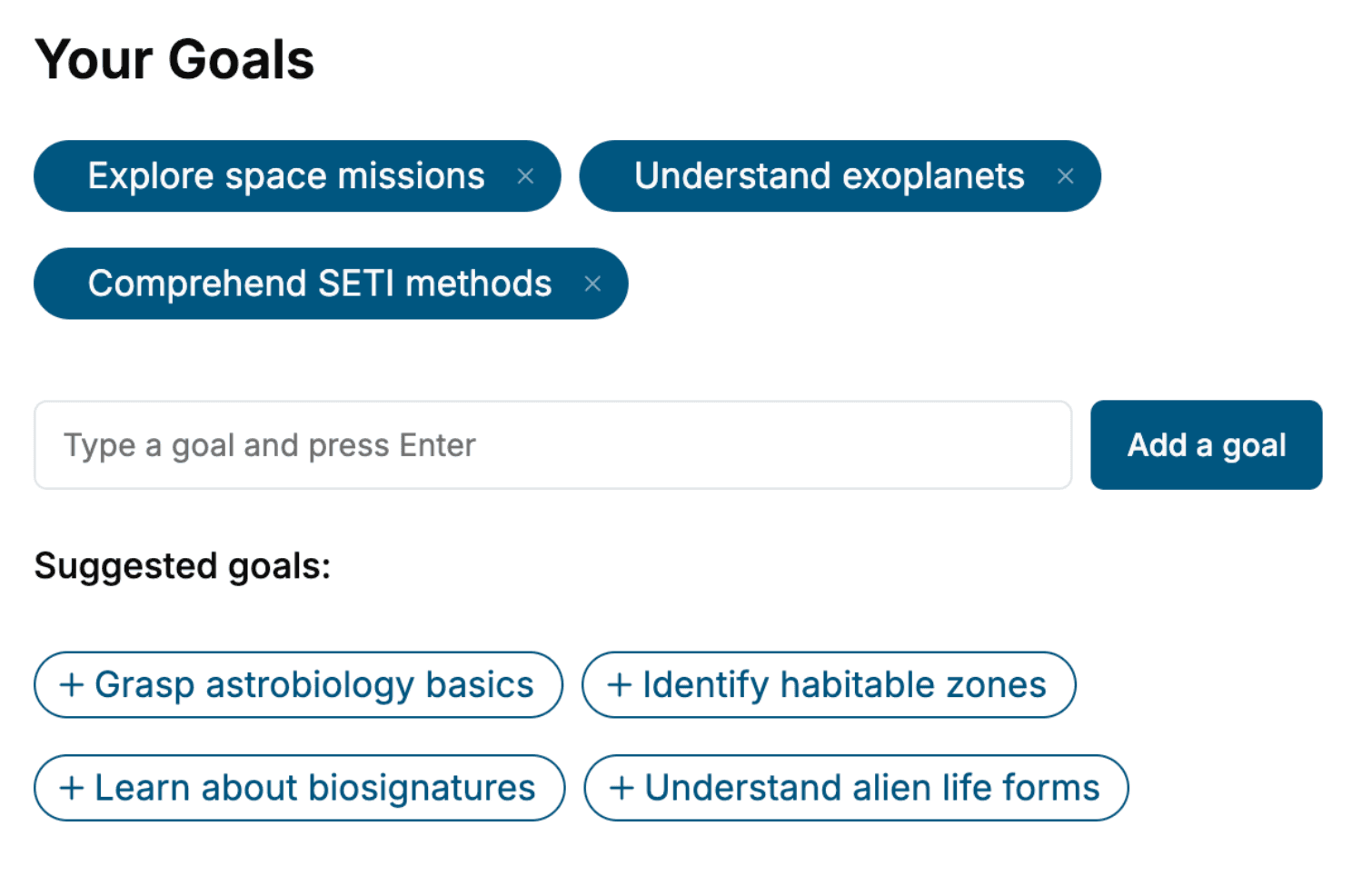
Tell us your goals and background, and get your own version of this book, tailored by AI to you.


Benefits of AI-tailored books
- Read one book, not ten:
- all the Cardiology knowledge you need, gathered into a single book.
- Save days of learning:
- tell us your goals, choose your topics, and the whole book is built around them.
- Learn effortlessly:
- Cardiology book written for your specific background and expertise.
- Understand at a glance:
- includes charts, tables, timelines, checklists, decision trees, and more.
- Stay current:
- written the day you order it, not years ago.
- Trust what you read:
- every book is checked for accuracy, with real research when your subject needs it.
Create your unique book in 3 steps
- 1. Select your focus
Select the focus of your Cardiology book and share your background
- 2. Personalize your book
Specify your goals and choose sub-topics to include
- 3. Get your tailored book
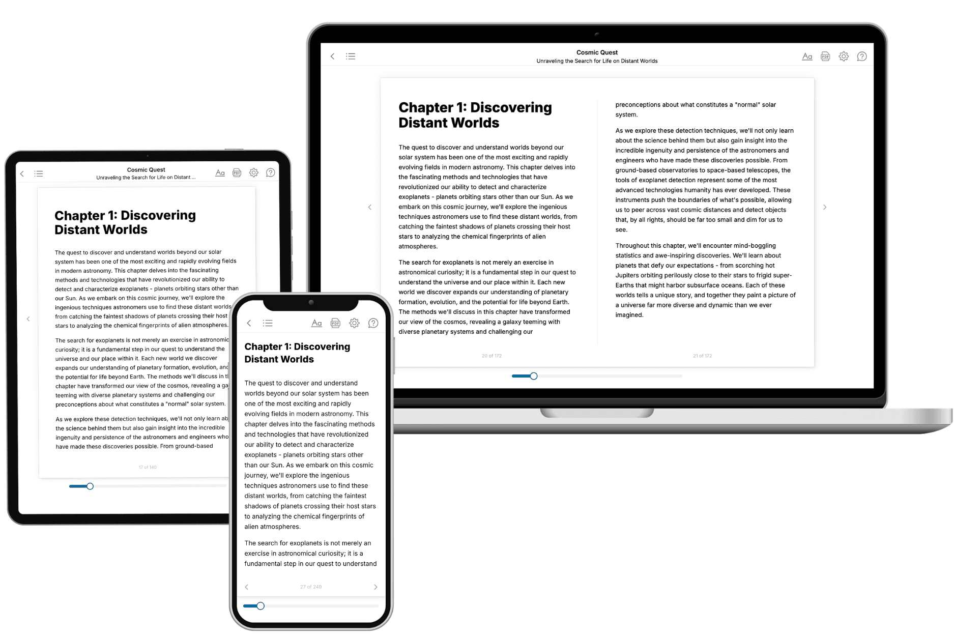
Your book is ready in 10 minutes. Read it online, download as EPUB or PDF, or send to Kindle.

Frequently asked questions
- What is TailoredRead?
TailoredRead creates complete, full-length nonfiction ebooks, written specifically for you. Tell us what you want to learn and why, and TailoredRead writes you a complete ebook on it, with charts, tables, and diagrams throughout. Read it online, on Kindle, or on any e-reader, or download it as a PDF or EPUB. Every book is written fresh the day you order it.
- How is the book tailored to me?
Before anything is written, you answer a few short questions about your goals, your background, and the topics you care about. Your book is then planned around your answers: written at your level, focused on your goals, and covering the topics you chose, instead of the one-size-fits-all book a store sells everyone.
- Will it read like generic AI content?
No. Your book isn't a chat transcript: it's planned chapter by chapter before a single word is written, told in a clear book voice, and explains ideas visually with charts, tables, and diagrams. After writing, every book goes through an editorial pass that polishes chapters so the book reads consistent from start to finish. Judge for yourself: browse thousands of sample books on our Explore Books page.
- How accurate is the content?
Our AI first checks whether your subject needs live research. Niche and fast-moving subjects are researched before writing, and the book plan and chapters are grounded in what it finds. After writing, every book goes through a quality pass that checks its facts and consistency, and researched books list their sources in a Notes section at the back.
- How long is the book?
You choose the length: Comprehensive (250-300 pages), Detailed (150-200 pages), Essential (70-100 pages), or Short (30-50 pages).
- Does the book include visuals?
Yes. Every book includes charts, tables, and diagrams: timelines, checklists, step-by-step guides, decision trees, comparison tables, matrices, and bar charts. A typical book includes dozens of these visual elements.
- Can I read it on Kindle? What formats can I download?
Yes. Every book can be read in our online reader or downloaded as a PDF or EPUB. The EPUB can be opened with Kindle, Apple Books, Kobo, and any e-reader. The Commercial and Author licenses also include an editable Word (DOCX) and Markdown download.
- How much does it cost?
About the price of a regular ebook. The exact price depends on your book's length and complexity, from $2 for the shortest books to $35 for the most comprehensive, and it's shown when you review your book, before you commit to anything. Each book is a one-time purchase, yours to keep, no subscription. Licenses that let you share, teach, or publish your book range from $35.99 to $49.99.
- Can I preview the book before purchasing?
Before you pay, you'll see what your book will cover: the title, a detailed description, the estimated length, and the complete chapter-by-chapter table of contents, so you know what you're getting before you decide. The full text itself is written after you purchase.
- How long does it take to create a book?
About 10 minutes. You can start reading the opening chapters while the rest is still being written.