Environmental Scanning Electron Microscopy
Master the Science and Practice of ESEM for Advanced Materials Analysis
Many researchers struggle with sample preparation limitations and artifact introduction when using conventional electron microscopy.
222 pages10 chaptersFeb 23, 2026Microscopy
About this book
Environmental Scanning Electron Microscopy (ESEM) solves this problem by allowing direct observation of specimens in their natural or near-natural state without extensive preparation. This comprehensive guide takes you from fundamental principles through advanced applications, covering everything you need to master ESEM technology. You'll learn how ESEM's unique pressure-controlled environment enables imaging of hydrated, non-conductive, and delicate materials while maintaining superior resolution. Discover practical techniques for optimizing imaging parameters, interpreting complex signals, and troubleshooting common challenges. Whether you're analyzing biological tissues, geological samples, polymers, or nanomaterials, this book provides the knowledge and strategies to extract maximum value from your ESEM system. Gain confidence in experimental design, data interpretation, and leveraging ESEM's dynamic observation capabilities for breakthrough research insights.
Table of contents
- IntroductionWhy ESEM Matters in Modern Research, How to Use This Book, The Current ESEM LandscapeRead below
- Chapter 1: Fundamentals of Electron MicroscopyElectron Behavior and Wave-Particle Duality, Core Components of Electron Microscopes, Conventional SEM vs. ESEM: Key Differences
- Chapter 2: ESEM Technology and Operating PrinciplesPressure Control and Vacuum Dynamics, Working in a Gaseous Environment, Specimen-Gas Interactions and Charging Mitigation, Unique Advantages of ESEM Technology
- Chapter 3: Sample Preparation for ESEMMinimal Preparation Strategies, Handling Hydrated and Wet Specimens, Working with Non-Conductive Materials
- Chapter 4: Optimizing Imaging ParametersAccelerating Voltage and Beam Current Selection, Magnification, Resolution, and Depth of Field, Scan Speed, Dwell Time, and Signal-to-Noise Optimization
- Chapter 5: Detectors and Signal GenerationTypes of Detectors in ESEM, Understanding Signal Generation Mechanisms, Achieving Optimal Image Contrast and Clarity, Selecting the Right Detector for Your Application
- Chapter 6: Analytical Techniques and Quantitative AnalysisEnergy Dispersive Spectroscopy (EDS) for Elemental Mapping, Electron Backscatter Diffraction (EBSD) for Crystal Structure, Combining Multiple Analytical Techniques
- Chapter 7: ESEM Applications in Materials ScienceAnalyzing Metals, Alloys, and Composites, Examining Polymers and Organic Materials, Studying Ceramics, Minerals, and Geological Samples
- Chapter 8: ESEM in Biological and Life SciencesImaging Biological Tissues and Cells, Preserving Hydration and Native Structure, Real-Time Observation of Biological Processes
- Chapter 9: Advanced ESEM Techniques and Dynamic StudiesIn-Situ Heating and Cooling Experiments, Environmental Control and Phase Transitions, Time-Resolved Imaging and Dynamic Behavior, Specialized Specimen Holders and Accessories
- Chapter 10: Troubleshooting and Problem SolvingAddressing Image Quality and Contrast Problems, Managing Charging Effects and Artifacts, Resolving Vacuum and Pressure-Related Issues
- ConclusionYour Path to ESEM Mastery, Future Directions in ESEM Technology
- Glossary of ESEM Terms
- Further Reading and Resources
Introduction
Electron microscopy has transformed how scientists and engineers see the world at the micro and nanoscale. For decades, conventional scanning electron microscopy (SEM) has delivered stunning images of surfaces, revealing details invisible to the naked eye or even the best optical microscopes. But conventional SEM comes with a significant constraint: specimens must be dry, conductive, and stable under high vacuum. That requirement has excluded entire categories of materials from direct observation, forcing researchers to prepare, coat, and alter their samples in ways that can introduce artifacts and obscure the very features they are trying to study.
Environmental Scanning Electron Microscopy, or ESEM, was developed specifically to break through those constraints. By maintaining a controlled gaseous environment inside the specimen chamber, ESEM allows imaging at pressures far above the high-vacuum conditions required by conventional SEM. This seemingly simple change has profound consequences. Hydrated biological tissues can be imaged without drying. Non-conductive polymers and ceramics can be examined without a conductive coating. Delicate geological samples can be studied in their natural state. The technology does not just expand what can be imaged; it fundamentally changes the quality and authenticity of the information that can be extracted.
This book is written for scientists, engineers, and researchers who already have some familiarity with electron microscopy and want to develop real, practical expertise in ESEM. Whether you are transitioning from conventional SEM, setting up an ESEM workflow for the first time, or looking to push your existing ESEM practice further, the chapters ahead are designed to give you both the conceptual foundation and the hands-on knowledge you need. The goal is not just to explain how ESEM works, but to help you use it confidently and effectively in your own research and analytical work.
The journey from understanding ESEM's principles to producing reliable, publication-quality data requires navigating a range of technical decisions: choosing the right pressure and gas composition, selecting appropriate detectors, interpreting signals correctly, and troubleshooting the inevitable challenges that arise. Each of these areas is covered in depth throughout this book, with concrete examples drawn from real-world applications in materials science, biology, geology, and beyond. By the time you reach the final chapter, you will have a comprehensive toolkit for tackling virtually any ESEM challenge your work presents.
Why ESEM Matters in Modern Research
The limitations of conventional SEM have always been a source of frustration for researchers working with materials that do not fit neatly into the high-vacuum, conductive-sample paradigm. Consider a biomedical researcher studying the surface structure of cartilage tissue. In conventional SEM, that tissue must be chemically fixed, dehydrated through a series of solvent washes, dried using critical point drying or freeze-drying, and then sputter-coated with a thin layer of gold or carbon. Each of those steps takes time, introduces potential artifacts, and moves the sample further from its natural biological state. ESEM eliminates most of those steps entirely, allowing the tissue to be imaged in a hydrated or partially hydrated condition that far better reflects its real structure.[1]
The impact of ESEM extends well beyond biology. In materials science, polymers and composite materials are notoriously difficult to image with conventional SEM because they are electrically insulating. Without a conductive coating, charge builds up on the surface under the electron beam, distorting the image and sometimes damaging the sample. ESEM's gaseous environment provides a natural charge-dissipation mechanism, allowing these materials to be imaged without any surface modification. This is particularly valuable in quality control settings, where altering a sample before inspection could mask the very defects being investigated.
Geological and environmental science applications benefit enormously from ESEM as well. Soil samples, clay minerals, and sedimentary rocks often contain moisture and complex mixtures of conductive and non-conductive phases. Imaging them in their natural state reveals information about pore structure, mineral distribution, and surface chemistry that would be obscured or destroyed by conventional preparation. Researchers studying cement hydration, for example, can watch the formation of calcium silicate hydrate phases in real time under ESEM conditions, something that is simply impossible with conventional SEM.[2]
One of the most compelling aspects of ESEM is its capacity for dynamic observation. Because the specimen chamber can maintain controlled temperature and humidity, researchers can design experiments that observe how a material responds to changing environmental conditions in real time. A polymer film can be watched as it absorbs moisture and swells. A metal surface can be observed as it corrodes under a controlled atmosphere. A biological cell can be monitored as it desiccates or rehydrates. These kinds of time-resolved observations provide insights into material behavior and biological processes that static imaging simply cannot deliver.
The analytical power of ESEM is further amplified when combined with spectroscopic techniques. Energy Dispersive Spectroscopy (EDS) can be performed under ESEM conditions to map elemental composition across a sample surface, even when that surface is wet or non-conductive. Electron Backscatter Diffraction (EBSD) can provide crystallographic information from materials that would be difficult to prepare for conventional analysis. These combined capabilities make ESEM one of the most versatile analytical platforms available in a modern materials characterization laboratory.
It is worth being clear about what ESEM does not do. It is not a replacement for all forms of electron microscopy. High-resolution transmission electron microscopy (TEM) still offers superior resolution for atomic-scale imaging. Conventional SEM remains the right choice for many dry, conductive samples where its simpler operation and lower cost are advantages. ESEM occupies a specific and important niche: it is the tool of choice when sample integrity, natural state preservation, or dynamic observation are priorities. Understanding where ESEM fits in the broader landscape of analytical techniques is the first step toward using it effectively.
The growing accessibility of ESEM technology has also changed the research landscape. Instruments that were once found only in specialized national facilities are now common in university departments, industrial laboratories, and research institutes around the world. As the user base has expanded, so has the body of knowledge about best practices, optimal parameters, and creative applications. This book draws on that accumulated knowledge to give you a practical, up-to-date guide to getting the most from your ESEM system.
Key insight: ESEM does not just make electron microscopy more convenient. It makes certain types of research possible that were previously out of reach. The ability to observe hydrated, non-conductive, and dynamically changing specimens in their near-natural state represents a genuine expansion of what science can see and measure.
How to Use This Book
This book is organized to take you from foundational principles through to advanced applications in a logical, progressive sequence. However, it is also designed so that experienced users can navigate directly to the chapters most relevant to their immediate needs. Whether you read it cover to cover or use it as a reference guide, the structure is intended to support both approaches.
The first two chapters establish the scientific foundation. Chapter 1 covers the fundamentals of electron microscopy, including electron behavior, the core components of electron microscopes, and the key differences between conventional SEM and ESEM. If you already have a solid grounding in SEM principles, you may move through this chapter quickly, using it mainly to identify the specific ways ESEM diverges from what you already know. Chapter 2 then dives into ESEM-specific technology: how pressure control works, what happens when electrons travel through a gaseous environment, and how the specimen-gas interaction mitigates charging. These two chapters together give you the conceptual framework needed to make sense of everything that follows.
Chapters 3 through 5 focus on practical operation. Chapter 3 addresses sample preparation, with specific guidance for hydrated specimens, non-conductive materials, and situations where minimal preparation is the goal. Chapter 4 covers imaging parameter optimization, including accelerating voltage, beam current, magnification, and scan speed. Chapter 5 explains detectors and signal generation in detail, helping you understand which detector to use for a given application and how to interpret the signals you receive. These three chapters are the core of day-to-day ESEM practice, and you will likely return to them frequently as reference material.
The analytical techniques covered in Chapter 6 extend ESEM's capabilities beyond imaging into quantitative characterization. EDS elemental mapping and EBSD crystallographic analysis are both explained with practical guidance on how to set up, run, and interpret these analyses under ESEM conditions. Chapters 7 and 8 then apply all of this knowledge to specific domains: materials science applications in Chapter 7, and biological and life science applications in Chapter 8. Each of these chapters includes worked examples and specific parameter recommendations for common specimen types.
Chapter 9 covers advanced techniques, including in-situ heating and cooling experiments, environmental control for phase transition studies, and time-resolved imaging. These are the capabilities that truly set ESEM apart from other microscopy platforms, and this chapter provides the guidance needed to design and execute these more complex experiments. Chapter 10 is dedicated to troubleshooting, addressing the most common problems encountered in ESEM practice: image quality issues, charging artifacts, and vacuum or pressure-related problems. A systematic approach to diagnosing and resolving these issues is presented, along with practical tips drawn from real laboratory experience.
Throughout the book, you will find several recurring features designed to make the content more actionable:
- Worked examples that walk through specific imaging scenarios with recommended parameters and expected outcomes
- Comparison tables that help you choose between options, such as detector types or gas compositions, for different applications
- Troubleshooting checklists that provide a structured approach to diagnosing common problems
- Key insight boxes that highlight the most important takeaways from each section
- Footnotes and references pointing to primary literature for readers who want to explore specific topics in greater depth
A glossary at the end of the book defines all key technical terms, and a further reading section points to important papers, textbooks, and online resources for continued learning. If you encounter an unfamiliar term while reading, the glossary is a quick reference that will keep you moving without losing the thread of the main text.
One practical suggestion: before working through the application-specific chapters (7 and 8), make sure you are comfortable with the material in Chapters 3 through 5. The application chapters assume familiarity with sample preparation strategies, imaging parameter selection, and detector choice. Readers who skip ahead sometimes find themselves confused by references to concepts that were explained in earlier chapters. The foundational material is not just background; it is the toolkit you will actively use when working through the application examples.
Finally, this book is most valuable when used alongside actual ESEM practice. Reading about pressure control and signal generation is useful, but the concepts become truly clear when you are sitting at the instrument, adjusting parameters, and watching the image respond. Wherever possible, try to connect what you read here to your own hands-on experience. The combination of conceptual understanding and practical experimentation is what builds genuine expertise.
The Current ESEM Landscape
ESEM technology has evolved considerably since its commercial introduction in the late 1980s. The original instruments, developed by Electroscan Corporation and later acquired by FEI Company (now part of Thermo Fisher Scientific), established the core principles that still underpin modern ESEM systems.[3] Over the following decades, improvements in detector design, vacuum system engineering, and digital image processing have dramatically expanded what these instruments can do. Today's ESEM systems offer capabilities that would have seemed extraordinary to the researchers who first worked with the technology.
The major instrument manufacturers currently offering ESEM or variable pressure SEM (VP-SEM) systems include Thermo Fisher Scientific, ZEISS, JEOL, and Hitachi. While the specific implementations differ between manufacturers, the underlying principles are consistent, and the knowledge in this book applies broadly across platforms. It is worth noting the distinction between true ESEM and variable pressure SEM. True ESEM systems, such as the Thermo Fisher Quanta series, use a specialized gaseous secondary electron detector (GSED) and can operate at pressures up to approximately 4,000 Pa. Variable pressure SEM systems operate at lower pressures (typically up to 300 Pa) and use a backscattered electron detector as their primary imaging mode. Both types are covered in this book, with specific notes where the guidance differs between them.
The research community using ESEM has grown substantially over the past two decades. A search of the scientific literature reveals thousands of published studies using ESEM across disciplines ranging from food science to semiconductor manufacturing. This breadth of application reflects the technology's genuine versatility. At the same time, it means that best practices and optimal parameters vary considerably depending on the specific application. One of the goals of this book is to help you navigate that diversity by providing clear principles that can be adapted to your specific needs, rather than a single rigid protocol that may not fit your situation.
Instrument software has also advanced significantly. Modern ESEM systems typically include automated routines for common tasks such as focus optimization, stigmation correction, and EDS spectrum acquisition. These automation features can accelerate routine work, but they can also create a false sense of security if users do not understand the underlying principles well enough to recognize when the automated system is making a poor decision. A recurring theme in this book is the importance of understanding why the instrument behaves as it does, not just how to operate the controls. That understanding is what allows you to override automated defaults when necessary and to diagnose problems when they arise.
The integration of ESEM with other analytical platforms is an active area of development. Correlative microscopy workflows, in which the same specimen is imaged by multiple techniques to build a more complete picture, are increasingly common. ESEM is well suited to correlative workflows because its minimal sample preparation requirements mean that specimens can often be transferred between instruments without significant alteration. For example, a biological tissue sample might be first imaged by ESEM to assess overall morphology, then transferred to a confocal fluorescence microscope for protein localization, and finally prepared for TEM to examine ultrastructure. The ESEM step in this workflow provides context and guides the subsequent, more demanding analyses.
Cryo-ESEM is another growing area that deserves mention. By combining ESEM's environmental control capabilities with cryogenic specimen stages, researchers can image frozen-hydrated specimens at temperatures below 0°C. This approach is particularly valuable for biological samples and soft matter systems where even partial dehydration would alter the structure of interest. Cryo-ESEM occupies a middle ground between conventional cryo-SEM and room-temperature ESEM, offering unique capabilities for studying ice formation, frozen biological structures, and low-temperature phase behavior in materials.[4]
Artificial intelligence and machine learning are beginning to influence ESEM practice as well. Automated image analysis tools can now segment features, classify phases, and measure morphological parameters from ESEM images with increasing accuracy. Some instrument manufacturers are integrating AI-assisted focusing and drift correction directly into their software. While these tools are still maturing, they represent a significant shift in how ESEM data will be acquired and analyzed in the coming years. This book focuses primarily on the established principles and practices that form the foundation of ESEM expertise, but it acknowledges these emerging developments where they are relevant to current practice.
The cost and accessibility of ESEM have also changed the landscape of who uses the technology. Shared facility models, where a single instrument serves multiple research groups or departments, are now the norm in many institutions. This means that ESEM users often work within a shared resource environment, where instrument time is scheduled, protocols must be documented, and results need to be reproducible across different operators. The practical guidance in this book is written with that reality in mind. Clear, documented workflows and a systematic approach to parameter selection are not just good scientific practice; they are essential for working effectively in a shared facility environment.
Understanding where ESEM sits within the broader ecosystem of characterization tools is important for using it wisely. The table below summarizes how ESEM compares to related techniques across several key dimensions:
- Resolution: ESEM achieves 1–20 nm resolution depending on conditions, better than optical microscopy but generally below high-vacuum SEM at equivalent accelerating voltages
- Sample requirements: Minimal preparation needed; hydrated, non-conductive, and delicate samples are all accessible
- Analytical capability: EDS and EBSD are both compatible with ESEM conditions, enabling compositional and crystallographic analysis
- Dynamic observation: Real-time imaging of environmental responses is a unique strength not available in most competing techniques
- Throughput: Faster than TEM for most applications; comparable to conventional SEM for dry samples; faster than conventional SEM for samples that would otherwise require extensive preparation
- Cost and complexity: Higher instrument cost and greater operational complexity than conventional SEM, but lower than TEM or focused ion beam (FIB) systems
The ESEM landscape today is one of mature, reliable technology with an expanding range of applications and a growing community of expert users. The fundamentals are well established, the instrumentation is robust, and the scientific literature provides a rich body of reference material. What is sometimes lacking is a clear, practical guide that bridges the gap between the technical literature and the day-to-day reality of operating an ESEM system. That is precisely the gap this book is designed to fill. The chapters ahead will take you from first principles to advanced practice, giving you the knowledge and confidence to make ESEM a powerful tool in your research arsenal.
Notes
Introduction
- 1. D. J. Stokes, Principles and Practice of Variable Pressure/Environmental Scanning Electron Microscopy (VP-ESEM), Wiley-Blackwell, 2008
- 2. A. M. Donald, "The use of environmental scanning electron microscopy for imaging wet and insulating materials," Nature Materials, 2003
- 3. G. D. Danilatos, Foundations of environmental scanning electron microscopy, Advances in Electronics and Electron Physics, 1988
- 4. P. Echlin, Low-Temperature Microscopy and Analysis, Plenum Press, New York, 1992
This is the end of the free sample. The full book has 10 chapters.
Want this book written for you?
Tell us your goals and background, and get your own version of this book, tailored by AI to you.


Benefits of AI-tailored books
- Read one book, not ten:
- all the Microscopy knowledge you need, gathered into a single book.
- Save days of learning:
- tell us your goals, choose your topics, and the whole book is built around them.
- Learn effortlessly:
- Microscopy book written for your specific background and expertise.
- Understand at a glance:
- includes charts, tables, timelines, checklists, decision trees, and more.
- Stay current:
- written the day you order it, not years ago.
- Trust what you read:
- every book is checked for accuracy, with real research when your subject needs it.
Create your unique book in 3 steps
- 1. Select your focus
Select the focus of your Microscopy book and share your background
- 2. Personalize your book
Specify your goals and choose sub-topics to include
- 3. Get your tailored book
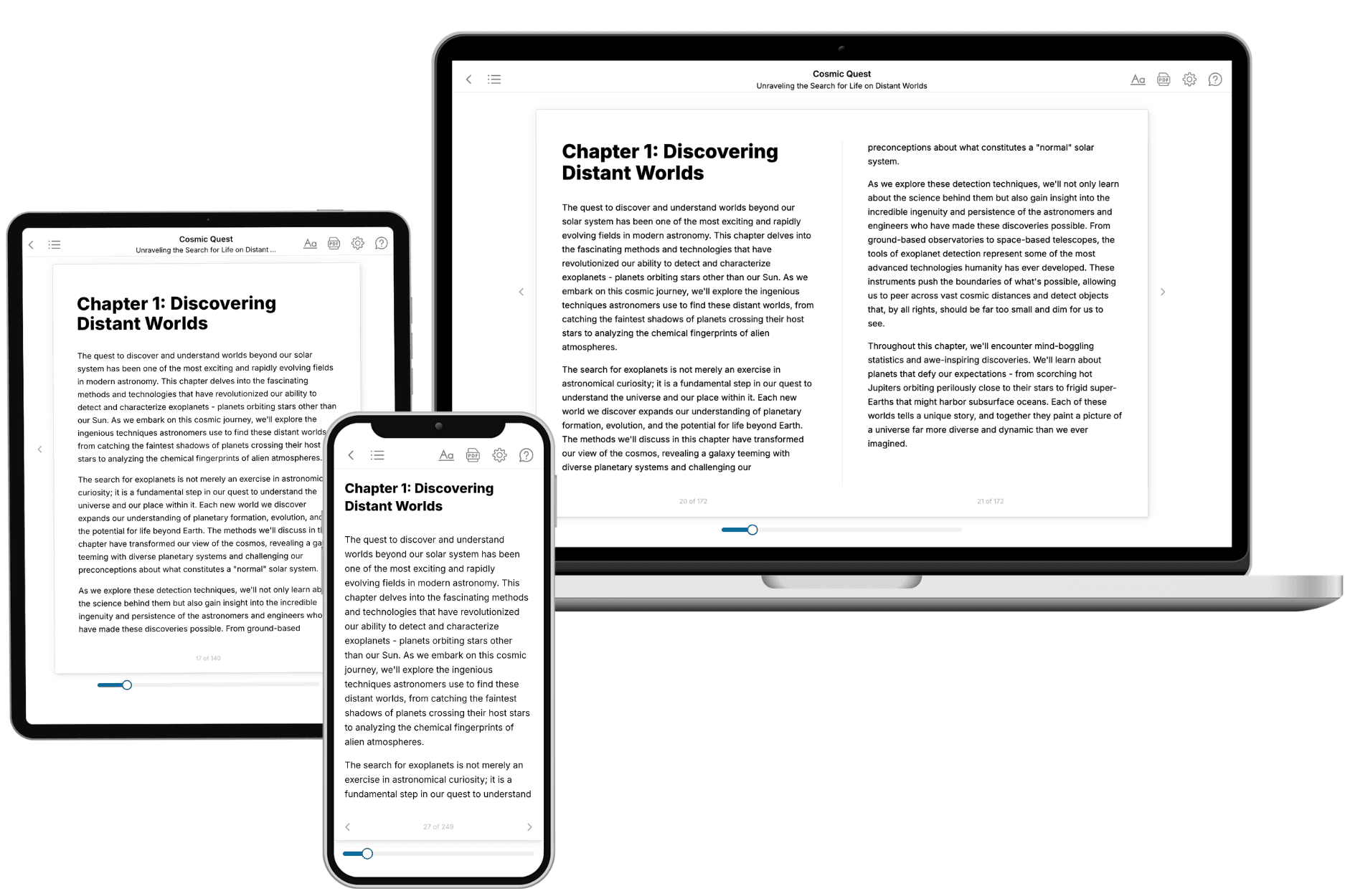
Your book is ready in 10 minutes. Read it online, download as EPUB or PDF, or send to Kindle.
Frequently asked questions
- What is TailoredRead?
TailoredRead creates complete, full-length nonfiction ebooks, written specifically for you. Tell us what you want to learn and why, and TailoredRead writes you a complete ebook on it, with charts, tables, and diagrams throughout. Read it online, on Kindle, or on any e-reader, or download it as a PDF or EPUB. Every book is written fresh the day you order it.
- How is the book tailored to me?
Before anything is written, you answer a few short questions about your goals, your background, and the topics you care about. Your book is then planned around your answers: written at your level, focused on your goals, and covering the topics you chose, instead of the one-size-fits-all book a store sells everyone.
- Will it read like generic AI content?
No. Your book isn't a chat transcript: it's planned chapter by chapter before a single word is written, told in a clear book voice, and explains ideas visually with charts, tables, and diagrams. After writing, every book goes through an editorial pass that polishes chapters so the book reads consistent from start to finish. Judge for yourself: browse thousands of sample books on our Explore Books page.
- How accurate is the content?
Our AI first checks whether your subject needs live research. Niche and fast-moving subjects are researched before writing, and the book plan and chapters are grounded in what it finds. After writing, every book goes through a quality pass that checks its facts and consistency, and researched books list their sources in a Notes section at the back.
- How long is the book?
You choose the length: Comprehensive (250-300 pages), Detailed (150-200 pages), Essential (70-100 pages), or Short (30-50 pages).
- Does the book include visuals?
Yes. Every book includes charts, tables, and diagrams: timelines, checklists, step-by-step guides, decision trees, comparison tables, matrices, and bar charts. A typical book includes dozens of these visual elements.
- Can I read it on Kindle? What formats can I download?
Yes. Every book can be read in our online reader or downloaded as a PDF or EPUB. The EPUB can be opened with Kindle, Apple Books, Kobo, and any e-reader. The Commercial and Author licenses also include an editable Word (DOCX) and Markdown download.
- How much does it cost?
About the price of a regular ebook. The exact price depends on your book's length and complexity, from $2 for the shortest books to $35 for the most comprehensive, and it's shown when you review your book, before you commit to anything. Each book is a one-time purchase, yours to keep, no subscription. Licenses that let you share, teach, or publish your book range from $35.99 to $49.99.
- Can I preview the book before purchasing?
Before you pay, you'll see what your book will cover: the title, a detailed description, the estimated length, and the complete chapter-by-chapter table of contents, so you know what you're getting before you decide. The full text itself is written after you purchase.
- How long does it take to create a book?
About 10 minutes. You can start reading the opening chapters while the rest is still being written.