The Heart Anatomy Coloring Guide
Interactive Learning Through Visual Discovery and Hands-On Exploration
Dive deep into the fascinating world of cardiac anatomy through an innovative blend of scientific accuracy and interactive learning.
269 pages13 chaptersOct 13, 2025Anatomy
About this book
This comprehensive guide transforms the complex study of heart structure into an engaging, hands-on experience that makes even the most intricate details accessible and memorable. You'll explore every aspect of cardiac anatomy, from the four chambers and their specialized functions to the intricate network of blood vessels that keep your heart beating. Each carefully designed illustration serves as both an educational tool and a creative outlet, allowing you to absorb information through multiple sensory channels while building a thorough understanding of how structure relates to function. The systematic approach guides you through increasingly complex concepts, starting with basic heart orientation and progressing through detailed examinations of valves, conduction pathways, and coronary circulation. Every coloring exercise is strategically designed to reinforce key anatomical relationships and help you visualize the dynamic processes that occur within this remarkable organ. Whether you're strengthening your existing knowledge or building upon foundational concepts, this interactive approach will deepen your appreciation for the heart's elegant design while providing you with lasting visual memories of its intricate architecture.
Table of contents
- IntroductionThe Science Behind Coloring for Learning, How to Use This Guide Effectively, Recommended Materials and Coloring Techniques, Basic Anatomical Orientation and TerminologyRead below
- Chapter 1: Heart Overview and PositionHeart Location in the Thoracic Cavity, Heart Size, Shape, and Orientation, Surrounding Structures and Relationships, Heart Surfaces and Borders
- Chapter 2: Heart Wall Layers and StructureThe Pericardium: Protective Outer Layer, Epicardium: The Heart's Outer Surface, Myocardium: The Muscular Heart Wall, Endocardium: The Inner Heart Lining
- Chapter 3: The Four Heart ChambersRight Atrium: Structure and Features, Left Atrium: Structure and Features, Right Ventricle: Structure and Features, Left Ventricle: Structure and Features, Chamber Relationships and Connections
- Chapter 4: Heart Valves and Their FunctionTricuspid Valve: Right Atrioventricular Valve, Pulmonary Valve: Right Semilunar Valve, Mitral Valve: Left Atrioventricular Valve, Aortic Valve: Left Semilunar Valve, Valve Opening and Closing Mechanisms
- Chapter 5: Blood Flow Through the HeartVenous Return to the Right Atrium, Pulmonary Circulation Pathway, Systemic Circulation Return, Complete Circulation Cycle
- Chapter 6: Major Blood Vessels of the HeartSuperior and Inferior Vena Cavae, Pulmonary Arteries and Veins, Aorta and Its Major Branches, Vessel Connections to Heart Chambers
- Chapter 7: Coronary Circulation SystemRight and Left Coronary Arteries, Major Coronary Arterial Branches, Coronary Venous System, Coronary Circulation Patterns and Variations
- Chapter 8: Electrical Conduction SystemSinoatrial Node: The Heart's Pacemaker, Atrioventricular Node and Conduction, Bundle of His and Bundle Branches, Purkinje Fiber Network, Complete Electrical Conduction Pathway
- Chapter 9: The Cardiac CycleAtrial Systole: Atrial Contraction Phase, Ventricular Systole: Ventricular Contraction, Diastole: Heart Relaxation Phase, Pressure Changes Throughout the Cycle
- Chapter 10: Heart Development and FormationEarly Embryonic Heart Formation, Chamber and Septum Development, Heart Valve Formation, Fetal Circulation Patterns
- Chapter 11: Anatomical Variations and AdaptationsNormal Anatomical Variations, Age-Related Heart Changes, Exercise-Induced Heart Adaptations, Comparative Heart Anatomy Across Species
- Chapter 12: Microscopic Heart StructureCardiac Muscle Cell Structure, Intercalated Discs and Cell Connections, Different Cardiac Tissue Types, Cellular Organization and Arrangement
- Chapter 13: Heart Integration with Body SystemsNervous System Control of Heart Function, Hormonal Influences on Heart Activity, Heart-Respiratory System Interactions, Coordination with Other Body Systems
- Conclusion
- Glossary
- Further Reading
Introduction
The human heart stands as one of nature's most remarkable engineering marvels, beating approximately 100,000 times each day and pumping over 2,000 gallons of blood throughout your body. This extraordinary organ, no larger than your fist, maintains the continuous circulation that sustains every cell in your body. Understanding its intricate anatomy requires more than memorizing names and locations—it demands a deep appreciation for how structure and function work together in perfect harmony. This guide transforms the traditional approach to learning cardiac anatomy by combining scientific accuracy with interactive coloring exercises that engage multiple learning pathways simultaneously.
Traditional anatomy education often relies heavily on passive reading and static diagrams, which can make complex three-dimensional structures difficult to visualize and remember. The heart's anatomy presents particular challenges because of its intricate internal architecture, overlapping structures, and the dynamic relationships between its various components. When you actively engage with anatomical illustrations through coloring, you create stronger neural connections that enhance both understanding and retention. This hands-on approach allows you to explore each structure methodically while building a comprehensive mental map of cardiac anatomy that will serve as a foundation for deeper physiological concepts.
Your journey through this guide will take you from basic heart orientation and positioning within the chest cavity to the microscopic details of cardiac muscle cells and their specialized connections. Each chapter builds upon previous knowledge while introducing increasingly sophisticated concepts about heart structure and function. The carefully designed coloring exercises serve as both learning tools and assessment opportunities, allowing you to identify areas that need additional attention while reinforcing your growing understanding of cardiac anatomy. By the time you complete this guide, you will have developed a thorough, visual understanding of the heart that goes far beyond surface-level memorization.
The Science Behind Coloring for Learning
Neuroscience research has consistently demonstrated that active learning techniques produce superior retention and understanding compared to passive methods. When you color anatomical structures, your brain engages multiple processing centers simultaneously, creating what researchers call multimodal learning. This approach activates visual processing areas responsible for spatial relationships, motor cortex regions that control hand movements, and cognitive centers that process color associations and pattern recognition. The result is a rich network of neural connections that makes information more accessible and memorable than traditional study methods alone[1].
The act of coloring requires sustained attention and focus, which promotes what psychologists term deep processing. Unlike superficial reading where information may pass through working memory without forming lasting connections, coloring demands that you examine each structure carefully, consider its relationships to surrounding elements, and make deliberate decisions about color choices. This level of engagement forces your brain to process information more thoroughly, creating stronger memory traces that persist longer than those formed through passive study methods. Research in educational psychology shows that students who engage in active learning techniques demonstrate 23% better performance on assessments compared to those using traditional methods[2].
Color coding serves as a powerful organizational tool that helps your brain categorize and retrieve information more efficiently. When you consistently use specific colors for related structures—such as blue for venous blood, red for arterial blood, or yellow for nervous tissue—you create visual mnemonics that strengthen memory formation. This systematic approach to color application mirrors the way your brain naturally organizes information into categories and hierarchies. The visual associations you create through consistent coloring patterns become retrieval cues that help you access stored information more quickly and accurately during examinations or practical applications.
The kinesthetic component of coloring engages your motor cortex and creates what researchers call embodied learning. The physical movements involved in coloring create additional memory pathways that complement visual and cognitive processing. This is particularly valuable for understanding three-dimensional relationships in anatomy, where spatial awareness plays a crucial role in comprehension. Studies have shown that students who engage in hands-on learning activities demonstrate improved spatial reasoning skills and better understanding of complex structural relationships[3]. The combination of visual, motor, and cognitive engagement creates a comprehensive learning experience that addresses different learning styles and preferences.
Coloring also provides immediate feedback about your understanding of anatomical relationships. As you work through each illustration, you naturally identify areas where your knowledge is strong and regions that require additional attention. This self-assessment capability allows you to adjust your study approach in real-time, spending more time on challenging concepts while reinforcing areas of strength. The process of selecting appropriate colors for different structures requires you to recall and apply anatomical knowledge actively, transforming what might otherwise be passive recognition into active recall practice.
The meditative quality of coloring can reduce stress and anxiety often associated with learning complex material. When your stress levels decrease, your brain's capacity for forming new memories and making connections between concepts improves significantly. The rhythmic, repetitive motions involved in coloring activate the parasympathetic nervous system, promoting a relaxed state that is optimal for learning and retention. This stress-reduction benefit is particularly valuable when studying challenging subjects like anatomy, where information density and complexity can sometimes feel overwhelming.
Research in cognitive psychology has identified the generation effect, which demonstrates that information you actively create or manipulate is remembered better than information you simply read or observe. When you color anatomical structures, you are essentially generating your own visual representation of the material, which creates stronger memory traces than viewing pre-colored illustrations. This active creation process engages higher-order thinking skills and promotes deeper understanding of the relationships between different anatomical components. The personal investment you make in creating each colored illustration becomes part of the learning experience itself.
The systematic progression through coloring exercises also supports scaffolded learning, where each new concept builds upon previously established knowledge. As you color increasingly complex structures, you reinforce earlier learning while adding new layers of understanding. This cumulative approach mirrors the way anatomical knowledge naturally develops, from basic structural identification to sophisticated understanding of functional relationships. The visual record you create through your coloring work serves as a personalized reference that reflects your learning journey and can be revisited for review and reinforcement.
How to Use This Guide Effectively
Maximizing your learning experience with this guide requires a systematic approach that balances thorough exploration with efficient progress. Begin each chapter by reading the introductory material completely before attempting any coloring exercises. This preliminary reading provides essential context and introduces key terminology that will guide your coloring decisions. Pay particular attention to the learning objectives outlined at the beginning of each section, as these will help you focus your attention on the most important concepts and relationships. Take notes on unfamiliar terms or concepts that seem particularly challenging, as these areas will likely require additional attention during the coloring process.
Approach each coloring exercise with a clear strategy rather than randomly selecting colors for different structures. Start by examining the entire illustration to understand the overall organization and identify major structural groups. Create a color key for each chapter that assigns specific colors to related structures—for example, use different shades of red for arterial structures, blues for venous components, and yellows for nervous tissue. This systematic approach will help you maintain consistency throughout the guide and create visual patterns that reinforce anatomical relationships. Keep your color key visible while working and refer to it regularly to ensure consistency across different illustrations.
Work through the coloring exercises in the order presented, as each builds upon knowledge from previous sections. Resist the temptation to skip ahead to more interesting or familiar topics, as this can create gaps in your foundational understanding that will make later concepts more difficult to grasp. If you encounter a structure or concept that seems unclear, return to the relevant text sections for clarification before continuing. The sequential organization of this guide is designed to support progressive learning, where each new concept integrates with previously established knowledge to create a comprehensive understanding of cardiac anatomy.
Allocate sufficient time for each coloring session to work thoughtfully rather than rushing through exercises. Plan for 45-60 minutes per major section, allowing time for careful reading, thoughtful coloring, and brief review of completed work. This pacing ensures that you can maintain focus and attention throughout each session while avoiding the fatigue that can interfere with learning. Take short breaks every 20-30 minutes to rest your eyes and hands, as this will help maintain the quality of your work and prevent the mechanical aspects of coloring from becoming tedious or counterproductive.
Use the coloring process as an opportunity for active self-testing and review. Before coloring each structure, try to recall its name, function, and relationships to surrounding components. This practice transforms the coloring exercise into a form of retrieval practice, which is one of the most effective methods for strengthening memory and understanding. If you cannot recall information about a particular structure, refer back to the text before coloring it, then test yourself again on the same structure in subsequent illustrations. This approach ensures that coloring reinforces accurate knowledge rather than simply creating attractive pictures.
Create your own annotations and labels as you work through each illustration. While the guide provides comprehensive labeling, adding your own notes about function, clinical significance, or personal memory aids will personalize the learning experience and create additional retrieval cues. Use the margins around each illustration to write brief descriptions of key functions, note important relationships between structures, or record questions that arise during your study. These personal annotations become valuable review materials that reflect your individual learning process and areas of particular interest or difficulty.
Regularly review completed sections to reinforce learning and identify areas that need additional attention. Schedule weekly review sessions where you examine previously colored illustrations and test your ability to identify and describe major structures without referring to labels or text. This spaced repetition approach helps transfer information from short-term to long-term memory and identifies concepts that may need additional reinforcement. During review sessions, pay particular attention to structures or relationships that you find difficult to remember, as these areas may benefit from additional coloring practice or supplementary study materials.
Consider working with study partners or groups to enhance your learning experience through discussion and collaborative problem-solving. Share your colored illustrations with others and explain the structures and relationships you have learned. Teaching others is one of the most effective ways to identify gaps in your own understanding and reinforce correct knowledge. Organize study sessions where group members take turns explaining different anatomical systems or quiz each other on structure identification and function. This social dimension of learning can make the study process more engaging while providing valuable feedback about your progress.
Keep a learning journal throughout your work with this guide to track your progress, record insights, and note areas for improvement. Document challenging concepts, successful learning strategies, and connections you discover between different anatomical systems. This reflective practice helps you become more aware of your own learning process and develop more effective study strategies. Review your journal entries periodically to identify patterns in your learning and adjust your approach as needed. The journal also serves as a valuable resource for final review sessions and can help you prepare for examinations or practical applications of your anatomical knowledge.
Recommended Materials and Coloring Techniques
Selecting appropriate coloring materials significantly impacts both the quality of your finished work and the effectiveness of your learning experience. Colored pencils represent the optimal choice for anatomical coloring because they offer precise control, allow for layering and blending, and can create both fine details and broader areas of color. High-quality colored pencils with soft, waxy cores provide smooth color application and rich pigmentation that makes structural details clearly visible. Avoid hard, scratchy pencils that require excessive pressure, as these can damage the paper and make extended coloring sessions uncomfortable. A set of 24-36 colored pencils provides sufficient color variety for most anatomical illustrations while maintaining a manageable selection that won't overwhelm your decision-making process.
Organize your colored pencils systematically to support consistent color coding throughout the guide. Group related colors together—reds and pinks for arterial structures, blues and purples for venous components, yellows and oranges for nervous tissue, and greens for lymphatic structures. This organization makes color selection more efficient and helps maintain consistency across different illustrations. Consider using a pencil case with individual slots or a desktop organizer that keeps your pencils visible and easily accessible. Label your pencils or create a reference chart that shows which specific colors you use for different types of anatomical structures.
Master basic coloring techniques that enhance the educational value of your work while creating visually appealing results. Light, even strokes in one direction create smooth, professional-looking color application that doesn't obscure important structural details. Apply color in thin layers, building up intensity gradually rather than pressing hard to achieve dark colors immediately. This layering technique allows you to create subtle variations in tone that can represent different tissue types or structural depths. Practice maintaining consistent pressure and stroke direction to develop muscle memory that makes the coloring process more automatic and less distracting from the learning objectives.
Use color intensity strategically to emphasize important structures and create visual hierarchy in your illustrations. Apply darker, more saturated colors to major structures that require primary attention, while using lighter tones for supporting elements or background structures. This technique helps direct your visual attention to the most important learning objectives while maintaining awareness of the broader anatomical context. For example, when studying heart chambers, use bold colors for the chamber walls while applying lighter tones to surrounding structures like blood vessels or pericardial layers.
Develop a systematic approach to directional shading that enhances the three-dimensional appearance of anatomical structures. Imagine a consistent light source coming from the upper left of each illustration, and apply lighter colors to surfaces that would face this light source while using darker tones for areas that would be in shadow. This shading technique helps you visualize the spatial relationships between structures and reinforces understanding of anatomical depth and positioning. Practice this technique on simple shapes before applying it to complex anatomical illustrations.
Learn to use color blending techniques that create smooth transitions between different tissue types or structural regions. Overlap colors slightly at boundaries between different structures, using light pressure to create gradual transitions rather than sharp lines. This blending approach reflects the reality of anatomical structures, where tissues often transition gradually rather than ending abruptly. Use a colorless blender pencil or light-colored pencil to smooth transitions and create more realistic representations of anatomical relationships.
Maintain your coloring materials properly to ensure consistent performance throughout your study period. Keep pencil points sharp using a quality sharpener or mechanical pencil system, as dull points make precise work difficult and can create frustrating coloring experiences. Store your pencils in a way that protects the points from damage and keeps them organized for easy access. Clean your pencils periodically with a soft cloth to remove accumulated debris that can interfere with smooth color application. Replace worn or damaged pencils promptly to maintain the quality of your work.
Consider supplementary materials that can enhance your coloring experience and learning outcomes. A kneaded eraser allows you to lighten areas that become too dark or remove small mistakes without damaging the paper. A blending stump or tortillon can help create smooth color transitions and subtle shading effects. A ruler or straight edge ensures clean, precise lines when adding your own labels or annotations. Good lighting is essential for accurate color perception and comfortable extended work sessions—use a desk lamp with daylight-balanced bulbs to reduce eye strain and ensure color accuracy.
Experiment with different coloring techniques to find approaches that work best for your learning style and artistic preferences. Some learners prefer stippling techniques that use small dots of color to build up areas gradually, while others work better with cross-hatching methods that use overlapping lines to create texture and depth. Try different approaches on practice sheets before applying them to important illustrations. Remember that the goal is educational effectiveness rather than artistic perfection—focus on techniques that enhance your understanding of anatomical relationships rather than creating museum-quality artwork.
Basic Anatomical Orientation and Terminology
Understanding anatomical position and directional terminology forms the foundation for all anatomical study and communication. The standard anatomical position serves as the universal reference point for describing body structures and their relationships. In this position, the body stands upright with feet parallel and flat on the floor, arms at the sides with palms facing forward, and head facing straight ahead. All anatomical descriptions assume this standard position, regardless of the actual position of the body being examined. This consistency ensures that anatomical terms have the same meaning for all healthcare professionals and students worldwide, preventing confusion that could arise from varying reference points.
Directional terms provide precise language for describing the location of anatomical structures relative to one another. Superior means toward the head or upper part of the body, while inferior indicates toward the feet or lower part. Anterior (or ventral) refers to the front of the body, and posterior (or dorsal) indicates the back. Medial describes structures closer to the midline of the body, while lateral refers to structures farther from the midline. These terms allow you to describe cardiac anatomy with precision—for example, the right ventricle is anterior to the left ventricle, and the superior vena cava enters the heart from a superior position relative to the inferior vena cava.
Anatomical planes provide standardized ways to section and view the body for study and analysis. The sagittal plane divides the body into right and left portions, with the midsagittal plane creating equal halves and parasagittal planes creating unequal divisions. The frontal (or coronal) plane separates the body into anterior and posterior sections, while the transverse (or horizontal) plane creates superior and inferior divisions. Understanding these planes is crucial for interpreting anatomical illustrations and medical imaging, as most views of the heart represent specific planar sections that reveal different structural relationships.
Regional terminology helps organize anatomical study by grouping related structures within specific body areas. The thoracic region contains the heart and represents your primary area of focus in this guide. Within the thoracic region, the mediastinum is the central compartment that houses the heart, great vessels, and other vital structures. The heart occupies most of the middle mediastinum, with specific relationships to surrounding structures that influence its position and function. Understanding these regional relationships helps you appreciate how the heart fits within the broader context of thoracic anatomy and how it interacts with neighboring structures.
Cardiac-specific terminology builds upon general anatomical language to describe heart structures and relationships with greater precision. The heart's apex represents its pointed inferior tip, while the base refers to its broader superior portion where the great vessels attach. The heart's surfaces include the anterior (sternocostal) surface, the inferior (diaphragmatic) surface, and the posterior (vertebral) surface. Each surface has specific relationships to surrounding structures that influence the heart's position and the clinical approaches used for examination and treatment.
Understanding the concept of anatomical variation is essential for developing realistic expectations about cardiac anatomy. While textbook illustrations show idealized representations of anatomical structures, real hearts exhibit considerable variation in size, shape, position, and detailed structure. These variations are usually normal and reflect the natural diversity found in human anatomy. Some variations are more common in certain populations or age groups, while others represent unique individual characteristics. Recognizing that variation is normal helps you develop a more flexible and realistic understanding of cardiac anatomy that will serve you well in clinical or research applications.
Clinical correlation terminology connects anatomical knowledge to practical applications in healthcare and research. Understanding how anatomical structures relate to surface landmarks helps you locate and examine the heart in living subjects. The heart's position relative to ribs, sternum, and other palpable structures provides reference points for physical examination, diagnostic procedures, and therapeutic interventions. Learning to think about anatomy in clinical terms from the beginning of your studies helps you develop the practical perspective that makes anatomical knowledge useful in real-world applications.
Developmental terminology describes how anatomical structures change throughout life, from embryonic formation through aging processes. The heart undergoes dramatic changes during embryonic and fetal development, with structures that are essential during fetal life becoming modified or obsolete after birth. Understanding developmental terminology helps you appreciate why certain anatomical features exist and how they relate to the heart's functional requirements at different life stages. This developmental perspective also helps explain some of the anatomical variations you may encounter in different age groups.
Comparative terminology allows you to understand cardiac anatomy within the broader context of vertebrate evolution and adaptation. While this guide focuses on human cardiac anatomy, understanding how the human heart relates to cardiac structures in other species provides valuable perspective on the fundamental principles of cardiac design and function. Comparative terminology helps you recognize which features of cardiac anatomy are universal among vertebrates and which represent specific adaptations to human physiological requirements. This broader perspective enhances your appreciation for the elegant solutions that evolution has developed for the challenges of circulation and cardiac function.
Notes
Introduction
- 1. W. R. Klemm, "Computer slide shows," College Teaching, 2007
- 2. S. Freeman and others, "Active learning increases student performance in science, engineering, and mathematics," Proceedings of the National Academy of Sciences, 2014
- 3. D. H. Uttal and others, "The malleability of spatial skills," Psychological Bulletin, 2013
This is the end of the free sample. The full book has 13 chapters.
Want this book written for you?
Tell us your goals and background, and get your own version of this book, tailored by AI to you.


Benefits of AI-tailored books
- Read one book, not ten:
- all the Anatomy knowledge you need, gathered into a single book.
- Save days of learning:
- tell us your goals, choose your topics, and the whole book is built around them.
- Learn effortlessly:
- Anatomy book written for your specific background and expertise.
- Understand at a glance:
- includes charts, tables, timelines, checklists, decision trees, and more.
- Stay current:
- written the day you order it, not years ago.
- Trust what you read:
- every book is checked for accuracy, with real research when your subject needs it.
Create your unique book in 3 steps
- 1. Select your focus
Select the focus of your Anatomy book and share your background
- 2. Personalize your book
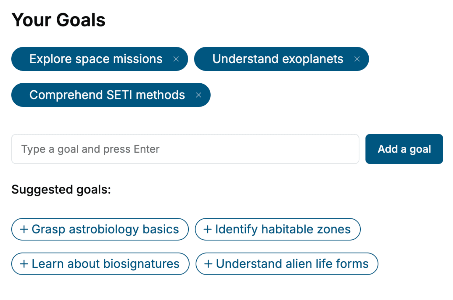
Specify your goals and choose sub-topics to include
- 3. Get your tailored book
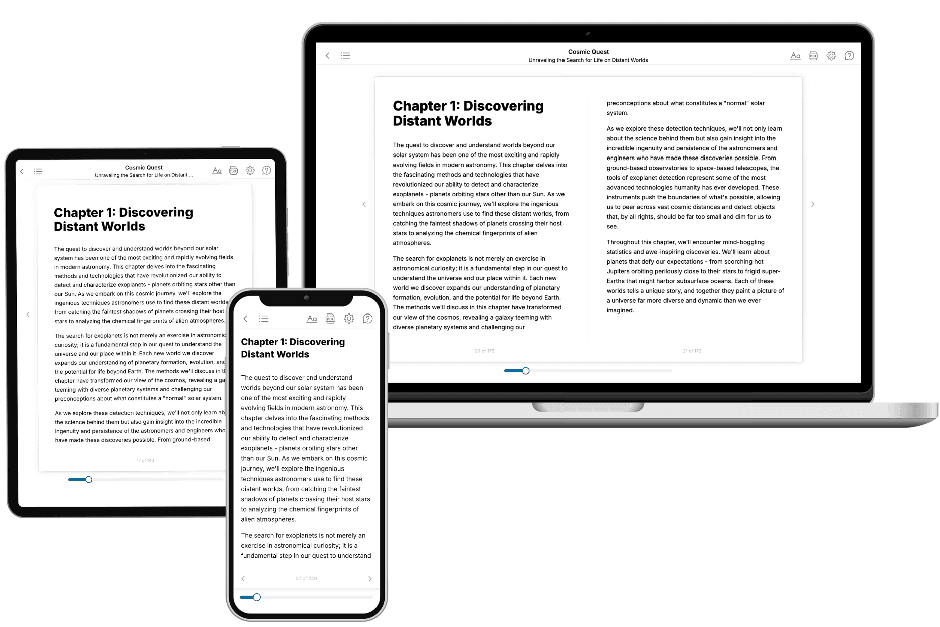
Your book is ready in 10 minutes. Read it online, download as EPUB or PDF, or send to Kindle.

Frequently asked questions
- What is TailoredRead?
TailoredRead creates complete, full-length nonfiction ebooks, written specifically for you. Tell us what you want to learn and why, and TailoredRead writes you a complete ebook on it, with charts, tables, and diagrams throughout. Read it online, on Kindle, or on any e-reader, or download it as a PDF or EPUB. Every book is written fresh the day you order it.
- How is the book tailored to me?
Before anything is written, you answer a few short questions about your goals, your background, and the topics you care about. Your book is then planned around your answers: written at your level, focused on your goals, and covering the topics you chose, instead of the one-size-fits-all book a store sells everyone.
- Will it read like generic AI content?
No. Your book isn't a chat transcript: it's planned chapter by chapter before a single word is written, told in a clear book voice, and explains ideas visually with charts, tables, and diagrams. After writing, every book goes through an editorial pass that polishes chapters so the book reads consistent from start to finish. Judge for yourself: browse thousands of sample books on our Explore Books page.
- How accurate is the content?
Our AI first checks whether your subject needs live research. Niche and fast-moving subjects are researched before writing, and the book plan and chapters are grounded in what it finds. After writing, every book goes through a quality pass that checks its facts and consistency, and researched books list their sources in a Notes section at the back.
- How long is the book?
You choose the length: Comprehensive (250-300 pages), Detailed (150-200 pages), Essential (70-100 pages), or Short (30-50 pages).
- Does the book include visuals?
Yes. Every book includes charts, tables, and diagrams: timelines, checklists, step-by-step guides, decision trees, comparison tables, matrices, and bar charts. A typical book includes dozens of these visual elements.
- Can I read it on Kindle? What formats can I download?
Yes. Every book can be read in our online reader or downloaded as a PDF or EPUB. The EPUB can be opened with Kindle, Apple Books, Kobo, and any e-reader. The Commercial and Author licenses also include an editable Word (DOCX) and Markdown download.
- How much does it cost?
About the price of a regular ebook. The exact price depends on your book's length and complexity, from $2 for the shortest books to $35 for the most comprehensive, and it's shown when you review your book, before you commit to anything. Each book is a one-time purchase, yours to keep, no subscription. Licenses that let you share, teach, or publish your book range from $35.99 to $49.99.
- Can I preview the book before purchasing?
Before you pay, you'll see what your book will cover: the title, a detailed description, the estimated length, and the complete chapter-by-chapter table of contents, so you know what you're getting before you decide. The full text itself is written after you purchase.
- How long does it take to create a book?
About 10 minutes. You can start reading the opening chapters while the rest is still being written.