The Heart Unveiled
A Comprehensive Coloring Guide to Cardiovascular Anatomy
Discover how the human heart and circulatory system work through hands-on exploration and artistic engagement.
207 pages9 chaptersJul 21, 2026Anatomy
About this book
This coloring guide takes you on a visual journey through cardiovascular anatomy, starting with the heart's basic structure and progressing to the complex networks of vessels that sustain life. Each chapter combines detailed anatomical illustrations with color-coding activities designed to strengthen your understanding and memory. You'll explore the chambers and valves of the heart, trace blood pathways through pulmonary and systemic circulation, examine the coronary arteries that nourish the heart itself, and investigate the electrical conduction system that coordinates every heartbeat. By actively coloring and labeling structures, you engage multiple learning pathways simultaneously—visual, kinesthetic, and cognitive—making complex anatomical concepts accessible and memorable. Whether you're a student, healthcare professional, or anatomy enthusiast, this interactive approach transforms passive reading into active learning, helping you build a comprehensive mental model of how your cardiovascular system keeps you alive.
Table of contents
- IntroductionWhy Cardiovascular Anatomy Matters, How Coloring Enhances Anatomical Learning, How to Use This Book, Recommended Color Palette and MarkersRead below
- Chapter 1: The Heart's External StructureLocation and Position in the Thorax, The Three Layers: Epicardium, Myocardium, and Endocardium, The Pericardium and Its Protective Role
- Chapter 2: The Heart's Internal Chambers and ValvesThe Four Chambers: Atria and Ventricles, The Four Valves and Their Functions, The Septum: Dividing Left and Right Sides
- Chapter 3: Pulmonary CirculationThe Pulmonary Arteries and Lungs, The Pulmonary Veins and Oxygenation, Tracing the Complete Pulmonary Circulation Pathway
- Chapter 4: Systemic CirculationThe Aorta and Its Major Branches, Major Systemic Arteries to the Body, Major Systemic Veins Returning to the Heart
- Chapter 5: The Coronary CirculationThe Left and Right Coronary Arteries, Major Branches and Their Distribution, Coronary Veins and the Coronary Sinus
- Chapter 6: Blood Vessel Structure and FunctionArtery Structure: Elastic and Muscular Layers, Vein Structure and Valves, Capillaries and Gas Exchange
- Chapter 7: The Cardiac Conduction SystemThe Sinoatrial Node: The Heart's Natural Pacemaker, The Conduction Pathway Through the Heart, How Electrical Signals Coordinate the Heartbeat
- Chapter 8: Blood Flow and Pressure DynamicsPressure Gradients and Blood Movement, The Cardiac Cycle: Systole and Diastole, Understanding Blood Pressure Throughout the System
- Chapter 9: Special Circulations and VariationsFetal Circulation and Shunts, The Cerebral Circulation and Circle of Willis, The Hepatic Portal Circulation
- ConclusionIntegrating Your Cardiovascular Knowledge, Next Steps in Your Anatomical Learning
- Glossary of Cardiovascular Terms
- Further Reading and Resources
Introduction
The human heart beats roughly 100,000 times a day, pushing about 2,000 gallons of blood through nearly 60,000 miles of blood vessels.[1] Those numbers are staggering, but they only become meaningful when you can picture the structures behind them: the chambers that fill and empty, the valves that snap shut to prevent backflow, the vessels that branch into networks finer than a human hair. Anatomy is not just memorization. It is spatial understanding, and spatial understanding comes from seeing, tracing, and engaging with structures directly.
This guide was built around a simple but well-supported idea: active engagement with anatomical diagrams produces deeper, more durable learning than passive reading alone. When you pick up a colored pencil and decide which shade of red belongs to an artery versus a vein, you are making a decision. That decision forces you to recall, compare, and commit. The act of coloring is not decoration. It is a learning mechanism, and the chapters ahead are structured to take full advantage of it.
The journey through this book moves from the outside of the heart inward, then outward again through the body's vast circulatory network. You will start with the heart's physical structure and protective layers, move through its chambers and valves, trace blood through the pulmonary and systemic loops, examine the coronary vessels that feed the heart muscle itself, and finish with the electrical system that keeps every beat precisely timed. Each chapter builds on the last, so the knowledge accumulates rather than sitting in isolated pockets.
Before diving into the anatomy itself, this introduction covers four things that will make every subsequent chapter more productive: why cardiovascular anatomy deserves serious attention, how the coloring method actually works in the brain, how to navigate this book's structure, and which tools will give you the best results on the page.
Why Cardiovascular Anatomy Matters
Cardiovascular disease remains the leading cause of death globally, accounting for an estimated 17.9 million deaths per year.[2] That statistic is not meant to alarm. It is meant to establish context. The cardiovascular system is not a background process. It is the central infrastructure of human life, and understanding how it works has direct consequences for how healthcare professionals diagnose problems, how students pass licensing exams, and how anyone with a body makes sense of their own health.
For students in nursing, medicine, physical therapy, or any allied health field, cardiovascular anatomy is foundational. A nurse who understands why the Mitral Valve sits between the Left Atrium and Left Ventricle can reason through the consequences of mitral stenosis without memorizing a separate list of symptoms. A physical therapist who grasps how the coronary arteries supply the myocardium can better interpret a patient's exercise limitations. Anatomy knowledge does not stay in the anatomy classroom. It travels with you into every clinical encounter.
Beyond clinical settings, cardiovascular anatomy matters for anyone trying to understand how the body responds to exercise, stress, diet, or disease. When a physician explains that a patient has a blockage in the Left Anterior Descending Artery, that phrase is meaningless without a mental map of where that vessel runs and what tissue it supplies. Building that mental map is exactly what this guide is designed to help you do.
The cardiovascular system is also one of the most elegantly integrated systems in the body. Nothing in it works in isolation. The pressure generated by the Left Ventricle depends on the resistance in the systemic vessels. The timing of valve opening depends on the electrical signals from the conduction system. The oxygen content of blood leaving the lungs depends on the efficiency of gas exchange at the capillary level. Understanding one part well makes every other part easier to grasp, which is why a structured, progressive approach to learning this system pays off so reliably.
There is also a practical motivation that applies specifically to visual learners and hands-on learners: cardiovascular anatomy is genuinely difficult to retain through text alone. The structures are three-dimensional, they move, and they are deeply interconnected. A written description of how blood flows from the Right Ventricle through the Pulmonary Trunk to the lungs and back through the Pulmonary Veins to the Left Atrium is accurate, but it does not stick the way a traced, colored diagram does. The visual and kinesthetic engagement this guide provides is not a supplement to real learning. For many people, it is the most effective form of it.
Finally, cardiovascular anatomy is a subject where small confusions compound quickly. Mixing up which side of the heart handles oxygenated versus deoxygenated blood, or misremembering which valve sits at which junction, creates cascading errors in understanding. Getting the foundations right, and anchoring them visually, prevents those compounding mistakes before they form.
How Coloring Enhances Anatomical Learning
The use of color-coding in anatomical education has a long history, and the reasoning behind it is grounded in how memory actually works. When you assign a specific color to a specific structure, you are creating what cognitive scientists call a dual-coding memory trace: the information is stored both as a verbal label and as a visual image.[2] Two memory traces are harder to lose than one. When you later try to recall the structure, either pathway can trigger the memory, which makes retrieval faster and more reliable.
Coloring also forces a level of attention that passive reading does not. When you read a label on a diagram, your eyes can slide past it without your brain fully processing it. When you have to decide which color to apply to the Tunica Media of an artery versus the Tunica Intima, you cannot skip the distinction. You have to understand it well enough to act on it. That moment of decision is where learning happens.
Research in educational psychology supports the idea that active retrieval and generation of information produces stronger long-term retention than passive exposure.[3] Coloring is a form of generation: you are producing something, not just consuming it. Each time you complete a diagram, you are essentially testing yourself on the spatial relationships and identities of the structures involved.
The kinesthetic component matters too. The physical act of moving a pencil or marker across a page engages motor memory, which is a separate and durable memory system. Students who have colored a diagram of the cardiac conduction system often report that they can mentally "trace" the pathway during an exam, almost as if their hand remembers the route. That is not coincidence. It is the motor memory system doing its job.
Color also creates visual anchors that help you organize information spatially. If you consistently use blue for deoxygenated blood and red for oxygenated blood throughout this guide, those colors become a shorthand that your brain can use to quickly orient itself in any new diagram. You stop having to re-read every label and start reading the image as a whole. That shift from effortful decoding to fluent recognition is the hallmark of genuine expertise.
One practical note: the benefits of coloring are strongest when you make your own decisions about color assignments rather than simply following a rigid prescription. This guide offers recommendations, but the goal is always your understanding. If a color choice helps you distinguish two structures more clearly, use it. The coloring is in service of the anatomy, not the other way around.
A note on the process: Before coloring any diagram, spend two minutes reading the accompanying text and identifying each labeled structure. Then color with the labels covered if possible. This small habit turns each coloring session into a low-stakes self-test, which dramatically increases retention compared to coloring while reading the labels simultaneously.
The combination of visual, kinesthetic, and cognitive engagement that coloring provides is sometimes called multimodal learning. The evidence consistently shows that engaging multiple sensory and cognitive channels during learning produces better outcomes than engaging just one.[4] This guide is designed to activate all three channels simultaneously, which is why the format works even for learners who have struggled with traditional anatomy textbooks.
How to Use This Book
The chapters in this guide follow a deliberate sequence. Starting with the heart's external structure and moving progressively inward, then outward through the circulatory system, each chapter assumes familiarity with the material that came before it. Working through the chapters in order is strongly recommended, especially on a first pass. Jumping ahead to the cardiac conduction system before understanding the chambers it coordinates will make the later material harder to absorb.
Each chapter is organized around the same basic structure:
- Explanatory text that introduces the structures and their functions in plain language
- Labeled diagrams ready for coloring, with structures identified by name
- Coloring instructions that suggest which structures to color and in what sequence
- Review questions at the end of each chapter to test your recall before moving on
The explanatory text is not meant to be exhaustive. It gives you enough context to understand what you are looking at and why it matters. For deeper clinical or physiological detail, the Further Reading section at the back of the book points toward resources that go further. The goal here is comprehension and retention of core anatomy, not a complete physiology course.
When you sit down to work through a chapter, a productive sequence looks like this:
- Read the explanatory text once through without coloring anything
- Look at the unlabeled or lightly labeled diagram and try to identify structures from memory
- Read the coloring instructions and assign colors to each structure before you begin
- Color the diagram, working from larger structures to smaller ones
- After coloring, cover the labels and try to name each structure you colored
- Answer the review questions at the end of the chapter
This sequence takes more time than simply reading and coloring simultaneously, but the extra steps are where most of the learning happens. The self-testing moments, specifically steps two and five, are particularly valuable. Research consistently shows that attempting to recall information before you are certain you know it accelerates learning more than additional study time does.[5]
If you are using this guide as part of a course or study group, the diagrams work well as discussion tools. Comparing color choices with a study partner and explaining why you assigned a particular color to a particular structure is a form of active recall that reinforces learning for both people in the conversation. Teaching a concept, even informally, is one of the most effective ways to consolidate your own understanding of it.
For learners returning to a chapter after some time away, a quick re-coloring of a diagram from memory, without looking at the labels first, is an efficient way to identify gaps. If you hesitate over a structure, that hesitation tells you exactly where to focus your review. Use the diagrams as diagnostic tools, not just as finished products to admire.
The Glossary at the back of the book lists every canonical term used throughout the guide with a brief definition. When a term appears in the text that feels unfamiliar, the Glossary is the fastest place to check. The canonical terms used in this guide are consistent throughout, so a term you learn in one chapter will appear in exactly the same form in every subsequent chapter.
Recommended Color Palette and Markers
Choosing the right tools makes a meaningful difference in the quality and usefulness of your colored diagrams. The paper in anatomy coloring books is typically thinner than standard drawing paper, so tools that bleed through or require heavy pressure can damage diagrams on the reverse side of the page. Starting with the right materials saves frustration and keeps your diagrams clean and readable.
The most versatile tools for this type of work are colored pencils. They offer precise control, do not bleed through thin paper, and can be layered to create gradations that help distinguish closely related structures. Look for pencils with soft, pigment-rich cores rather than hard, waxy ones. Brands with good reputations for anatomical coloring work include Prismacolor Premier and Faber-Castell Polychromos, though many mid-range options work well too.
If you prefer markers, fine-tip felt markers with water-based ink are a reasonable choice, but test them on a spare page first to check for bleed-through. Alcohol-based markers like Copic or Prismacolor markers produce vivid color but almost always bleed through thin paper. They work best if you place a blank sheet behind the page you are coloring. Avoid broad-tip markers for any diagram with fine structural detail, as they make it impossible to stay within the boundaries of small structures like valve leaflets or vessel walls.
A recommended base palette for cardiovascular anatomy coloring includes the following color assignments, which are used consistently throughout this guide:
- Bright red for oxygenated blood and the vessels carrying it (systemic arteries, Pulmonary Veins)
- Dark blue or blue-purple for deoxygenated blood and the vessels carrying it (systemic veins, Pulmonary Arteries, Pulmonary Trunk)
- Pink or light red for the heart muscle layers (Myocardium, Epicardium)
- Yellow for the Pericardium and connective tissue structures
- Orange for the cardiac conduction system components (Sinoatrial Node, Atrioventricular Node, Bundle of His, Purkinje Fibers)
- Green for lymphatic structures and nerve tissue where labeled
- Light blue or gray for the chambers of the heart when shown empty or in cross-section
- Brown or tan for valve structures (Tricuspid Valve, Mitral Valve, Pulmonary Valve, Aortic Valve, Chordae Tendineae)
These assignments are recommendations, not rules. The most important principle is internal consistency: whatever color you assign to a structure in the first chapter, use the same color for that structure in every subsequent chapter. Consistency is what allows the colors to function as a visual shorthand. If bright red means oxygenated blood in one diagram and means the Myocardium in another, the color loses its mnemonic value entirely.
Before starting each chapter, take a moment to lay out the colors you plan to use and match them to the structures listed in that chapter's coloring instructions. This brief planning step prevents the frustrating situation of reaching a small, detailed structure and realizing you have already used your best contrasting color on something else. A simple color key drawn in the margin of each diagram, matching color swatches to structure names, takes about two minutes and makes the finished diagram far more useful as a study reference.
For learners who prefer a digital approach, tablet-based coloring apps can work well with this guide if you photograph or scan the diagrams. Apps that support layers are particularly useful because you can color on a separate layer from the labels, making it easy to hide labels for self-testing. The same color consistency principles apply regardless of whether you are working digitally or on paper.
One final practical note: keep a fine-tip black pen or a sharp dark pencil nearby for adding annotations. As you color and learn, you will encounter connections between structures that the diagrams do not explicitly show. Writing a brief note in the margin, such as a reminder that the Left Anterior Descending Artery supplies the anterior wall of the Left Ventricle, turns your coloring guide into a personalized reference document that reflects your own learning process. Those personal annotations are often the most memorable parts of any study resource.
Notes
Introduction
- 1. American Heart Association, "Heart and Stroke Statistics," American Heart Association, 2023
- 2. World Health Organization, "Cardiovascular Diseases," World Health Organization, 2021
- 2. Allan Paivio, Mental Representations, Oxford University Press, 1986
- 3. Henry Roediger and Jeffrey Karpicke, "Test-Enhanced Learning," Psychological Science, 2006
- 4. Richard Mayer, Multimedia Learning, Cambridge University Press, 2001
- 5. Henry Roediger and Jeffrey Karpicke, "Test-Enhanced Learning," Psychological Science, 2006
This is the end of the free sample. The full book has 9 chapters.
Want this book written for you?
Tell us your goals and background, and get your own version of this book, tailored by AI to you.


Benefits of AI-tailored books
- Read one book, not ten:
- all the Anatomy knowledge you need, gathered into a single book.
- Save days of learning:
- tell us your goals, choose your topics, and the whole book is built around them.
- Learn effortlessly:
- Anatomy book written for your specific background and expertise.
- Understand at a glance:
- includes charts, tables, timelines, checklists, decision trees, and more.
- Stay current:
- written the day you order it, not years ago.
- Trust what you read:
- every book is checked for accuracy, with real research when your subject needs it.
Create your unique book in 3 steps
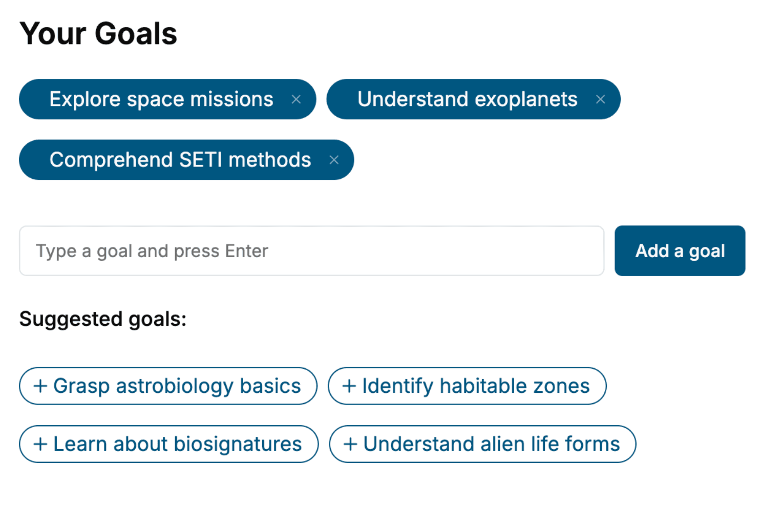
- 1. Select your focus
Select the focus of your Anatomy book and share your background
- 2. Personalize your book
Specify your goals and choose sub-topics to include
- 3. Get your tailored book
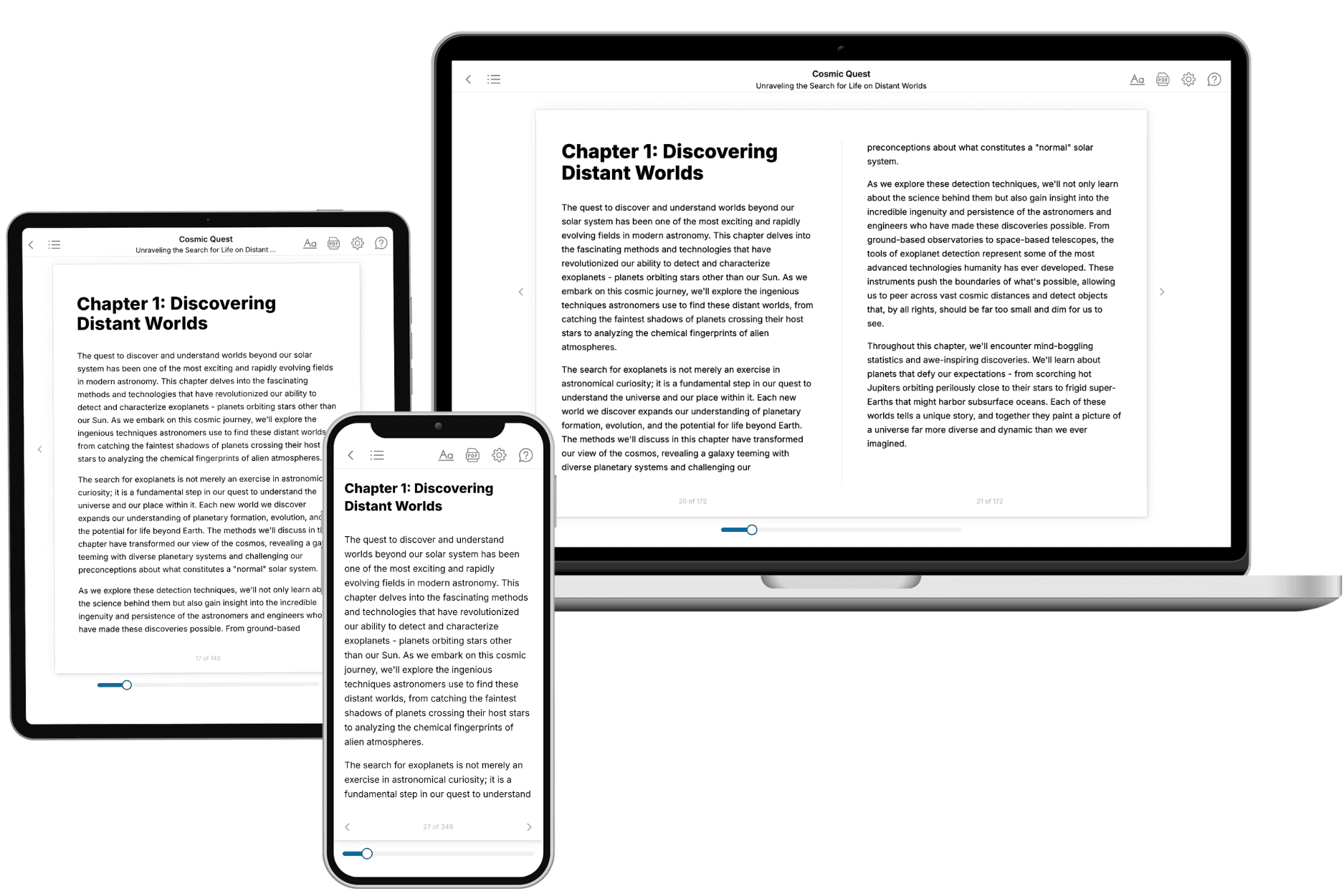
Your book is ready in 10 minutes. Read it online, download as EPUB or PDF, or send to Kindle.

Frequently asked questions
- What is TailoredRead?
TailoredRead creates complete, full-length nonfiction ebooks, written specifically for you. Tell us what you want to learn and why, and TailoredRead writes you a complete ebook on it, with charts, tables, and diagrams throughout. Read it online, on Kindle, or on any e-reader, or download it as a PDF or EPUB. Every book is written fresh the day you order it.
- How is the book tailored to me?
Before anything is written, you answer a few short questions about your goals, your background, and the topics you care about. Your book is then planned around your answers: written at your level, focused on your goals, and covering the topics you chose, instead of the one-size-fits-all book a store sells everyone.
- Will it read like generic AI content?
No. Your book isn't a chat transcript: it's planned chapter by chapter before a single word is written, told in a clear book voice, and explains ideas visually with charts, tables, and diagrams. After writing, every book goes through an editorial pass that polishes chapters so the book reads consistent from start to finish. Judge for yourself: browse thousands of sample books on our Explore Books page.
- How accurate is the content?
Our AI first checks whether your subject needs live research. Niche and fast-moving subjects are researched before writing, and the book plan and chapters are grounded in what it finds. After writing, every book goes through a quality pass that checks its facts and consistency, and researched books list their sources in a Notes section at the back.
- How long is the book?
You choose the length: Comprehensive (250-300 pages), Detailed (150-200 pages), Essential (70-100 pages), or Short (30-50 pages).
- Does the book include visuals?
Yes. Every book includes charts, tables, and diagrams: timelines, checklists, step-by-step guides, decision trees, comparison tables, matrices, and bar charts. A typical book includes dozens of these visual elements.
- Can I read it on Kindle? What formats can I download?
Yes. Every book can be read in our online reader or downloaded as a PDF or EPUB. The EPUB can be opened with Kindle, Apple Books, Kobo, and any e-reader. The Commercial and Author licenses also include an editable Word (DOCX) and Markdown download.
- How much does it cost?
About the price of a regular ebook. The exact price depends on your book's length and complexity, from $2 for the shortest books to $35 for the most comprehensive, and it's shown when you review your book, before you commit to anything. Each book is a one-time purchase, yours to keep, no subscription. Licenses that let you share, teach, or publish your book range from $35.99 to $49.99.
- Can I preview the book before purchasing?
Before you pay, you'll see what your book will cover: the title, a detailed description, the estimated length, and the complete chapter-by-chapter table of contents, so you know what you're getting before you decide. The full text itself is written after you purchase.
- How long does it take to create a book?
About 10 minutes. You can start reading the opening chapters while the rest is still being written.