Histology Mastery
Advanced Techniques for Cell Identification and Tissue Analysis
Navigating the intricate world of histology can be challenging, even for seasoned professionals.
300 pages15 chaptersMay 13, 2025Medical ScienceCreated by Seth
About this book
You face the constant need to stay updated with cutting-edge techniques while maintaining a deep understanding of fundamental concepts. "Histology Mastery: Advanced Techniques for Cell Identification and Tissue Analysis" is your comprehensive guide to overcoming these obstacles. This book delves into the most advanced aspects of histological analysis, from epithelial tissue types to digital pathology. You'll gain in-depth knowledge of staining techniques, including H&E and special stains, as well as cutting-edge methods like confocal microscopy and immunohistochemistry. The text also covers crucial topics such as histological fixation, tissue sectioning, and the intricate details of the extracellular matrix. Whether you're developing teaching materials or writing a histology manual, this book provides the authoritative content you need. Each chapter offers clear, accessible explanations of complex concepts, coupled with practical applications that will enhance your professional skills. By the end of this book, you'll have the confidence and expertise to tackle any histological challenge, elevating your work in cell identification and tissue analysis to new heights.
Table of contents
- IntroductionOverview of Advanced Histology, How to Use This Book, The Importance of Histology in Modern Science, Technological Advances in Histological AnalysisRead below
- Chapter 1: Advanced Epithelial Tissue AnalysisEpithelial Tissue Types and Their Characteristics, Functional Aspects of Epithelial Tissues, Advanced Techniques for Epithelial Cell Identification, Common Pathological Changes in Epithelial Tissues
- Chapter 2: Connective Tissue Matrix ExplorationComponents of the Connective Tissue Matrix, Functional Roles of the Extracellular Matrix, Analytical Methods for Matrix Examination, Pathological Alterations in Connective Tissue Matrix
- Chapter 3: Muscle Fiber Structure and FunctionTypes of Muscle Tissue and Their Characteristics, Ultrastructure of Muscle Fibers, Histological Aspects of Muscle Contraction, Histopathological Changes in Muscle Tissue
- Chapter 4: Nervous Tissue Analysis TechniquesNeuronal Structure and Classification, Types and Functions of Glial Cells, Specialized Staining Techniques for Nervous Tissue, Histopathological Analysis of Nervous Tissue
- Chapter 5: Advanced Staining TechniquesH&E Staining: Advanced Applications and Interpretation, Special Stains for Specific Tissue Components, Fluorescence Staining Methods, Troubleshooting Common Staining Issues
- Chapter 6: Basement Membrane: Structure and FunctionMolecular Components of the Basement Membrane, Functional Roles in Various Tissues, Techniques for Visualizing Basement Membranes, Pathological Changes in Basement Membranes
- Chapter 7: Fundamentals of HistopathologyPrinciples of Histopathological Analysis, Common Pathological Changes in Tissues, Histological Criteria for Diagnosis, Standardized Reporting in Histopathology
- Chapter 8: Digital Pathology and Image AnalysisDigital Imaging in Histology, Computational Image Analysis Techniques, Artificial Intelligence in Histological Analysis, Challenges and Future Directions in Digital Pathology
- Chapter 9: Advanced Histology TerminologyCellular Structure Terminology, Tissue-Specific Terminology, Histopathological Terms and Definitions, Molecular and Genetic Terminology in Histology
- Chapter 10: Confocal Microscopy in HistologyPrinciples of Confocal Microscopy, Sample Preparation for Confocal Imaging, Applications in Histological Research, Interpreting Confocal Microscopy Images
- Chapter 11: Advanced Histological Fixation MethodsPrinciples of Tissue Fixation, Types of Fixatives and Their Applications, Optimizing Fixation Protocols for Different Tissues, Recognizing and Avoiding Fixation Artifacts
- Chapter 12: Extracellular Matrix in DepthMolecular Components of the Extracellular Matrix, Functional Roles in Tissue Homeostasis, Cell-Matrix Interactions, Extracellular Matrix in Pathological Conditions
- Chapter 13: Cell Adhesion Molecules and JunctionsTypes of Cell Adhesion Molecules, Structure and Function of Cellular Junctions, Techniques for Visualizing Cell Adhesion, Cell Adhesion in Pathological Processes
- Chapter 14: Advanced Tissue Sectioning TechniquesMethods of Tissue Sectioning, Cryosectioning and Its Applications, Ultrathin Sectioning for Electron Microscopy, Troubleshooting Sectioning Artifacts
- Chapter 15: Advanced ImmunohistochemistryPrinciples of Immunohistochemistry, Antibody Selection and Validation, Multiplex Immunohistochemistry Techniques, Quantification in Immunohistochemistry
- ConclusionFuture Directions in Histology, Emerging Technologies in Tissue Analysis
- Glossary
- Further Reading
Introduction
Welcome to the world of advanced histology, where the intricate structures of life reveal themselves under the scrutiny of cutting-edge techniques and technologies. As you embark on this journey through "Histology Mastery: Advanced Techniques for Cell Identification and Tissue Analysis," you are poised to deepen your understanding and refine your skills in one of the most fundamental and evolving fields of biological science. This introduction serves as your gateway to the complex and fascinating realm of cellular and tissue architecture, setting the stage for the in-depth exploration that lies ahead in subsequent chapters.
Histology, the study of the microscopic structure of tissues, has come a long way since its inception. From the early days of simple light microscopy to today's advanced digital imaging and molecular techniques, the field has continuously evolved, offering ever more precise and detailed insights into the building blocks of life. As an advanced practitioner with a strong foundation in anatomy and neuroscience, you are well-positioned to appreciate the nuances and complexities that this book will unveil. The knowledge and techniques presented here will not only enhance your professional capabilities but also empower you to contribute meaningfully to the creation of educational materials and the development of comprehensive histology manuals.
Throughout this book, you will encounter a blend of time-honored principles and cutting-edge methodologies. From the classic hematoxylin and eosin (H&E) staining to the latest developments in immunohistochemistry and digital pathology, each chapter is designed to build upon your existing knowledge and push the boundaries of your expertise. The goal is to equip you with a comprehensive toolkit that will enable you to tackle the most challenging aspects of histological analysis with confidence and precision.
As we delve into the various topics, keep in mind that histology is not just about identifying structures; it's about understanding the intricate relationships between cells, tissues, and organs. This holistic approach will be emphasized throughout the book, helping you to see beyond individual components and appreciate the complex interplay that defines biological systems. Whether you're examining the delicate balance of the extracellular matrix or unraveling the mysteries of neural tissue, the insights gained here will prove invaluable in your professional endeavors.
Now, let's begin our exploration of advanced histology by examining the core components of this introduction: an overview of advanced histology, guidance on how to use this book effectively, the importance of histology in modern science, and the technological advances that are shaping the future of histological analysis.
Overview of Advanced Histology
Advanced histology represents the pinnacle of tissue analysis, combining traditional microscopic examination with sophisticated molecular and imaging techniques. At this level, histology transcends mere description and enters the realm of functional understanding and predictive analysis. The field encompasses a wide array of methodologies, each designed to reveal specific aspects of cellular and tissue biology that are crucial for both research and clinical applications.
One of the hallmarks of advanced histology is its interdisciplinary nature. It draws upon principles from cell biology, biochemistry, physics, and even computer science to provide a comprehensive view of tissue structure and function. This integration of diverse scientific disciplines allows for a more nuanced interpretation of histological data, enabling researchers and clinicians to draw meaningful conclusions about tissue health, disease processes, and potential therapeutic interventions.
At the core of advanced histology lies the ability to visualize and interpret cellular and subcellular structures with unprecedented clarity and specificity. This is achieved through a combination of advanced staining techniques, high-resolution microscopy, and sophisticated image analysis tools. For instance, immunohistochemistry allows for the precise localization of specific proteins within tissues, providing insights into cellular function and pathological changes that would be impossible to discern with traditional staining methods alone.
Another key aspect of advanced histology is the emphasis on quantitative analysis. Modern histological techniques go beyond qualitative descriptions, offering precise measurements of cellular features, protein expression levels, and tissue architecture. This quantitative approach enables more objective comparisons between samples and facilitates the detection of subtle changes that might be missed by conventional histological examination.
The field of advanced histology also encompasses the study of dynamic cellular processes. Techniques such as live cell imaging and intravital microscopy allow researchers to observe cellular behaviors in real-time, providing insights into processes like cell migration, division, and interactions within living tissues. These dynamic studies complement traditional fixed-tissue histology, offering a more complete picture of tissue function and pathology.
Furthermore, advanced histology has embraced the digital revolution, with digital pathology and whole slide imaging becoming increasingly prevalent. These technologies not only facilitate the storage and sharing of histological data but also open up new possibilities for automated analysis and machine learning applications. The integration of artificial intelligence into histological analysis promises to enhance diagnostic accuracy and efficiency, potentially revolutionizing both research and clinical practice.
As you progress through this book, you will explore these advanced concepts in detail, gaining a deeper understanding of how they can be applied to solve complex biological questions and improve diagnostic accuracy. The knowledge and skills you acquire will enable you to push the boundaries of histological analysis, contributing to advancements in both basic science and clinical medicine.
It's important to note that while advanced histology offers powerful tools for tissue analysis, it also requires a critical and analytical mindset. The interpretation of complex histological data demands a thorough understanding of both the strengths and limitations of each technique. Throughout this book, you will be encouraged to develop this critical perspective, ensuring that you can apply advanced histological methods effectively and interpret results accurately.
In the following sections, we will delve into specific advanced histological techniques, exploring their principles, applications, and potential pitfalls. By the end of this book, you will have a comprehensive understanding of the state-of-the-art in histological analysis, equipping you to tackle the most challenging questions in tissue biology and pathology.
How to Use This Book
"Histology Mastery: Advanced Techniques for Cell Identification and Tissue Analysis" is designed to be a comprehensive resource for advanced practitioners in the field of histology. To maximize your learning and ensure that you can effectively apply the knowledge gained from this book, it's important to understand its structure and how to navigate through its contents. This section will guide you on how to make the most of this educational resource, whether you're using it for self-study, as a reference in your professional work, or as a basis for creating educational materials.
The book is organized into fifteen main chapters, each focusing on a specific aspect of advanced histology. These chapters are designed to build upon each other, starting with fundamental concepts and progressing to more complex and specialized topics. However, each chapter is also self-contained, allowing you to focus on specific areas of interest or to use the book as a reference guide for particular techniques or tissue types.
At the beginning of each chapter, you'll find a brief overview that outlines the key concepts and techniques that will be covered. This overview serves as a roadmap for the chapter, helping you to understand the context and significance of the material that follows. Pay close attention to these introductions, as they often highlight the connections between different topics and provide insights into how the chapter's content fits into the broader field of histology.
Throughout the book, you'll encounter detailed explanations of advanced histological techniques, accompanied by high-quality images and diagrams. These visual aids are crucial for understanding the complex structures and processes discussed in the text. Take the time to study these images carefully, relating them to the accompanying descriptions. This practice will enhance your ability to recognize and interpret histological features in your own work.
Each chapter also includes practical tips and troubleshooting advice, drawn from years of experience in histological research and practice. These insights are invaluable for avoiding common pitfalls and optimizing your histological techniques. Pay special attention to these sections, as they often contain the kind of practical knowledge that can make a significant difference in the quality and reliability of your histological analyses.
To reinforce your learning and help you apply the concepts covered, each chapter concludes with a set of review questions and practical exercises. These are designed to challenge your understanding and encourage you to think critically about the material. Don't skip these sections – they are an excellent way to assess your grasp of the content and to identify areas where you might need further study.
The book also includes extensive references and suggestions for further reading at the end of each chapter. These resources are carefully curated to provide you with additional depth on specific topics and to keep you informed about the latest developments in the field. Make use of these references to expand your knowledge beyond the scope of this book and to stay current with emerging trends in histology.
For those using this book as a basis for creating educational materials or writing a histology manual, pay particular attention to the way concepts are presented and explained. The structure of each chapter can serve as a model for organizing your own educational content. The balance between theoretical explanations and practical applications demonstrated throughout the book can guide you in creating comprehensive and engaging learning materials.
As you work through the book, it's recommended that you take notes and highlight key points. This active engagement with the material will help reinforce your learning and make it easier to review important concepts later. Consider creating your own summary sheets or mind maps for each chapter, synthesizing the main ideas and techniques in a format that's easy for you to review and reference.
Remember that mastery of advanced histology techniques requires both theoretical understanding and practical experience. While this book provides a wealth of knowledge, it's essential to complement your reading with hands-on practice whenever possible. Apply the techniques and principles discussed in the book to your own histological samples, and don't hesitate to experiment with different methods to see how they affect your results.
Finally, keep in mind that histology is a rapidly evolving field. While this book covers the most up-to-date techniques and knowledge available at the time of writing, new developments are constantly emerging. Use this book as a foundation, but stay curious and continue to seek out new information and techniques as they become available. The skills and knowledge you gain from this book will provide you with the critical thinking and analytical abilities needed to evaluate and incorporate new advances in histology throughout your career.
The Importance of Histology in Modern Science
Histology, the study of the microscopic structure of tissues, stands as a cornerstone of modern biological and medical sciences. Its importance cannot be overstated, as it provides the fundamental knowledge necessary for understanding the complex interplay between structure and function in living organisms. In an era of rapid scientific advancement, histology continues to play a crucial role in bridging the gap between molecular biology and gross anatomy, offering unique insights that are essential for both basic research and clinical applications.
At its core, histology allows us to visualize and analyze the basic units of life – cells and tissues – in their native context. This contextual understanding is critical for interpreting the complex processes that occur within organisms. While molecular biology techniques can provide detailed information about specific genes or proteins, histology offers a holistic view of how these components interact within the structural framework of tissues and organs. This integrative approach is essential for understanding physiological processes, developmental biology, and the mechanisms underlying various diseases[1].
In the realm of medical science, histology serves as the foundation for pathology, the study of disease processes. Histopathological examination remains the gold standard for diagnosing many diseases, particularly cancer. The ability to identify abnormal cellular and tissue structures is crucial for accurate diagnosis, prognosis, and treatment planning. Advanced histological techniques, such as immunohistochemistry and in situ hybridization, have further enhanced the diagnostic power of histology, allowing for the detection of specific molecular markers associated with various pathological conditions[2].
Moreover, histology plays a vital role in drug development and toxicology studies. The evaluation of tissue responses to new therapeutic agents or potential toxins relies heavily on histological analysis. This is crucial for assessing both the efficacy and safety of new drugs, as well as understanding the mechanisms of drug action and potential side effects at the tissue level. The ability to visualize and quantify changes in tissue structure and cellular composition provides invaluable information that complements biochemical and molecular data in preclinical and clinical studies[3].
In the field of regenerative medicine and tissue engineering, histology is indispensable for evaluating the success of tissue reconstruction and stem cell therapies. The ability to assess the integration of engineered tissues, the differentiation of stem cells, and the restoration of normal tissue architecture relies heavily on histological techniques. These analyses are crucial for advancing our ability to repair or replace damaged tissues and organs, offering hope for treating a wide range of degenerative diseases and injuries[4].
The importance of histology extends to evolutionary biology and comparative anatomy as well. By studying the histological features of different species, researchers can gain insights into evolutionary adaptations and the development of specialized tissues and organs. This comparative approach not only enhances our understanding of biological diversity but also provides valuable insights into the evolutionary origins of human anatomy and physiology[5].
In neuroscience, histology remains a critical tool for understanding brain structure and function. Advanced histological techniques have revolutionized our ability to map neural circuits, identify specific cell types, and visualize the distribution of neurotransmitters and receptors in the nervous system. These insights are fundamental to unraveling the complexities of brain function and developing new treatments for neurological and psychiatric disorders[6].
Furthermore, the integration of histology with other cutting-edge technologies has opened up new avenues for research and diagnosis. For instance, the combination of histological techniques with advanced imaging modalities, such as confocal microscopy and electron microscopy, allows for unprecedented visualization of cellular and subcellular structures. Similarly, the integration of histology with molecular biology techniques, such as laser capture microdissection and single-cell sequencing, enables the correlation of tissue morphology with gene expression profiles at a resolution previously unattainable[7].
In conclusion, histology remains a vital and dynamic field in modern science, continually adapting to incorporate new technologies and methodologies. Its importance lies not only in its ability to provide detailed structural information but also in its capacity to bridge multiple disciplines, from molecular biology to clinical medicine. As you delve deeper into the advanced techniques and applications covered in this book, remember that the knowledge and skills you gain will contribute to this broader scientific landscape, enabling you to make significant contributions to our understanding of life at its most fundamental level.
Technological Advances in Histological Analysis
The field of histology has undergone a remarkable transformation in recent years, driven by rapid advancements in technology. These innovations have not only enhanced the precision and efficiency of traditional histological techniques but have also opened up entirely new avenues for tissue analysis. As you embark on your journey through advanced histology, it's crucial to understand these technological advances and their impact on the field. This section will explore some of the most significant developments that are shaping the future of histological analysis.
One of the most revolutionary advances in histology has been the development of digital pathology and whole slide imaging (WSI) systems. These technologies allow for the digitization of entire histological slides at high resolution, creating virtual slides that can be viewed, analyzed, and shared electronically. Digital pathology has transformed the way histological specimens are examined, enabling remote consultation, automated image analysis, and the creation of large-scale digital archives. The advantages of this technology are numerous, including improved workflow efficiency, enhanced collaboration among pathologists, and the potential for applying artificial intelligence algorithms to assist in diagnosis[8].
Artificial intelligence (AI) and machine learning algorithms are increasingly being applied to histological image analysis. These computational approaches can assist in tasks such as cell counting, tissue segmentation, and the identification of specific histological features. AI-powered systems have shown promise in improving the accuracy and consistency of histological assessments, particularly in areas such as cancer diagnosis and grading. As these technologies continue to evolve, they are likely to become integral tools in both research and clinical histology, augmenting the capabilities of human experts[9].
Advanced microscopy techniques have greatly expanded the capabilities of histological analysis. Confocal microscopy, for instance, allows for the visualization of thick tissue sections with high resolution and the ability to create three-dimensional reconstructions. This technique has been particularly valuable in neuroscience and developmental biology studies. Similarly, multiphoton microscopy enables deep tissue imaging with minimal photodamage, making it ideal for intravital imaging of living tissues. These advanced microscopy methods provide unprecedented insights into tissue architecture and cellular interactions in both fixed and living specimens[10].
The field of immunohistochemistry has also seen significant advancements. Multiplex immunohistochemistry techniques now allow for the simultaneous detection of multiple antigens within a single tissue section. This capability is particularly valuable for studying complex cellular interactions and heterogeneous tissue environments, such as in tumor microenvironments. Coupled with spectral imaging systems, these techniques enable the visualization and quantification of numerous markers simultaneously, providing a more comprehensive view of tissue composition and cellular phenotypes[11].
Advancements in tissue clearing techniques have revolutionized the study of three-dimensional tissue architecture. Methods such as CLARITY, CUBIC, and iDISCO+ allow for the rendering of entire organs optically transparent while preserving their molecular and cellular architecture. When combined with light-sheet microscopy, these techniques enable the visualization of intact organs at cellular resolution, providing unprecedented insights into complex anatomical structures and cellular distributions[12].
The integration of histology with molecular biology techniques has led to the development of in situ sequencing and spatial transcriptomics methods. These technologies allow for the visualization and quantification of gene expression patterns directly within tissue sections, preserving spatial information. This integration of histological and molecular data provides a powerful tool for understanding gene function in the context of tissue architecture and has applications in fields ranging from developmental biology to cancer research[13].
Advances in sample preparation techniques have also contributed significantly to the field. Cryogenic electron microscopy (cryo-EM) techniques, for instance, allow for the visualization of biological specimens in their near-native state at near-atomic resolution. While traditionally used for studying isolated macromolecules, cryo-EM is increasingly being applied to tissue sections, offering new insights into cellular ultrastructure[14].
The field of nanotechnology has also made inroads into histological analysis. Nanoparticle-based probes and sensors can be used to detect specific molecules or cellular processes within tissues with high sensitivity and specificity. These tools offer new possibilities for studying dynamic cellular processes and molecular interactions in both fixed and living tissues[15].
As you progress through this book and your career in histology, it's important to stay abreast of these technological advances and their applications. The integration of these new tools and techniques with traditional histological methods will undoubtedly lead to new discoveries and insights into tissue biology and pathology. By mastering both the fundamental principles of histology and these cutting-edge technologies, you will be well-equipped to contribute to the ongoing evolution of this vital field.
Notes
Introduction
- 1. A. L. Mescher, Junqueira's Basic Histology, McGraw-Hill Education, 2018
- 2. V. Kumar and others, Robbins Basic Pathology, 10th Edition, Elsevier, 2017
- 3. B. Bolon and R. Ochoa, Fundamentals of Toxicologic Pathology, 3rd Edition, Academic Press, 2018
- 4. A. Atala and R. Lanza, Handbook of Tissue Engineering Scaffolds, Woodhead Publishing, 2018
- 5. K. V. Kardong, Vertebrates: Comparative Anatomy, Function, Evolution, 8th Edition, McGraw-Hill Education, 2018
- 6. G. Paxinos and K. B. J. Franklin, Paxinos and Franklin's the Mouse Brain in Stereotaxic Coordinates, 5th Edition, Academic Press, 2019
- 7. W. Pawlina and M. H. Ross, Histology: A Text and Atlas: With Correlated Cell and Molecular Biology, 8th Edition, Wolters Kluwer, 2018
- 8. L. Pantanowitz and others, "Digital pathology," Journal of Pathology Informatics, 2018
- 9. K. Bera and others, "Artificial intelligence in digital pathology - new tools for diagnosis and precision oncology," Nature Reviews Clinical Oncology, 2019
- 10. J. Jonkman and C. M. Brown, "Any Way You Slice It-A Comparison of Confocal Microscopy Techniques," Journal of Biomolecular Techniques, 2015
- 11. W. C. C. Tan and others, Overview of multiplex immunohistochemistry/immunofluorescence techniques in the era of cancer immunotherapy, Cancer Communications, 2020
- 12. H. R. Ueda and others, "Tissue clearing and its applications in neuroscience," Nature Reviews Neuroscience, 2020
- 13. P. L. Ståhl and others, "Visualization and analysis of gene expression in tissue sections by spatial transcriptomics," Science, 2016
- 14. R. I. Koning and others, "Cryo-electron tomography," Biochemical Society Transactions, 2018
- 15. O. S. Wolfbeis, "An overview of nanoparticles commonly used in fluorescent bioimaging," Chemical Society Reviews, 2015
This is the end of the free sample. The full book has 15 chapters.
Want this book written for you?
Tell us your goals and background, and get your own version of this book, tailored by AI to you.


Benefits of AI-tailored books
- Read one book, not ten:
- all the Medical Science knowledge you need, gathered into a single book.
- Save days of learning:
- tell us your goals, choose your topics, and the whole book is built around them.
- Learn effortlessly:
- Medical Science book written for your specific background and expertise.
- Understand at a glance:
- includes charts, tables, timelines, checklists, decision trees, and more.
- Stay current:
- written the day you order it, not years ago.
- Trust what you read:
- every book is checked for accuracy, with real research when your subject needs it.
Create your unique book in 3 steps
- 1. Select your focus
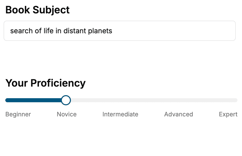
Select the focus of your Medical Science book and share your background
- 2. Personalize your book
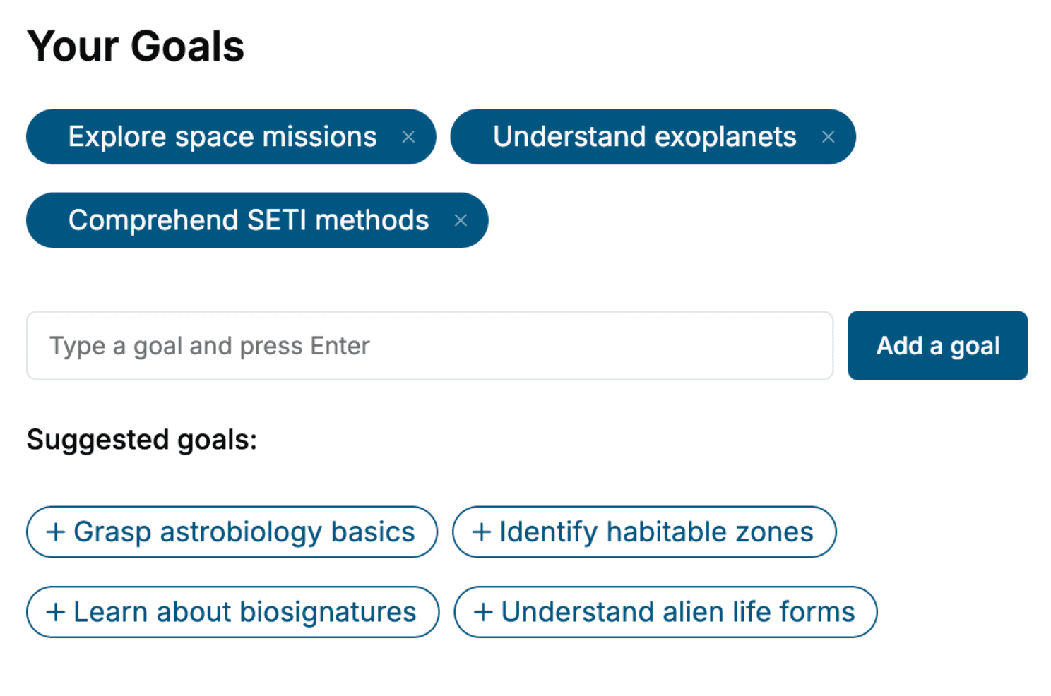
Specify your goals and choose sub-topics to include
- 3. Get your tailored book
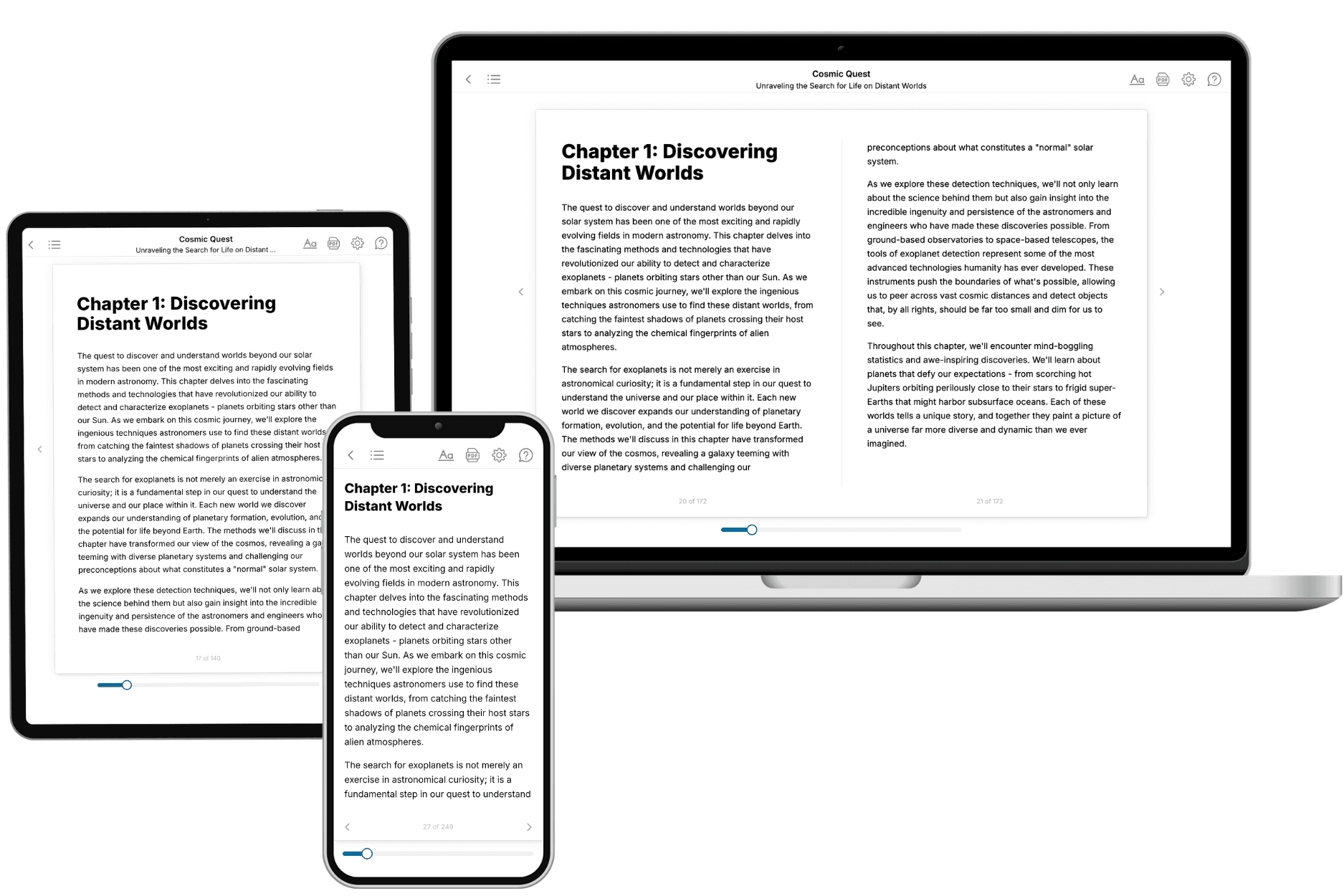
Your book is ready in 10 minutes. Read it online, download as EPUB or PDF, or send to Kindle.
Frequently asked questions
- What is TailoredRead?
TailoredRead creates complete, full-length nonfiction ebooks, written specifically for you. Tell us what you want to learn and why, and TailoredRead writes you a complete ebook on it, with charts, tables, and diagrams throughout. Read it online, on Kindle, or on any e-reader, or download it as a PDF or EPUB. Every book is written fresh the day you order it.
- How is the book tailored to me?
Before anything is written, you answer a few short questions about your goals, your background, and the topics you care about. Your book is then planned around your answers: written at your level, focused on your goals, and covering the topics you chose, instead of the one-size-fits-all book a store sells everyone.
- Will it read like generic AI content?
No. Your book isn't a chat transcript: it's planned chapter by chapter before a single word is written, told in a clear book voice, and explains ideas visually with charts, tables, and diagrams. After writing, every book goes through an editorial pass that polishes chapters so the book reads consistent from start to finish. Judge for yourself: browse thousands of sample books on our Explore Books page.
- How accurate is the content?
Our AI first checks whether your subject needs live research. Niche and fast-moving subjects are researched before writing, and the book plan and chapters are grounded in what it finds. After writing, every book goes through a quality pass that checks its facts and consistency, and researched books list their sources in a Notes section at the back.
- How long is the book?
You choose the length: Comprehensive (250-300 pages), Detailed (150-200 pages), Essential (70-100 pages), or Short (30-50 pages).
- Does the book include visuals?
Yes. Every book includes charts, tables, and diagrams: timelines, checklists, step-by-step guides, decision trees, comparison tables, matrices, and bar charts. A typical book includes dozens of these visual elements.
- Can I read it on Kindle? What formats can I download?
Yes. Every book can be read in our online reader or downloaded as a PDF or EPUB. The EPUB can be opened with Kindle, Apple Books, Kobo, and any e-reader. The Commercial and Author licenses also include an editable Word (DOCX) and Markdown download.
- How much does it cost?
About the price of a regular ebook. The exact price depends on your book's length and complexity, from $2 for the shortest books to $35 for the most comprehensive, and it's shown when you review your book, before you commit to anything. Each book is a one-time purchase, yours to keep, no subscription. Licenses that let you share, teach, or publish your book range from $35.99 to $49.99.
- Can I preview the book before purchasing?
Before you pay, you'll see what your book will cover: the title, a detailed description, the estimated length, and the complete chapter-by-chapter table of contents, so you know what you're getting before you decide. The full text itself is written after you purchase.
- How long does it take to create a book?
About 10 minutes. You can start reading the opening chapters while the rest is still being written.