The Kidney Coloring Atlas
A Visual Journey Through Renal Anatomy and Function
Have you ever struggled to visualize how the kidney's complex structures fit together and function as an integrated system?
207 pages11 chaptersOct 27, 2025Anatomy Coloring
About this book
Traditional anatomy textbooks present static images that fail to engage your mind in the active learning process necessary for true mastery. This coloring atlas transforms your approach to understanding renal anatomy by combining detailed illustrations with the proven cognitive benefits of hands-on coloring. You'll explore every level of kidney structure—from the external capsule to the microscopic glomerulus—while actively reinforcing your knowledge through color-coding and visual association. Each illustration is carefully designed to highlight spatial relationships, blood flow patterns, and functional connections that bring the kidney to life. Whether you're preparing for exams, refreshing your anatomical knowledge, or simply fascinated by how this remarkable organ filters blood and maintains homeostasis, this atlas provides the perfect balance of scientific accuracy and engaging learning methodology. By the time you complete this atlas, you'll possess a comprehensive, three-dimensional understanding of renal anatomy that static study methods simply cannot achieve.
Table of contents
- IntroductionWhy Coloring Enhances Anatomical Learning, How to Use This Atlas Effectively, Recommended Coloring Tools and Techniques, The Kidney: An Overview of Form and FunctionRead below
- Chapter 1: Gross Anatomy of the KidneyExternal Features and Surface Anatomy, Anatomical Position and Surrounding Structures, Renal Capsule and Protective Layers, The Renal Hilum and Its Contents
- Chapter 2: Internal ArchitectureCortex and Medulla: Regional Organization, Renal Pyramids and Columns, Lobes and Lobules of the Kidney
- Chapter 3: The Renal Collecting SystemMinor Calyces and Renal Papillae, Major Calyces and Their Arrangement, The Renal Pelvis and Ureter Connection, Pathway of Urine Flow
- Chapter 4: Renal Blood SupplyRenal Artery and Its Major Branches, Segmental and Interlobar Arteries, Arcuate and Interlobular Vessels, Venous Drainage Patterns
- Chapter 5: The Nephron: Functional UnitCortical and Juxtamedullary Nephrons, Components of the Nephron, Spatial Distribution Within the Kidney
- Chapter 6: The Renal CorpuscleGlomerular Capillary Network, Bowman's Capsule: Parietal and Visceral Layers, The Filtration Barrier and Its Components, Mesangial Cells and Matrix
- Chapter 7: The Tubular SystemProximal Convoluted Tubule Structure, Loop of Henle: Descending and Ascending Limbs, Distal Convoluted Tubule, Collecting Ducts and Papillary Ducts
- Chapter 8: The Juxtaglomerular ApparatusJuxtaglomerular Cells and Renin Secretion, Macula Densa and Tubular Feedback, Extraglomerular Mesangial Cells
- Chapter 9: Renal MicrovasculatureAfferent and Efferent Arterioles, Peritubular Capillary Network, Vasa Recta and Medullary Circulation
- Chapter 10: Renal Interstitium and Supporting StructuresCortical Interstitial Components, Medullary Interstitial Cells and Matrix, Lymphatic Drainage of the Kidney
- Chapter 11: Clinical Anatomical CorrelationsCommon Anatomical Variations, How Diseases Affect Renal Structure, Kidney Anatomy in Medical Imaging
- ConclusionIntegrating Structure and Function, Continuing Your Anatomical Studies
Introduction
The kidney stands as one of the most architecturally sophisticated organs in the human body, a masterpiece of biological engineering that performs over a million filtration operations simultaneously every moment of your life. Yet for many students and healthcare professionals, understanding its intricate three-dimensional structure remains frustratingly elusive. Traditional anatomy textbooks present static, two-dimensional images that fail to capture the spatial relationships between structures or engage the active learning processes necessary for deep comprehension. This atlas takes a fundamentally different approach, one grounded in the neuroscience of learning and the proven benefits of hands-on engagement with anatomical material.
Active learning transforms passive observation into dynamic understanding. When you color an anatomical structure, you're not simply filling in spaces on a page—you're creating neural pathways that connect visual information with spatial reasoning, functional knowledge, and long-term memory formation. Research in educational psychology consistently demonstrates that learners who actively manipulate information retain significantly more than those who merely read or observe[1]. The act of choosing colors, tracing boundaries, and following structures through multiple illustrations engages your motor cortex, visual processing centers, and prefrontal planning regions simultaneously, creating a rich, multisensory learning experience that static study cannot replicate.
The kidney's complexity demands this kind of active engagement. From the smooth outer capsule to the microscopic podocytes wrapping around glomerular capillaries, renal anatomy spans multiple scales and organizational levels. Blood enters through a single renal artery but branches into over one million glomerular capillary networks, each serving as a filtration unit. Urine begins its formation in these microscopic structures, then travels through an elaborate collecting system that converges from millions of nephrons into a single ureter. Understanding how these components fit together—how cortical structures relate to medullary structures, how blood flow patterns enable filtration, how the juxtaglomerular apparatus monitors and regulates kidney function—requires more than memorization. It requires visualization, spatial reasoning, and the kind of deep processing that coloring facilitates.
This atlas guides you through every level of renal organization with carefully designed illustrations that build progressively from gross anatomy to microscopic detail. Each chapter focuses on a specific anatomical region or functional system, presenting multiple views and cross-sections that reveal how structures appear from different perspectives. The illustrations are intentionally designed for coloring, with clear boundaries, consistent labeling, and strategic use of white space that invites your active participation. As you work through the atlas, you'll develop an increasingly sophisticated mental model of kidney structure, one that integrates form with function and connects individual components into a coherent, three-dimensional whole.
Why Coloring Enhances Anatomical Learning
The cognitive benefits of coloring anatomical structures extend far beyond simple engagement or enjoyment. When you color a renal structure, you activate multiple brain regions simultaneously, creating what neuroscientists call elaborative encoding—the process of connecting new information to existing knowledge through multiple pathways. Your visual cortex processes the shape and boundaries of structures, your motor cortex controls the precise movements of coloring, your language centers process the anatomical terms, and your prefrontal cortex makes decisions about color choices and relationships between structures. This multisensory engagement creates stronger, more durable memories than passive reading or observation alone.
Coloring forces you to pay attention to boundaries and relationships in ways that casual observation does not. Consider the renal corpuscle, where Bowman's capsule surrounds the glomerular capillary tuft. When you color these structures, you must carefully trace where the visceral layer of Bowman's capsule meets the parietal layer, where the capillaries enter and exit, and how the urinary space separates these components. This detailed attention to boundaries creates precise mental representations of spatial relationships. You cannot color carelessly and still understand the anatomy—the act of coloring demands the same precision that anatomical understanding requires. This alignment between the learning activity and the learning goal makes coloring uniquely effective for anatomy education.
Color-coding creates visual associations that enhance memory retrieval. When you consistently use specific colors for related structures—perhaps red for arteries, blue for veins, yellow for tubular structures—you create a visual language that helps organize complex information. Later, when you need to recall the pathway of blood through the kidney, the color associations you created while coloring serve as retrieval cues. Research on memory demonstrates that information encoded with multiple cues (visual, motor, semantic) is more easily retrieved than information encoded through a single modality[2]. Your color choices become part of the memory itself, providing additional pathways for accessing stored information.
The active decision-making involved in coloring promotes deeper cognitive processing than passive study methods. Each time you choose a color for a structure, you must consider its function, its relationships to surrounding structures, and its role in the larger system. Should the proximal convoluted tubule be the same color as the distal convoluted tubule, or different colors to emphasize their distinct functions? Should cortical and juxtamedullary nephrons use the same color scheme, or different shades to highlight their anatomical differences? These decisions require you to think critically about anatomical relationships and functional significance, transforming coloring from a mechanical task into an analytical exercise that deepens understanding.
Coloring also provides immediate feedback about your understanding. If you cannot decide what color to use for a structure, or if you're uncertain where one structure ends and another begins, you've identified a gap in your knowledge. This metacognitive awareness—knowing what you don't know—is crucial for effective learning. Unlike passive reading, where you might move past confusing material without realizing you don't understand it, coloring makes gaps in understanding immediately apparent. You cannot color what you cannot identify or distinguish from surrounding structures. This built-in feedback mechanism helps you focus your study efforts on areas that need additional attention.
The repetitive nature of coloring reinforces learning through spaced practice. As you color multiple views of the same structure—perhaps seeing the renal artery in a gross anatomical view, then in a coronal section, then in a detailed view of its branches—you encounter the same information multiple times in different contexts. This spaced repetition, combined with the varied perspectives, strengthens memory consolidation and helps you develop a flexible, three-dimensional understanding of renal anatomy. Each encounter with a structure deepens your mental model, adding new details and connections that integrate with previously learned information.
Finally, coloring creates a personalized study resource that reflects your own learning process. The atlas you complete becomes uniquely yours, marked by your color choices, your notes, and your emphasis on structures you found particularly important or challenging. This personalization increases the value of the atlas as a review tool. When you return to your colored illustrations before an exam or clinical application, you're not just seeing generic anatomical drawings—you're seeing the product of your own cognitive engagement, complete with the associations and insights you developed during the coloring process. This personal connection to the material enhances both motivation and memory retention.
How to Use This Atlas Effectively
Maximizing the educational value of this atlas requires a strategic approach that goes beyond simply coloring illustrations in sequence. Begin each chapter by reading the introductory text carefully, paying attention to the key concepts and relationships that will be illustrated. This preview activates your prior knowledge and creates a mental framework for organizing the new information you'll encounter. Understanding the functional significance of structures before you color them helps you make more meaningful decisions about color choices and focus your attention on the most important anatomical relationships. Think of the reading as creating a scaffold upon which you'll build detailed visual knowledge through coloring.
Approach each illustration systematically rather than randomly. Start by identifying all labeled structures before you begin coloring, ensuring you understand what each term refers to and how it relates to surrounding structures. If you encounter unfamiliar terms, consult the accompanying text or external resources before proceeding. This preparation prevents the common mistake of coloring mechanically without understanding what you're coloring. Once you've identified all structures, plan your color scheme before applying any color. Consider which structures should share colors to emphasize relationships, and which should contrast to highlight distinctions. This planning phase engages your analytical thinking and ensures your color choices support rather than obscure anatomical understanding.
Use a consistent color-coding system throughout the atlas to reinforce relationships across chapters. Establishing conventions early—such as always using red for arterial structures, blue for venous structures, and yellow for tubular components—creates visual continuity that helps you recognize structures in different contexts. However, don't let consistency become rigidity. When you need to distinguish between similar structures, such as different segments of the nephron tubule, use variations in shade or intensity rather than completely different colors. This approach maintains the overall organizational scheme while allowing for necessary distinctions. Consider creating a color key on the inside cover of your atlas to maintain consistency as you work through different chapters.
Layer your learning by working through the atlas in the intended sequence, which progresses from gross anatomy to increasingly detailed microscopic structures. This organization mirrors the way anatomical knowledge naturally builds, with each chapter providing foundation for the next. The gross anatomy chapters establish the overall organization of the kidney and its major regions. The vascular chapters show how blood reaches every part of the organ. The nephron chapters reveal the microscopic functional units that perform filtration. By following this progression, you develop a hierarchical understanding that connects large-scale organization with microscopic detail. Resist the temptation to skip ahead to chapters that seem more interesting—the sequential approach ensures you have the necessary context for understanding complex structures.
Engage actively with the text accompanying each illustration. Don't simply color and move on—read the descriptions, study the relationships explained in the text, and verify that your colored illustration reflects the anatomical relationships described. Use the text to check your understanding, asking yourself questions like:
- Can I identify this structure in my colored illustration?
- Do my color choices reflect the functional relationships described?
- Can I trace the pathway described in the text through my colored structures?
- Do I understand why these structures are positioned in this relationship?
Create additional learning tools as you work through the atlas. Consider making flashcards for structures you find challenging, with the structure name on one side and its location, function, and relationships on the other. Draw simplified diagrams in a separate notebook that show key relationships without all the detail of the atlas illustrations. Write summary paragraphs explaining the pathway of blood through the kidney or the journey of filtrate through the nephron. These supplementary activities reinforce your learning through additional encoding pathways and provide varied review materials for later study. The atlas serves as your primary visual resource, but these additional tools help consolidate and organize the information in different formats.
Review your colored illustrations regularly using spaced repetition principles. Don't wait until you've completed the entire atlas to review earlier chapters. Instead, periodically return to previously colored illustrations and test your recall by covering the labels and attempting to identify structures from memory. This retrieval practice—actively recalling information rather than passively reviewing it—is one of the most effective learning strategies identified by cognitive psychology research[3]. Use your colored atlas as a self-testing tool, progressively increasing the difficulty by trying to recall not just structure names but also their functions, relationships, and clinical significance.
Consider working through the atlas with a study partner or group. Collaborative learning adds social and verbal dimensions to the visual and motor engagement of coloring. Discuss your color choices and the reasoning behind them. Quiz each other on structure identification. Explain anatomical relationships to each other in your own words. Teaching material to others is one of the most powerful learning strategies available, forcing you to organize information clearly and identify gaps in your own understanding. Even if you prefer to do the actual coloring independently, regular study sessions where you compare colored illustrations and discuss anatomical concepts can significantly enhance learning outcomes.
Recommended Coloring Tools and Techniques
Selecting appropriate coloring tools significantly impacts both the quality of your finished illustrations and the effectiveness of the learning process. Colored pencils are generally the most versatile and recommended option for anatomical coloring. They allow for precise control, can be layered to create different intensities, and don't bleed through paper. Choose a set with at least 24 colors to provide sufficient variety for distinguishing similar structures. High-quality colored pencils with soft, blendable cores work best for anatomical illustration, as they allow you to create smooth, even coverage and subtle gradations in shade. Brands designed for adult coloring or professional illustration typically offer better pigmentation and blendability than children's colored pencils.
Fine-tipped markers offer an alternative that produces vibrant, consistent colors with less effort than colored pencils. If you choose markers, select alcohol-based or gel-based varieties specifically designed to minimize bleed-through, and test them on a less important page before using them throughout the atlas. Use markers with fine tips (0.5mm or smaller) for precise coloring of small structures and boundaries. Consider using markers for larger areas and colored pencils for detail work, combining the efficiency of markers with the precision of pencils. Always place a protective sheet behind the page you're coloring to prevent any bleed-through from affecting subsequent pages.
Develop a systematic coloring technique that ensures clean, professional-looking results. Begin by outlining the boundaries of each structure with a sharp pencil or fine-tipped marker, carefully following the printed lines. This outlining step helps you understand the exact extent of each structure and prevents colors from bleeding into adjacent areas. After outlining, fill in the structure using light, consistent strokes in one direction. For colored pencils, apply light pressure initially and build up color intensity gradually through multiple layers. This layering technique provides better control than pressing hard with a single application and allows you to create subtle variations in shade that can indicate depth or different regions within a structure.
Use color intensity strategically to convey additional information beyond simple identification. For example, when coloring the renal vasculature, you might use darker shades of red for larger arteries and progressively lighter shades for smaller branches, visually representing the branching hierarchy. Similarly, you could use color intensity to indicate the concentration of certain substances, such as using darker yellow in the proximal tubule where reabsorption is most active and lighter yellow in more distal segments. These strategic variations in intensity add an additional layer of information to your illustrations without requiring additional colors or cluttering the image with extra labels.
Maintain sharp points on your colored pencils throughout the coloring process. Dull pencils produce imprecise edges and make it difficult to color small structures accurately. Keep a quality pencil sharpener nearby and sharpen frequently, especially when working on detailed microscopic structures. For very small structures or tight spaces, consider using the sharp edge of a colored pencil rather than the point, which allows for greater control in confined areas. Some anatomical colorists find that mechanical colored pencils with fine leads (0.5mm or 0.7mm) provide the most consistent precision for detailed work, though they typically offer a more limited color range than traditional colored pencils.
Create a personal color key that you'll use consistently throughout the atlas. Before you begin coloring the first chapter, decide on your color scheme for major categories of structures:
- Arterial structures: Shades of red, from dark red for large arteries to pink for capillaries
- Venous structures: Shades of blue, from dark blue for large veins to light blue for venules
- Tubular structures: Shades of yellow or orange, with variations for different nephron segments
- Collecting system: Shades of green to distinguish from nephron tubules
- Capsules and membranes: Neutral colors like gray or tan
- Interstitial spaces: Very light colors or leave uncolored to provide contrast
Consider using colored pencil techniques to create texture and depth in your illustrations. Hatching (parallel lines), cross-hatching (intersecting lines), and stippling (dots) can indicate different tissue types or regions within structures. For example, you might use light stippling in the renal cortex to suggest its granular appearance, while using smooth, even coloring in the medulla to represent its more homogeneous structure. These textural variations add visual interest and can help you remember the histological characteristics of different regions. However, use these techniques judiciously—the primary goal is clarity and learning, not artistic elaboration.
Protect your work as you progress through the atlas. Place a clean sheet of paper under your hand while coloring to prevent smudging and transfer of oils from your skin to the page. If you're using colored pencils, consider using a fixative spray on completed pages to prevent smudging, though test this first on a less important page to ensure it doesn't cause colors to bleed or change. Store your atlas flat or in a protective cover to prevent pages from rubbing against each other. The time and cognitive effort you invest in coloring these illustrations makes them valuable study resources worth protecting for future review.
The Kidney: An Overview of Form and Function
The kidneys are paired, bean-shaped organs located in the retroperitoneal space on either side of the vertebral column, typically spanning from the twelfth thoracic vertebra to the third lumbar vertebra. Each kidney measures approximately 10-12 centimeters in length, 5-7 centimeters in width, and 3 centimeters in thickness, with the left kidney typically slightly larger than the right. Despite their modest size—each kidney weighs only about 150 grams—these organs receive approximately 20-25% of the cardiac output, processing roughly 180 liters of blood plasma daily to produce 1-2 liters of urine. This extraordinary blood flow reflects the kidney's critical role in maintaining homeostasis through filtration, reabsorption, secretion, and endocrine functions.
The kidney's primary function is filtration of blood to remove metabolic wastes while retaining essential substances and regulating fluid and electrolyte balance. This process begins in approximately one million microscopic functional units called nephrons, each consisting of a renal corpuscle (where filtration occurs) and a tubular system (where reabsorption and secretion modify the filtrate). The nephrons are not randomly distributed but are organized in a precise architectural pattern that creates distinct cortical and medullary regions. This organization is not merely anatomical but functional—the spatial arrangement of nephrons and their associated blood vessels creates the osmotic gradients necessary for concentrating urine and conserving water.
Beyond filtration and urine formation, the kidneys perform several vital endocrine and metabolic functions. The juxtaglomerular apparatus secretes renin, initiating a hormonal cascade that regulates blood pressure and sodium balance. Specialized cells in the kidney produce erythropoietin, the hormone that stimulates red blood cell production in bone marrow. The kidneys also activate vitamin D, converting it to its active form (calcitriol) that regulates calcium absorption and bone metabolism. They perform gluconeogenesis during fasting states, producing glucose to maintain blood sugar levels. These diverse functions make the kidneys essential not just for waste removal but for cardiovascular regulation, oxygen delivery, bone health, and metabolic homeostasis.
The kidney's architecture reflects an elegant solution to the challenge of filtering large volumes of blood while precisely controlling what is retained and what is excreted. Blood enters through the renal artery, which branches into progressively smaller vessels that eventually form the glomerular capillaries where filtration occurs. The filtrate then travels through an elaborate tubular system where approximately 99% of the filtered water and valuable solutes are reabsorbed back into the bloodstream. The remaining 1% becomes urine, which flows through the collecting system to the renal pelvis and ureter. This process requires precise coordination between vascular and tubular structures, with each nephron's blood vessels and tubules arranged in specific spatial relationships that enable efficient exchange of substances.
Understanding renal anatomy requires appreciating structures at multiple scales, from the organ level down to the cellular level. At the gross anatomical level, you'll learn to identify the kidney's external features, its protective coverings, and its relationship to surrounding structures. At the regional level, you'll explore how the cortex and medulla are organized and how the collecting system channels urine toward the ureter. At the microscopic level, you'll examine the intricate structure of the nephron, the specialized cells that line different tubular segments, and the unique arrangement of blood vessels that enables both filtration and reabsorption. Each level of organization is essential for understanding how the kidney functions as an integrated system.
The kidney's blood supply follows a unique pattern unlike that of any other organ. After entering through the renal artery, blood passes through two capillary beds in series—first the glomerular capillaries where filtration occurs, then the peritubular capillaries that surround the tubules and enable reabsorption. This arrangement, with capillary beds separated by the efferent arteriole, allows for precise regulation of filtration pressure and creates the conditions necessary for efficient reabsorption. In the medulla, specialized vessels called vasa recta form hairpin loops that run parallel to the loops of Henle, creating and maintaining the osmotic gradient essential for concentrating urine. Understanding this vascular architecture is crucial for grasping how the kidney regulates blood pressure, controls filtration rate, and concentrates urine.
Clinical medicine frequently encounters kidney disease, making anatomical knowledge directly relevant to patient care. Conditions like glomerulonephritis affect the filtration barrier, polycystic kidney disease distorts the normal architecture with fluid-filled cysts, and renal artery stenosis reduces blood flow and triggers hypertension. Kidney stones form in the collecting system and can obstruct urine flow. Diabetic nephropathy progressively damages glomerular structures, leading to kidney failure. Understanding normal anatomy provides the foundation for recognizing pathological changes in imaging studies, interpreting laboratory findings, and understanding disease mechanisms. The anatomical knowledge you gain from this atlas translates directly into clinical competence, whether you're interpreting a CT scan, understanding a patient's symptoms, or explaining a diagnosis.
As you work through this atlas, you'll develop a progressively more sophisticated understanding of renal anatomy. Early chapters establish the overall organization and major structures. Middle chapters explore the vascular supply and the nephron in detail. Later chapters examine specialized structures like the juxtaglomerular apparatus and integrate anatomical knowledge with functional and clinical concepts. By the end, you'll possess a comprehensive, three-dimensional mental model of kidney structure that integrates form with function. This understanding will serve you whether you're a medical student preparing for exams, a healthcare professional refreshing your anatomical knowledge, or simply someone fascinated by the remarkable engineering of this vital organ. The journey begins with the first illustration and your first color choice—an active step toward mastery of renal anatomy.
Notes
Introduction
- 1. E. Dale, Audio-Visual Methods in Teaching, 3rd ed. Holt, Rinehart & Winston, 1969
- 2. F. I. Craik and R. S. Lockhart, "Levels of processing," Journal of Verbal Learning and Verbal Behavior, 1972
- 3. H. L. Roediger and A. C. Butler, "The critical role of retrieval practice in long-term retention," Trends in Cognitive Sciences, 2011
This is the end of the free sample. The full book has 11 chapters.
Want this book written for you?
Tell us your goals and background, and get your own version of this book, tailored by AI to you.


Benefits of AI-tailored books
- Read one book, not ten:
- all the Anatomy Coloring knowledge you need, gathered into a single book.
- Save days of learning:
- tell us your goals, choose your topics, and the whole book is built around them.
- Learn effortlessly:
- Anatomy Coloring book written for your specific background and expertise.
- Understand at a glance:
- includes charts, tables, timelines, checklists, decision trees, and more.
- Stay current:
- written the day you order it, not years ago.
- Trust what you read:
- every book is checked for accuracy, with real research when your subject needs it.
Create your unique book in 3 steps
- 1. Select your focus
Select the focus of your Anatomy Coloring book and share your background
- 2. Personalize your book
Specify your goals and choose sub-topics to include
- 3. Get your tailored book
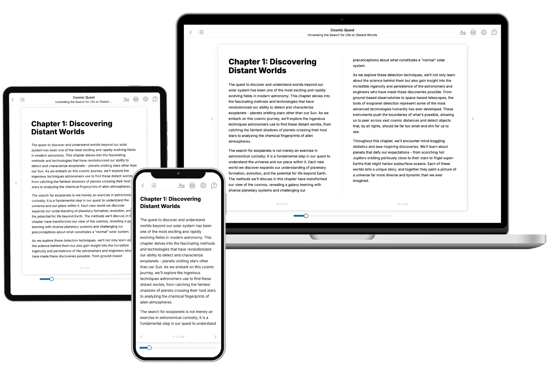
Your book is ready in 10 minutes. Read it online, download as EPUB or PDF, or send to Kindle.

Frequently asked questions
- What is TailoredRead?
TailoredRead creates complete, full-length nonfiction ebooks, written specifically for you. Tell us what you want to learn and why, and TailoredRead writes you a complete ebook on it, with charts, tables, and diagrams throughout. Read it online, on Kindle, or on any e-reader, or download it as a PDF or EPUB. Every book is written fresh the day you order it.
- How is the book tailored to me?
Before anything is written, you answer a few short questions about your goals, your background, and the topics you care about. Your book is then planned around your answers: written at your level, focused on your goals, and covering the topics you chose, instead of the one-size-fits-all book a store sells everyone.
- Will it read like generic AI content?
No. Your book isn't a chat transcript: it's planned chapter by chapter before a single word is written, told in a clear book voice, and explains ideas visually with charts, tables, and diagrams. After writing, every book goes through an editorial pass that polishes chapters so the book reads consistent from start to finish. Judge for yourself: browse thousands of sample books on our Explore Books page.
- How accurate is the content?
Our AI first checks whether your subject needs live research. Niche and fast-moving subjects are researched before writing, and the book plan and chapters are grounded in what it finds. After writing, every book goes through a quality pass that checks its facts and consistency, and researched books list their sources in a Notes section at the back.
- How long is the book?
You choose the length: Comprehensive (250-300 pages), Detailed (150-200 pages), Essential (70-100 pages), or Short (30-50 pages).
- Does the book include visuals?
Yes. Every book includes charts, tables, and diagrams: timelines, checklists, step-by-step guides, decision trees, comparison tables, matrices, and bar charts. A typical book includes dozens of these visual elements.
- Can I read it on Kindle? What formats can I download?
Yes. Every book can be read in our online reader or downloaded as a PDF or EPUB. The EPUB can be opened with Kindle, Apple Books, Kobo, and any e-reader. The Commercial and Author licenses also include an editable Word (DOCX) and Markdown download.
- How much does it cost?
About the price of a regular ebook. The exact price depends on your book's length and complexity, from $2 for the shortest books to $35 for the most comprehensive, and it's shown when you review your book, before you commit to anything. Each book is a one-time purchase, yours to keep, no subscription. Licenses that let you share, teach, or publish your book range from $35.99 to $49.99.
- Can I preview the book before purchasing?
Before you pay, you'll see what your book will cover: the title, a detailed description, the estimated length, and the complete chapter-by-chapter table of contents, so you know what you're getting before you decide. The full text itself is written after you purchase.
- How long does it take to create a book?
About 10 minutes. You can start reading the opening chapters while the rest is still being written.