The Liver Anatomy Coloring Book
Learn the Structure, Function, and Pathways of Your Body's Most Vital Organ Through Interactive Coloring
Imagine mastering liver anatomy so thoroughly that you can visualize its structure, trace its blood flow, and understand its function with complete confidence.
207 pages9 chaptersNov 10, 2025Anatomy
About this book
This interactive coloring book transforms how you learn about one of your body's most essential organs. Rather than passively reading dense anatomical descriptions, you'll actively engage with detailed, scientifically accurate illustrations that bring the liver's complexity to life. Each page guides you through a specific anatomical feature—from the liver's external lobes and ligaments to its intricate internal lobular architecture, from the dual blood supply system to the biliary pathways. As you color, you'll internalize spatial relationships, reinforce key structures, and build a comprehensive mental model of hepatic anatomy. Perfect for students, healthcare professionals, and curious learners, this book combines the proven benefits of visual learning with the cognitive advantages of hands-on activity. By the time you finish, you won't just know liver anatomy—you'll truly understand it.
Table of contents
- IntroductionWhy Liver Anatomy Matters, How Coloring Enhances Anatomical Learning, How to Use This Book, Color Coding Guide and SuppliesRead below
- Chapter 1: Liver Overview and External AnatomyPosition and Size in the Body, The Four Lobes: Right, Left, Caudate, and Quadrate, Surfaces and Borders, Ligaments and Peritoneal Attachments
- Chapter 2: Hepatic Blood Supply and CirculationThe Dual Blood Supply: Hepatic Artery and Portal Vein, The Hepatic Portal System and Nutrient Delivery, The Hepatic Artery and Oxygenation, Hepatic Veins and Venous Drainage
- Chapter 3: Internal Liver ArchitectureHepatic Lobules: The Functional Units, Hepatocytes and Their Arrangement, Sinusoids and Nutrient Exchange, Portal Triads and Central Veins
- Chapter 4: The Biliary SystemBile Production and Hepatocyte Function, Bile Canaliculi and Collection, Intrahepatic and Extrahepatic Bile Ducts, Connection to the Gallbladder and Intestines
- Chapter 5: Hepatic Segmentation and Surgical AnatomyThe Couinaud Classification System, Identifying Individual Segments, Surgical Planes and Resection Boundaries
- Chapter 6: Microscopic Liver StructureHepatocyte Organelles and Function, Kupffer Cells and Immune Function, Stellate Cells and Fibrosis, Sinusoidal Endothelial Cells
- Chapter 7: Liver Function and Metabolic PathwaysDetoxification and Drug Metabolism, Protein Synthesis and Clotting Factors, Carbohydrate Metabolism and Glucose Regulation, Lipid Metabolism and Cholesterol Management
- Chapter 8: Pathological Changes and Clinical RelevanceCirrhosis and Hepatic Fibrosis, Hepatitis and Inflammatory Changes, Fatty Liver Disease and Steatosis, How Pathology Appears in Clinical Imaging
- Chapter 9: Coloring Projects and Practice PagesComprehensive Multi-System Diagrams, Focused Structure Coloring Exercises, Labeling and Identification Practice
- ConclusionFrom Learning to Mastery, Next Steps in Your Anatomical Journey
- Glossary of Liver Anatomy Terms
- Further Reading and Resources
Introduction
The liver stands as one of the most remarkable organs in the human body, performing over five hundred distinct functions that keep you alive and healthy every single day. Weighing approximately three pounds and nestled beneath your right ribcage, this reddish-brown organ works tirelessly to filter toxins, produce essential proteins, regulate blood sugar, manufacture bile, and store vital nutrients. Despite its critical importance, the liver often remains poorly understood, even among those studying anatomy and physiology. Its complex three-dimensional structure, intricate vascular networks, and microscopic architecture can seem overwhelming when encountered through traditional textbook descriptions alone.
This coloring book offers a different approach to mastering liver anatomy—one that transforms passive reading into active, engaging learning. By combining scientifically accurate illustrations with hands-on coloring activities, you'll build a comprehensive understanding of hepatic structure and function that goes far beyond memorization. Each page has been carefully designed to guide you through specific anatomical features, from the organ's external surfaces and lobes to its internal lobular organization, from its dual blood supply system to the intricate biliary pathways that transport bile. As you select colors, trace boundaries, and fill in structures, you'll engage multiple learning pathways simultaneously, creating stronger neural connections and more durable memories.
Whether you're a medical student preparing for examinations, a nursing student learning clinical anatomy, a healthcare professional refreshing your knowledge, or simply someone fascinated by human biology, this book provides the tools you need to truly understand the liver. The following chapters will take you on a systematic journey through hepatic anatomy, starting with external features and progressing to microscopic structures, functional pathways, and clinical applications. By the time you complete this book, you won't just recognize liver structures—you'll understand how they relate to one another, how they function together, and why they matter for health and disease. Let's begin this visual learning adventure into one of your body's most vital organs.
Why Liver Anatomy Matters
Understanding liver anatomy provides the foundation for comprehending countless aspects of human health, disease, and medical treatment. The liver's strategic position in the body—receiving nutrient-rich blood directly from the digestive tract through the hepatic portal system—allows it to serve as the body's primary metabolic processing center. Every substance you ingest, whether food, medication, or toxin, passes through the liver for processing before entering general circulation. This anatomical arrangement explains why liver function is so critical and why liver disease can have such widespread effects throughout the body. Without a solid grasp of hepatic structure, you cannot fully understand how medications are metabolized, how nutrients are stored and released, or how toxins are neutralized.
For healthcare professionals, detailed knowledge of liver anatomy is essential for clinical practice across multiple specialties. Surgeons must understand the Couinaud segmentation system to plan hepatic resections and avoid damaging critical vascular structures. Radiologists need to recognize normal anatomical variations and identify pathological changes in imaging studies. Gastroenterologists must understand the biliary system to diagnose and treat conditions affecting bile flow. Even primary care providers benefit from understanding hepatic anatomy when interpreting liver function tests, prescribing medications that undergo hepatic metabolism, or counseling patients about alcohol consumption and liver health. The liver's anatomical complexity directly impacts clinical decision-making in countless scenarios.
The liver's unique dual blood supply system—receiving approximately seventy-five percent of its blood from the portal vein and twenty-five percent from the hepatic artery—creates anatomical relationships found nowhere else in the body[1]. This arrangement allows the liver to receive nutrient-rich blood from the intestines while also maintaining adequate oxygenation. Understanding this vascular anatomy helps explain why portal hypertension develops in liver disease, why certain tumors preferentially receive blood from one vessel versus another, and why surgical procedures must carefully preserve both blood supplies. The anatomical basis of hepatic circulation underlies much of hepatology and hepatobiliary surgery.
Beyond clinical applications, liver anatomy reveals fundamental principles of organ design and biological efficiency. The liver's lobular architecture, with hepatocytes arranged in plates radiating from central veins and surrounded by sinusoids, maximizes surface area for metabolic exchange. This microscopic organization allows approximately one hundred billion hepatocytes to process blood efficiently, with each cell exposed to nutrient-rich blood flowing from portal triads toward central veins. The elegant relationship between structure and function in the liver demonstrates how anatomical organization enables physiological capability. By studying hepatic anatomy, you gain insights into how complex organs achieve remarkable metabolic capacity within limited physical space.
The biliary system's anatomy—from microscopic bile canaliculi between hepatocytes to the common bile duct connecting to the duodenum—illustrates how the liver integrates with other digestive organs. Bile production and secretion depend entirely on this anatomical pathway, which allows the liver to eliminate waste products, excrete cholesterol, and deliver bile salts for fat digestion. When you understand the biliary tree's structure, you can better comprehend conditions like gallstones, cholestasis, and biliary obstruction. The anatomy of bile flow connects hepatic function to digestive processes, demonstrating how organ systems work together rather than in isolation.
Liver anatomy also provides context for understanding common pathological conditions that affect millions of people worldwide. Cirrhosis, which involves progressive fibrosis and architectural distortion, fundamentally alters the liver's normal structure. Fatty liver disease involves abnormal lipid accumulation within hepatocytes, changing the organ's microscopic appearance. Hepatitis causes inflammatory changes that affect sinusoidal architecture and hepatocyte integrity. By first learning normal anatomy, you create a reference point for recognizing and understanding these pathological changes. The transition from normal to diseased liver becomes comprehensible when you can visualize the structures being affected.
Finally, mastering liver anatomy builds confidence and competence that extends beyond this single organ. The skills you develop while learning hepatic structure—visualizing three-dimensional relationships, understanding vascular networks, recognizing functional units, and connecting structure to function—transfer to learning other organ systems. The liver's complexity makes it an excellent training ground for developing anatomical thinking. Once you've conquered hepatic anatomy, you'll find other organs more approachable. The investment you make in understanding the liver pays dividends throughout your anatomical education and professional practice.
How Coloring Enhances Anatomical Learning
Coloring transforms anatomy from an abstract intellectual exercise into a concrete, multisensory experience that engages your brain in fundamentally different ways than passive reading. When you color an anatomical structure, you must actively process its boundaries, relationships, and spatial extent. This requires sustained attention and deliberate decision-making about where one structure ends and another begins. Research in educational psychology demonstrates that active learning strategies—those requiring learners to manipulate, organize, or create content—produce significantly better retention than passive review[2]. Coloring represents a form of active learning that combines visual processing, fine motor control, and cognitive engagement.
The act of coloring creates what cognitive scientists call elaborative encoding—the process of connecting new information to existing knowledge through meaningful processing. As you select a color for the hepatic artery and carefully trace its branches throughout the liver, you're not just memorizing a name; you're building a mental model of how arterial blood reaches hepatic tissue. When you color the portal vein in a different shade and observe how it parallels the arterial system, you're encoding the relationship between these two vascular networks. This elaborative processing creates richer, more interconnected memories that are easier to retrieve later. The physical act of coloring forces you to engage with anatomical relationships in ways that reading alone cannot achieve.
Coloring also addresses the challenge of learning three-dimensional anatomy from two-dimensional images. The liver exists as a complex, three-dimensional organ with structures that overlap, interweave, and exist at different depths. By coloring multiple views of the same structure—anterior, posterior, inferior surfaces—you begin to construct a mental three-dimensional model. When you color the right lobe in one view and then find it again in a different perspective, you're building spatial understanding. This process of recognizing the same structure from multiple angles develops the spatial reasoning skills essential for clinical practice, where you'll need to interpret CT scans, ultrasounds, and surgical views of the liver.
The color-coding system itself serves as a powerful organizational tool that helps you categorize and remember anatomical information. When you consistently use red for arteries, blue for veins, and green for bile ducts across all illustrations, you're creating a visual schema that makes complex diagrams immediately interpretable. This color consistency reduces cognitive load—the mental effort required to process information—by providing instant visual cues about structure identity. Your brain can quickly scan a colored diagram and identify vascular versus biliary structures without reading every label. This efficiency allows you to focus mental resources on understanding relationships rather than identifying individual components.
Coloring activities also provide immediate feedback about your understanding. If you're uncertain where the boundary between the right and left hepatic lobes lies, you'll confront this uncertainty when deciding where to stop coloring with one color and start with another. This moment of uncertainty signals a gap in your knowledge that you can immediately address by consulting the text or labels. In contrast, passive reading can create an illusion of understanding—you might read about the falciform ligament dividing the liver without truly grasping its location until you try to color the structures it separates. The coloring process reveals what you actually understand versus what you only think you understand.
The repetitive, focused nature of coloring also promotes a meditative state that enhances learning. Unlike the scattered attention that often accompanies reading, coloring requires sustained focus on a single task. This focused attention state, sometimes called flow, is associated with optimal learning and memory formation. As you color, you enter a calm, concentrated mental state where distractions fade and your full cognitive resources engage with the anatomical content. Many learners find that they remember information better when it's associated with this focused, relaxed state. The combination of concentration and reduced stress creates ideal conditions for encoding new memories.
Finally, coloring creates a personalized study resource that reflects your own learning process. The completed pages in this book will become uniquely yours, marked by your color choices, your notes, and your engagement with the material. This personalization increases the value of the resource for future review. When you return to a page you've colored, you'll often remember the experience of coloring it—where you were, what you were thinking about, what questions arose. These contextual memories serve as retrieval cues that help you access the anatomical information. Your colored pages become more than just illustrations; they become memory anchors connected to your learning experience.
How to Use This Book
This book is designed to be used systematically, with each chapter building on knowledge from previous sections. Begin with Chapter 1, which covers external liver anatomy, before progressing to internal structures and microscopic organization. This sequence mirrors how anatomists traditionally approach organ study—starting with what you can observe externally before dissecting deeper. However, the book's modular design also allows you to focus on specific topics of interest or return to particular chapters for review. If you're preparing for an examination on hepatic vasculature, you might spend extra time on Chapter 2. If you're studying liver pathology, Chapter 8 will provide essential anatomical context for understanding disease processes.
Each chapter follows a consistent structure designed to maximize learning efficiency:
- Introductory text that provides context and explains why the anatomical structures matter
- Detailed descriptions of each structure, including location, relationships, and function
- Coloring instructions that guide you through the illustration systematically
- Clinical correlations that connect anatomy to real-world medical applications
- Review questions that test your understanding and highlight key concepts
Approach each illustration methodically rather than randomly coloring whatever catches your eye. Read the accompanying text first to understand what you're about to color. Then, follow the suggested coloring sequence, which typically moves from larger structures to smaller details or from one functional system to another. For example, when coloring a diagram showing both vascular and biliary structures, you might color all arterial structures first, then venous structures, then biliary ducts. This systematic approach reinforces the organization of anatomical systems and prevents the confusion that can result from jumping randomly between unrelated structures.
Use the labels and leader lines as guides, but challenge yourself to identify structures before reading their labels. When you encounter a new illustration, spend a moment trying to recognize structures based on their position, shape, and relationships. Then check the labels to confirm your identifications. This self-testing approach, called retrieval practice, strengthens memory more effectively than simply reading labels[3]. Even when you're uncertain, the act of attempting to retrieve information from memory enhances learning. Don't worry about making mistakes during this process—errors followed by correction are powerful learning opportunities.
Consider creating a study schedule that spaces your coloring sessions over time rather than cramming all at once. Research on spaced repetition shows that distributing learning over multiple sessions produces better long-term retention than massed practice. You might color one or two sections per day, then return to previously colored pages for review. This approach allows time for initial memories to consolidate before you reinforce them through review. A suggested schedule might look like this:
- Day 1: Read and color Chapter 1, Section 1
- Day 2: Review Day 1 material, then complete Chapter 1, Section 2
- Day 3: Review Days 1-2, then complete Chapter 1, Section 3
- Day 4: Complete Chapter 1, Section 4, then review entire Chapter 1
Supplement your coloring with additional study techniques to maximize retention. After completing a chapter, try drawing key structures from memory without looking at the book. This retrieval practice will reveal what you've truly learned versus what you only recognized when seeing it. Create flashcards for structures you find challenging, using your colored illustrations as references. Explain anatomical concepts to a study partner or even to yourself out loud—teaching is one of the most effective ways to solidify understanding. Consider using online anatomical resources or three-dimensional models to view structures from angles not shown in this book, building a more complete spatial understanding.
Don't rush through the coloring process. While it might be tempting to quickly fill in colors to "finish" a page, the learning happens during the careful, thoughtful engagement with each structure. Take time to trace boundaries, observe relationships, and think about function while you color. If a particular structure confuses you, pause to reread the description or consult additional resources. The goal isn't to complete the book quickly—it's to build lasting, comprehensive understanding of liver anatomy. Quality of engagement matters far more than speed of completion. Some learners find that a single detailed illustration might take thirty to forty-five minutes to color thoughtfully, and this investment of time produces proportionally deeper learning.
Finally, use the completed book as an ongoing reference resource. After you've colored all the illustrations, the book becomes a personalized atlas that you can consult when studying related topics, preparing for examinations, or encountering liver-related concepts in clinical practice. The time you invest in coloring creates a resource that will serve you for years. Many healthcare professionals keep their anatomy coloring books throughout their careers, finding them more accessible and personally meaningful than standard atlases. Your colored pages represent not just anatomical information but also your learning journey—a tangible record of the effort you invested in mastering this complex organ.
Color Coding Guide and Supplies
Consistent color coding throughout this book will help you quickly identify structure types and understand complex illustrations at a glance. While you're free to use any colors you prefer, following the suggested color scheme provides several advantages. First, it creates visual consistency across all chapters, making it easier to recognize similar structures in different contexts. Second, it follows conventions commonly used in anatomical illustrations, preparing you to interpret standard medical diagrams. Third, it reduces decision fatigue—you won't need to decide what color to use for each structure because the system provides clear guidance. The recommended color scheme uses intuitive associations that make anatomical relationships memorable.
The suggested color coding system for this book includes:
- Red or pink: Arterial structures (hepatic artery and its branches)
- Blue or purple: Venous structures (portal vein, hepatic veins, and their tributaries)
- Green or yellow-green: Biliary structures (bile ducts, bile canaliculi, gallbladder)
- Yellow or light orange: Hepatocytes and liver parenchyma
- Light brown or tan: Connective tissue, capsule, and ligaments
- Light gray or silver: Sinusoids and spaces of Disse
- Orange: Different liver lobes or segments (using different shades to distinguish adjacent regions)
When coloring structures that overlap or exist at different depths, use lighter shades or colored pencil techniques to show layering. For example, if sinusoids run between plates of hepatocytes, you might color the hepatocytes in solid yellow and then lightly shade the sinusoids in gray, allowing the yellow to show through. This layering technique helps represent three-dimensional relationships on a two-dimensional page. Alternatively, you can use stippling or hatching patterns rather than solid colors for structures that lie beneath others. These techniques add visual depth and help you understand spatial relationships that might otherwise be unclear.
The choice of coloring supplies significantly affects your experience and results. Colored pencils offer the most control and are ideal for detailed anatomical work. They allow you to vary pressure for different shading intensities, blend colors smoothly, and color precisely within small areas. High-quality colored pencils with soft, blendable cores work best—brands designed for artists rather than children's pencils provide better results. A set of twenty-four to thirty-six colors gives you sufficient variety for anatomical coloring. Sharpen your pencils frequently to maintain precision, especially when coloring small structures like bile canaliculi or small blood vessels.
Fine-tipped markers provide vibrant, consistent colors and work well for larger structures, but they require more care to avoid bleeding through pages or coloring outside boundaries. If you choose markers, consider these guidelines:
- Use alcohol-based markers designed for detailed work rather than broad-tipped highlighters
- Place a protective sheet behind each page to prevent bleed-through
- Color in one direction with smooth strokes rather than scribbling back and forth
- Test markers on a separate sheet first to ensure colors match your intentions
- Use lighter colors for large areas and save darker, more saturated colors for emphasis
Gel pens offer another option, particularly for adding fine details, labels, or notes to your colored illustrations. They work well for tracing vessel pathways, adding arrows to show flow direction, or highlighting specific features. However, gel pens are generally too fine-tipped for filling large areas efficiently. Consider using them as supplements to colored pencils or markers rather than as your primary coloring tool. A set of gel pens in the standard anatomical colors (red, blue, green) can be valuable for adding annotations and emphasis to your colored pages.
Organize your coloring supplies for efficient use during study sessions. Keep your materials in a pencil case or organizer with your book so everything is readily available when you're ready to work. Arrange colors in the order you'll typically use them—reds, blues, greens, yellows—so you can quickly grab the right color without searching. This organization might seem minor, but it reduces friction in your study process and helps maintain focus on learning rather than logistics. Some learners create a color key on an index card that they keep with their supplies, showing which specific pencil or marker corresponds to each anatomical structure type.
Finally, remember that the coloring supplies are tools for learning, not ends in themselves. You don't need expensive professional-grade materials to benefit from this book. Even basic colored pencils will serve the purpose if used thoughtfully and consistently. The value comes from your engagement with the anatomical content, not from the sophistication of your art supplies. That said, using materials you enjoy can make the learning process more pleasant and encourage regular study sessions. If you find that higher-quality supplies make you more likely to use the book consistently, they're worth the investment. Choose supplies that match your budget, preferences, and learning style, then focus your energy on the anatomy itself.
Notes
Introduction
- 1. Abdel-Misih and others, "Liver Anatomy," Surgical Clinics of North America, 2010
- 2. Scott Freeman and others, "Active learning increases student performance in science, engineering, and mathematics," Proceedings of the National Academy of Sciences, 2014
- 3. Henry L. Roediger and Andrew C. Butler, "The critical role of retrieval practice in long-term retention," Trends in Cognitive Sciences, 2011
This is the end of the free sample. The full book has 9 chapters.
Want this book written for you?
Tell us your goals and background, and get your own version of this book, tailored by AI to you.


Benefits of AI-tailored books
- Read one book, not ten:
- all the Anatomy knowledge you need, gathered into a single book.
- Save days of learning:
- tell us your goals, choose your topics, and the whole book is built around them.
- Learn effortlessly:
- Anatomy book written for your specific background and expertise.
- Understand at a glance:
- includes charts, tables, timelines, checklists, decision trees, and more.
- Stay current:
- written the day you order it, not years ago.
- Trust what you read:
- every book is checked for accuracy, with real research when your subject needs it.
Create your unique book in 3 steps
- 1. Select your focus
Select the focus of your Anatomy book and share your background
- 2. Personalize your book
Specify your goals and choose sub-topics to include
- 3. Get your tailored book
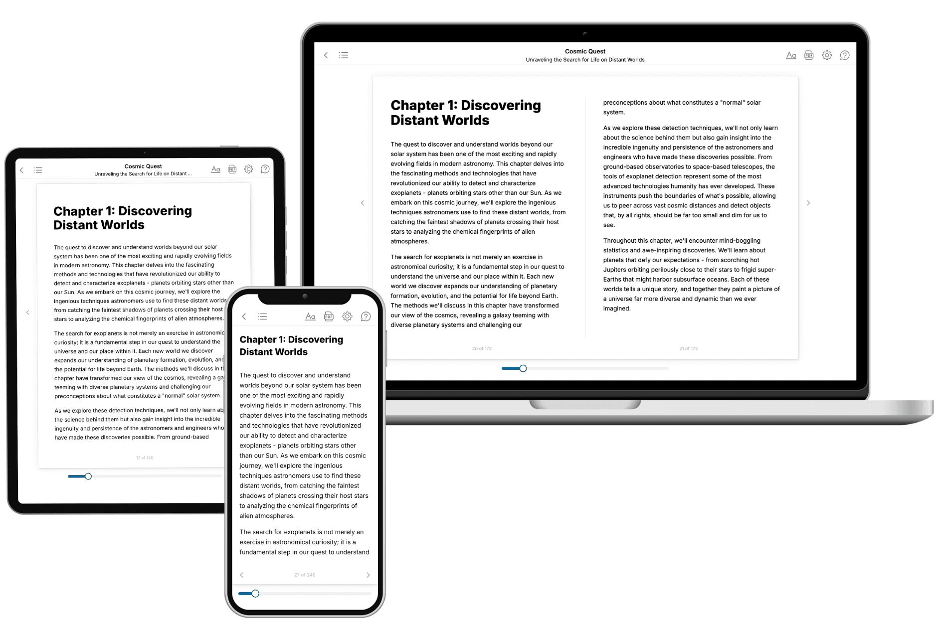
Your book is ready in 10 minutes. Read it online, download as EPUB or PDF, or send to Kindle.

Frequently asked questions
- What is TailoredRead?
TailoredRead creates complete, full-length nonfiction ebooks, written specifically for you. Tell us what you want to learn and why, and TailoredRead writes you a complete ebook on it, with charts, tables, and diagrams throughout. Read it online, on Kindle, or on any e-reader, or download it as a PDF or EPUB. Every book is written fresh the day you order it.
- How is the book tailored to me?
Before anything is written, you answer a few short questions about your goals, your background, and the topics you care about. Your book is then planned around your answers: written at your level, focused on your goals, and covering the topics you chose, instead of the one-size-fits-all book a store sells everyone.
- Will it read like generic AI content?
No. Your book isn't a chat transcript: it's planned chapter by chapter before a single word is written, told in a clear book voice, and explains ideas visually with charts, tables, and diagrams. After writing, every book goes through an editorial pass that polishes chapters so the book reads consistent from start to finish. Judge for yourself: browse thousands of sample books on our Explore Books page.
- How accurate is the content?
Our AI first checks whether your subject needs live research. Niche and fast-moving subjects are researched before writing, and the book plan and chapters are grounded in what it finds. After writing, every book goes through a quality pass that checks its facts and consistency, and researched books list their sources in a Notes section at the back.
- How long is the book?
You choose the length: Comprehensive (250-300 pages), Detailed (150-200 pages), Essential (70-100 pages), or Short (30-50 pages).
- Does the book include visuals?
Yes. Every book includes charts, tables, and diagrams: timelines, checklists, step-by-step guides, decision trees, comparison tables, matrices, and bar charts. A typical book includes dozens of these visual elements.
- Can I read it on Kindle? What formats can I download?
Yes. Every book can be read in our online reader or downloaded as a PDF or EPUB. The EPUB can be opened with Kindle, Apple Books, Kobo, and any e-reader. The Commercial and Author licenses also include an editable Word (DOCX) and Markdown download.
- How much does it cost?
About the price of a regular ebook. The exact price depends on your book's length and complexity, from $2 for the shortest books to $35 for the most comprehensive, and it's shown when you review your book, before you commit to anything. Each book is a one-time purchase, yours to keep, no subscription. Licenses that let you share, teach, or publish your book range from $35.99 to $49.99.
- Can I preview the book before purchasing?
Before you pay, you'll see what your book will cover: the title, a detailed description, the estimated length, and the complete chapter-by-chapter table of contents, so you know what you're getting before you decide. The full text itself is written after you purchase.
- How long does it take to create a book?
About 10 minutes. You can start reading the opening chapters while the rest is still being written.