The Lung Anatomy Coloring Book
Master Respiratory System Structure Through Interactive Learning
Elevate your understanding of respiratory anatomy with this comprehensive coloring guide designed for visual learners.
207 pages9 chaptersJan 19, 2026Anatomy
About this book
This book combines detailed, scientifically accurate illustrations with interactive coloring activities to help you master lung structure and function. Each page presents a different anatomical system or region, from the trachea and bronchial tree to the delicate alveoli where gas exchange occurs. By actively coloring and labeling structures, you'll develop a deeper, more intuitive grasp of how the respiratory system works. Perfect for students, healthcare professionals, educators, and anyone seeking to understand the intricate architecture of the lungs, this resource transforms passive reading into engaging, memorable learning. The combination of clear anatomical drawings and hands-on coloring creates multiple pathways for your brain to process and retain complex information, making this an invaluable tool for anyone serious about respiratory anatomy.
Table of contents
- IntroductionWhy Coloring Enhances Anatomical Learning, How to Use This Book, Overview of the Respiratory SystemRead below
- Chapter 1: The Respiratory Pathway: From Air to LungsThe Nasal Cavity and Upper Airways, The Pharynx and Larynx, The Trachea: Gateway to the Lungs
- Chapter 2: The Bronchial Tree: Branching AirwaysPrimary Bronchi and Lung Division, Secondary and Tertiary Bronchi, Bronchioles and Terminal Airways
- Chapter 3: Lung Lobes and SegmentsThe Right Lung: Three Lobes, The Left Lung: Two Lobes, Bronchopulmonary Segments
- Chapter 4: The Alveoli: Where Gas Exchange HappensAlveolar Structure and Organization, The Gas Exchange Process, Types of Alveolar Cells
- Chapter 5: Pulmonary Circulation and Blood SupplyPulmonary Arteries and Deoxygenated Blood, Pulmonary Veins and Oxygenated Blood, Bronchial Circulation and Lung Nutrition
- Chapter 6: The Pleura and Thoracic CavityVisceral and Parietal Pleura, The Pleural Space and Fluid, Thoracic Cavity Boundaries and Mediastinum
- Chapter 7: Muscles of RespirationThe Diaphragm: Primary Breathing Muscle, Intercostal Muscles and Rib Movement, Accessory Muscles of Respiration
- Chapter 8: Innervation and Nervous ControlThe Vagus Nerve and Parasympathetic Control, Sympathetic Nervous System Influence, Respiratory Control Centers in the Brain
- Chapter 9: Lymphatic Drainage and Immune FunctionPulmonary Lymph Nodes, Lymphatic Vessels of the Lungs, Immune Defense Mechanisms in the Lungs
- ConclusionIntegrating Your Anatomical Knowledge, Next Steps in Your Learning Journey
- Glossary of Terms
- Further Reading and Resources
Introduction
The human respiratory system represents one of the most elegant and essential biological systems in the body. Every breath you take involves a complex orchestration of structures working in perfect harmony—from the moment air enters your nostrils to the microscopic exchange of gases deep within your lungs. Understanding this intricate architecture requires more than passive reading; it demands active engagement with the material. This coloring book offers you a unique pathway to mastery, combining scientifically accurate illustrations with the proven learning benefits of hands-on interaction. By coloring and labeling anatomical structures, you'll create neural connections that transform abstract concepts into concrete, memorable knowledge that stays with you long after you close this book.
Traditional anatomy textbooks often present information in dense paragraphs accompanied by static images. While valuable, this approach can make it challenging to truly internalize the three-dimensional relationships between structures and understand how individual components fit into the larger system. The interactive nature of coloring changes this dynamic entirely. When you actively choose colors, trace boundaries, and physically engage with each illustration, you're not just observing anatomy—you're experiencing it. This multisensory approach activates different regions of your brain simultaneously, creating stronger memory pathways and deeper comprehension. Whether you're a nursing student preparing for exams, a respiratory therapist seeking to refresh your knowledge, or simply someone fascinated by human biology, this book provides the tools you need to build genuine expertise.
The lungs themselves are remarkable organs that perform the vital function of gas exchange, bringing oxygen into your bloodstream while removing carbon dioxide—a waste product of cellular metabolism. But the lungs don't work in isolation. They're supported by an extensive network of airways, blood vessels, nerves, muscles, and protective membranes, all housed within the thoracic cavity. Each component plays a specific role, and understanding these individual parts while grasping how they integrate into a functional whole is the key to true anatomical literacy. This introduction will prepare you for the journey ahead by explaining why coloring is such an effective learning tool, how to maximize your use of this book, and providing a foundational overview of the respiratory system that will serve as your roadmap through the detailed chapters that follow.
Why Coloring Enhances Anatomical Learning
The effectiveness of coloring as a learning tool is grounded in solid cognitive science. When you color an anatomical structure, you engage multiple learning modalities simultaneously—visual, kinesthetic, and cognitive. This multi-modal engagement creates what educational psychologists call elaborative encoding, where information is processed more deeply and connected to existing knowledge networks in your brain. Research has consistently shown that active learning strategies produce better retention than passive reading alone[1]. The physical act of coloring requires you to slow down, observe carefully, and make deliberate decisions about each structure, transforming what might be a quick glance at a diagram into a meaningful interaction that embeds knowledge more permanently.
Coloring also addresses a fundamental challenge in anatomy education: the overwhelming volume of information. The respiratory system alone contains hundreds of named structures, each with specific locations, relationships, and functions. When faced with this complexity, your brain needs organizational strategies to make sense of it all. Color-coding provides exactly this kind of organizational framework. By assigning different colors to related structures—perhaps using blue for all venous structures and red for arterial ones, or different shades for different lung lobes—you create visual categories that help your brain chunk information into manageable units. This categorical organization makes it easier to recall information later because you're not just remembering isolated facts; you're remembering patterns and relationships.
The benefits of coloring extend beyond simple memorization to support genuine understanding. When you color the branching pattern of the bronchial tree, for example, you're not just memorizing names—you're internalizing the hierarchical organization of the airways. You begin to see how the trachea divides into primary bronchi, which further subdivide into secondary and tertiary bronchi, and so on. This visual understanding of structural relationships helps you predict where structures should be located and understand why certain clinical conditions affect specific regions of the lungs. The spatial reasoning you develop through coloring translates directly into the kind of three-dimensional thinking essential for healthcare professionals who need to interpret chest X-rays, CT scans, or perform physical examinations.
Another powerful aspect of coloring is its ability to reduce cognitive load. When you're first learning anatomy, everything seems equally important and equally unfamiliar. This can lead to cognitive overload, where your working memory becomes saturated and learning efficiency drops. Coloring helps manage this by breaking learning into discrete, focused tasks. Instead of trying to absorb an entire page of information at once, you focus on one structure at a time—identifying it, understanding its boundaries, and coloring it carefully. This focused attention allows for deeper processing of each element before moving to the next. The result is more thorough learning with less mental fatigue.
The tactile engagement of coloring also creates what memory researchers call motor memory. Your hands learn the shapes and contours of anatomical structures through the physical act of coloring them. This creates an additional memory trace separate from visual or verbal memory. Many students report that during exams, they can recall information by remembering the physical sensation of coloring a particular structure. This phenomenon, sometimes called muscle memory,
provides yet another retrieval pathway for accessing stored information. When multiple memory systems encode the same information, recall becomes more reliable and automatic.
Coloring also promotes mindfulness and reduces anxiety associated with learning complex material. The repetitive, focused nature of coloring has a calming effect that can make study sessions more enjoyable and sustainable. When learning feels less stressful, you're more likely to engage with material regularly and for longer periods—both critical factors for mastery. Additionally, the creative aspect of choosing colors and creating visually appealing pages adds an element of personal expression to your learning. The pages you color become uniquely yours, which can increase your sense of ownership over the material and motivation to continue learning.
Finally, colored diagrams serve as powerful review tools. After you've completed coloring a page, that illustration becomes a personalized study aid. The colors you've chosen create visual associations that trigger recall of related information. When you review your colored pages before an exam or clinical rotation, the visual patterns you've created help you quickly reconstruct your understanding of complex anatomical relationships. Many learners find that they can close their eyes and visualize their colored diagrams, effectively creating mental images they can reference whenever needed. This visualization skill is invaluable not just for exams, but for real-world applications where you need to mentally picture anatomical structures during patient care or diagnostic reasoning.
How to Use This Book
To maximize your learning from this coloring book, approach it as an active learning tool rather than a passive coloring activity. Before you begin coloring any page, take time to read the accompanying text carefully. Understanding the function and relationships of structures before you color them provides context that makes the coloring process more meaningful. As you read, try to visualize where each structure might be located and how it connects to others. This pre-coloring preparation primes your brain to notice important details when you turn to the illustration. Consider keeping a notebook alongside this book where you can jot down questions, make additional notes, or sketch your own simplified diagrams to reinforce understanding.
When selecting colors, develop a consistent color-coding system and stick with it throughout the book. Consistency is crucial because it allows you to build associations that transfer from one chapter to the next. Here's a recommended approach that many anatomy students find effective:
- Arteries: Use red or pink shades to represent oxygenated blood in systemic circulation (though remember that pulmonary arteries carry deoxygenated blood)
- Veins: Use blue or purple shades for deoxygenated blood in systemic circulation (with pulmonary veins carrying oxygenated blood)
- Airways: Use yellow, orange, or light green for the bronchial tree and airways
- Muscles: Use brown, tan, or red-brown shades for muscular structures
- Nerves: Use bright yellow or gold for neural structures
- Lymphatic structures: Use light green or pale yellow-green
- Membranes and connective tissue: Use light gray, beige, or pale pink
As you color, work slowly and deliberately. This isn't a race, and rushing defeats the purpose of the exercise. Pay attention to the boundaries between structures, as these boundaries often represent important anatomical relationships. When coloring adjacent structures, use contrasting colors to make the boundaries clear. If a structure has multiple parts or regions, consider using different shades of the same color family to show that they're related while still distinguishing them. For example, you might use light blue for the visceral pleura and darker blue for the parietal pleura, immediately showing both their relationship and their distinction.
Label structures as you color them, even if labels are already provided. The act of writing reinforces memory through yet another sensory channel. Use a fine-point pen or pencil to add labels, and consider including additional information beyond just the name—perhaps a brief note about function or a clinical correlation. For instance, next to the diaphragm, you might note innervated by phrenic nerve (C3-C5)
or primary muscle of inspiration.
These annotations transform your coloring book into a comprehensive, personalized study guide that reflects your growing understanding.
Don't feel obligated to work through the book in strict sequential order, though the chapters are organized to build progressively from larger structures to smaller ones and from basic anatomy to more complex relationships. If you're currently studying a particular topic in a course, feel free to jump to the relevant chapter. However, if you're learning independently, following the book's sequence will provide a logical progression that mirrors how anatomists typically approach the respiratory system. Each chapter builds on concepts introduced in previous ones, so sequential reading provides the most coherent learning experience.
After completing each chapter, take time to review without looking at the text. Quiz yourself by covering the labels and trying to identify structures based on their location and appearance. Better yet, try to explain the function of each structure and how it relates to neighboring structures. This self-testing is one of the most effective learning strategies available[2]. If you struggle to recall something, that's valuable feedback—it tells you where to focus additional study. Return to the text, re-read the relevant section, and consider coloring that structure again on a blank sheet of paper to reinforce your memory.
Consider using this book collaboratively if possible. Study groups can enhance learning by allowing you to explain concepts to others and hear different perspectives. You might color individually but then come together to compare your work, discuss why you made certain color choices, and quiz each other on structure identification. Teaching others is one of the most powerful ways to solidify your own understanding. When you can explain the path of air through the bronchial tree or describe the boundaries of the pleural cavity to a peer, you've moved beyond memorization to genuine mastery.
Finally, supplement this coloring book with other learning resources. While coloring is highly effective, comprehensive understanding comes from multiple sources. Use anatomy atlases to see photographic images of real lungs, watch videos of dissections to understand three-dimensional relationships, and read clinical case studies to see how anatomical knowledge applies to patient care. This coloring book should be the centerpiece of your respiratory anatomy studies, but surrounding it with complementary resources will accelerate your learning and deepen your understanding. Keep track of particularly helpful resources in your notebook so you can return to them during review sessions.
Overview of the Respiratory System
The respiratory system serves one fundamental purpose: to facilitate gas exchange between the external environment and your bloodstream. Every cell in your body requires oxygen to produce energy through cellular respiration, and this process generates carbon dioxide as a waste product that must be eliminated. The respiratory system accomplishes this through two main processes: ventilation (the mechanical movement of air into and out of the lungs) and respiration (the exchange of gases between air and blood, and between blood and tissues). Understanding this system requires familiarity with both the conducting airways that transport air and the respiratory zones where gas exchange actually occurs.
The respiratory system is traditionally divided into two main sections: the upper respiratory tract and the lower respiratory tract. The upper respiratory tract includes the nose, nasal cavity, sinuses, pharynx, and larynx. These structures warm, humidify, and filter incoming air while also serving other functions like smell and speech production. The lower respiratory tract begins with the trachea and includes all the structures within the lungs themselves—the bronchi, bronchioles, and alveoli. This division isn't just anatomical; it has clinical significance as well. Infections of the upper respiratory tract (like the common cold) typically present differently than lower respiratory tract infections (like pneumonia), and treatments often differ accordingly.
Air enters the respiratory system through the nose or mouth and travels through a progressively branching network of airways. This branching pattern, often called the bronchial tree due to its resemblance to an inverted tree, is one of the most important structural features to understand. The trachea serves as the trunk, dividing into two primary bronchi (the main branches) that enter the left and right lungs. These primary bronchi further divide into secondary bronchi (also called lobar bronchi because each supplies a lobe of the lung), which divide into tertiary bronchi (segmental bronchi), which continue dividing into smaller and smaller bronchioles. This branching continues through approximately 23 generations of divisions, ending in tiny terminal bronchioles and respiratory bronchioles that lead to the alveolar sacs where gas exchange occurs.
The lungs themselves are paired organs located in the thoracic cavity, separated by the mediastinum—a central compartment containing the heart, great vessels, esophagus, and other structures. The right lung is larger and divided into three lobes (superior, middle, and inferior), while the left lung has only two lobes (superior and inferior) to accommodate the heart's position. Each lobe is further subdivided into bronchopulmonary segments, which are functionally independent units with their own bronchus and blood supply. Understanding this segmental organization is clinically important because disease processes often affect specific segments, and surgeons can sometimes remove diseased segments while preserving healthy lung tissue.
The functional units of the lungs are the alveoli—tiny, thin-walled air sacs where gas exchange occurs. Your lungs contain approximately 300 to 500 million alveoli, providing an enormous surface area for gas exchange (roughly 70 square meters, about the size of a tennis court)[3]. Each alveolus is surrounded by a dense network of capillaries, and the barrier between air and blood is incredibly thin—often less than 0.5 micrometers. This minimal distance allows oxygen to diffuse rapidly from the alveolar air into the blood while carbon dioxide diffuses in the opposite direction. The efficiency of this system is remarkable: during rest, your entire blood volume passes through the lungs and is reoxygenated approximately once per minute.
The pulmonary circulation system is unique in the body. Unlike systemic circulation, where arteries carry oxygenated blood and veins carry deoxygenated blood, the pulmonary system reverses this pattern. The pulmonary arteries carry deoxygenated blood from the right ventricle of the heart to the lungs, where it picks up oxygen. The pulmonary veins then return this newly oxygenated blood to the left atrium of the heart, from where it's pumped to the rest of the body. This distinction is crucial to understand and often confuses beginning students. Additionally, the lungs receive a separate blood supply through the bronchial arteries (branches of the aorta) that nourish the lung tissue itself rather than participating in gas exchange.
The lungs are enclosed by a double-layered membrane called the pleura. The visceral pleura adheres tightly to the lung surface, while the parietal pleura lines the inner chest wall. Between these layers is the pleural space (or pleural cavity), which contains a small amount of lubricating fluid. This fluid allows the layers to slide smoothly against each other during breathing while the surface tension keeps them from separating. The pleural space normally maintains a negative pressure relative to atmospheric pressure, which is essential for keeping the lungs expanded. Understanding pleural anatomy is critical for comprehending conditions like pneumothorax (collapsed lung) and pleural effusion (fluid accumulation in the pleural space).
Breathing is accomplished through the coordinated action of respiratory muscles. The diaphragm, a dome-shaped muscle separating the thoracic and abdominal cavities, is the primary muscle of inspiration. When it contracts, it flattens and moves downward, increasing the volume of the thoracic cavity. The external intercostal muscles between the ribs also contract during inspiration, lifting the rib cage upward and outward. These volume increases create negative pressure within the lungs, drawing air in. Expiration during quiet breathing is largely passive—the muscles relax, elastic recoil of the lungs and chest wall decreases thoracic volume, and air flows out. During forced breathing (like during exercise), additional accessory muscles assist with both inspiration and expiration.
The respiratory system doesn't operate autonomously; it's under sophisticated neural control. Respiratory centers in the brainstem (specifically the medulla oblongata and pons) generate the rhythmic pattern of breathing and adjust it based on the body's needs. These centers receive input from chemoreceptors that monitor blood levels of oxygen, carbon dioxide, and pH. When carbon dioxide levels rise (which happens during exercise or when holding your breath), the respiratory centers increase breathing rate and depth to eliminate the excess. The vagus nerve provides parasympathetic innervation to the airways, while sympathetic nerves also influence airway diameter and secretions. This neural control allows your breathing to adjust automatically to changing demands without conscious effort, though you can also voluntarily override the automatic pattern when needed.
Finally, the respiratory system includes important defense mechanisms that protect against inhaled pathogens and particles. The airways are lined with mucus-secreting cells and ciliated cells that trap particles and move them upward toward the throat where they can be swallowed or expelled. Immune cells patrol the airways and alveoli, ready to respond to infections. Lymphatic vessels drain fluid and transport immune cells throughout the lungs, with lymph nodes filtering this fluid and mounting immune responses when necessary. Understanding these protective mechanisms helps explain why the lungs, despite constant exposure to the external environment, usually remain healthy and why certain conditions (like smoking or immunosuppression) increase vulnerability to respiratory infections.
As you work through the chapters ahead, you'll explore each of these components in detail. You'll trace the path of air from the nasal cavity through the branching airways to the alveoli. You'll examine the structure of the lungs themselves, understanding how lobes and segments are organized. You'll study the blood vessels that deliver blood for oxygenation and the vessels that nourish lung tissue. You'll investigate the pleural membranes and thoracic cavity that house and protect the lungs. You'll learn about the muscles that power breathing and the nerves that control them. Each chapter builds on this foundational overview, adding layers of detail and understanding. By the time you complete this book, you'll have developed a comprehensive, integrated understanding of respiratory anatomy that will serve you well in whatever field you pursue.
Notes
Introduction
- 1. S. Freeman and others, "Active learning increases student performance in science, engineering, and mathematics," Proceedings of the National Academy of Sciences, 2014
- 2. H. L. Roediger and A. C. Butler, "The critical role of retrieval practice in long-term retention," Trends in Cognitive Sciences, 2011
- 3. M. Ochs and others, "The number of alveoli in the human lung," American Journal of Respiratory and Critical Care Medicine, 2004
This is the end of the free sample. The full book has 9 chapters.
Want this book written for you?
Tell us your goals and background, and get your own version of this book, tailored by AI to you.


Benefits of AI-tailored books
- Read one book, not ten:
- all the Anatomy knowledge you need, gathered into a single book.
- Save days of learning:
- tell us your goals, choose your topics, and the whole book is built around them.
- Learn effortlessly:
- Anatomy book written for your specific background and expertise.
- Understand at a glance:
- includes charts, tables, timelines, checklists, decision trees, and more.
- Stay current:
- written the day you order it, not years ago.
- Trust what you read:
- every book is checked for accuracy, with real research when your subject needs it.
Create your unique book in 3 steps
- 1. Select your focus
Select the focus of your Anatomy book and share your background
- 2. Personalize your book
Specify your goals and choose sub-topics to include
- 3. Get your tailored book
Your book is ready in 10 minutes. Read it online, download as EPUB or PDF, or send to Kindle.

Frequently asked questions
- What is TailoredRead?
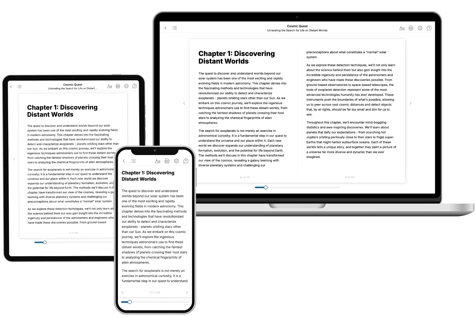
TailoredRead creates complete, full-length nonfiction ebooks, written specifically for you. Tell us what you want to learn and why, and TailoredRead writes you a complete ebook on it, with charts, tables, and diagrams throughout. Read it online, on Kindle, or on any e-reader, or download it as a PDF or EPUB. Every book is written fresh the day you order it.
- How is the book tailored to me?
Before anything is written, you answer a few short questions about your goals, your background, and the topics you care about. Your book is then planned around your answers: written at your level, focused on your goals, and covering the topics you chose, instead of the one-size-fits-all book a store sells everyone.
- Will it read like generic AI content?
No. Your book isn't a chat transcript: it's planned chapter by chapter before a single word is written, told in a clear book voice, and explains ideas visually with charts, tables, and diagrams. After writing, every book goes through an editorial pass that polishes chapters so the book reads consistent from start to finish. Judge for yourself: browse thousands of sample books on our Explore Books page.
- How accurate is the content?
Our AI first checks whether your subject needs live research. Niche and fast-moving subjects are researched before writing, and the book plan and chapters are grounded in what it finds. After writing, every book goes through a quality pass that checks its facts and consistency, and researched books list their sources in a Notes section at the back.
- How long is the book?
You choose the length: Comprehensive (250-300 pages), Detailed (150-200 pages), Essential (70-100 pages), or Short (30-50 pages).
- Does the book include visuals?
Yes. Every book includes charts, tables, and diagrams: timelines, checklists, step-by-step guides, decision trees, comparison tables, matrices, and bar charts. A typical book includes dozens of these visual elements.
- Can I read it on Kindle? What formats can I download?
Yes. Every book can be read in our online reader or downloaded as a PDF or EPUB. The EPUB can be opened with Kindle, Apple Books, Kobo, and any e-reader. The Commercial and Author licenses also include an editable Word (DOCX) and Markdown download.
- How much does it cost?
About the price of a regular ebook. The exact price depends on your book's length and complexity, from $2 for the shortest books to $35 for the most comprehensive, and it's shown when you review your book, before you commit to anything. Each book is a one-time purchase, yours to keep, no subscription. Licenses that let you share, teach, or publish your book range from $35.99 to $49.99.
- Can I preview the book before purchasing?
Before you pay, you'll see what your book will cover: the title, a detailed description, the estimated length, and the complete chapter-by-chapter table of contents, so you know what you're getting before you decide. The full text itself is written after you purchase.
- How long does it take to create a book?
About 10 minutes. You can start reading the opening chapters while the rest is still being written.