The Lymph Node Coloring Book
Master the Anatomy of Your Immune System Through Interactive Learning
Many people struggle to visualize how lymph nodes actually work because traditional textbooks present anatomy as static, two-dimensional information.
222 pages10 chaptersJun 9, 2026Anatomy
About this book
This interactive coloring book transforms your learning by engaging both sides of your brain—combining visual recognition with active participation. As you color detailed illustrations of lymph node structures, you'll develop a deeper, more intuitive understanding of how these vital immune organs function. Each chapter builds systematically from basic lymphatic system overview through regional lymph node groups, internal architecture, and cellular components. The hands-on coloring process helps cement anatomical details in your memory far more effectively than passive reading. Whether you're a healthcare student, medical professional, or anatomy enthusiast, this book provides a refreshing, engaging approach to mastering lymph node anatomy while creating a personalized study resource you'll reference for years to come.
Table of contents
- IntroductionWhy Lymph Node Anatomy Matters, How to Use This Coloring Book, The Science Behind Learning Through Coloring, What You'll Master in This BookRead below
- Chapter 1: The Lymphatic System OverviewComponents of the Lymphatic System, How Lymph Circulates Through Your Body, The Immune Function of Lymph Nodes
- Chapter 2: Lymph Node Structure and OrganizationBasic Lymph Node Anatomy, Size, Shape, and Location Characteristics, The Capsule and Trabeculae
- Chapter 3: Internal Architecture: The CortexUnderstanding the Cortex Region, Germinal Centers and B Cell Activity, Cortical Lymphocytes and Follicles
- Chapter 4: Internal Architecture: The Paracortex and MedullaThe Paracortex and T Cell Zones, Medullary Cords and Sinuses, Tracing Lymph Flow Through Internal Regions
- Chapter 5: Cellular Components of Lymph NodesB Lymphocytes and Antibody Production, T Lymphocytes and Cell-Mediated Immunity, Macrophages and Dendritic Cells
- Chapter 6: Cervical and Head Lymph NodesCervical Lymph Node Groups, Head and Neck Drainage Patterns, Clinical Significance of Cervical Nodes
- Chapter 7: Axillary and Upper Extremity Lymph NodesAxillary Lymph Node Anatomy, Upper Extremity Drainage Pathways, Breast Tissue Drainage Patterns
- Chapter 8: Inguinal and Lower Extremity Lymph NodesInguinal Lymph Node Groups, Lower Extremity Drainage Patterns, Pelvic Lymph Node Relationships
- Chapter 9: Thoracic and Abdominal Lymph NodesMediastinal and Thoracic Nodes, Mesenteric and Abdominal Nodes, Organ-Specific Drainage Patterns
- Chapter 10: Lymph Node Pathology and Clinical CorrelationUnderstanding Lymphadenopathy, Lymph Node Response to Infection, Clinical Examination and Palpation
- ConclusionYour Lymph Node Anatomy Mastery, Next Steps in Your Learning Journey
- Glossary of Terms
- Further Reading and Resources
Introduction
There is a quiet, remarkable system working inside your body right now. It filters fluid, hunts pathogens, and coordinates immune responses across every region of your body. Yet for most people, the lymphatic system remains one of anatomy's least understood networks. Lymph nodes, in particular, tend to get reduced to a single sentence in textbooks: small, bean-shaped organs that filter lymph. That description is technically accurate, but it barely scratches the surface of what these structures actually do or how they are organized.
The challenge with learning lymph node anatomy is not a lack of information. It is a lack of engagement. Traditional study methods ask you to read dense paragraphs, memorize labeled diagrams, and recall structures on demand. That approach works for some learners, but for most people, it produces knowledge that fades quickly after an exam. The structures never feel real. The relationships between regions never click into place. You can recite the layers of a lymph node without ever truly understanding how they connect to each other or to the broader immune system.
This book takes a different approach. Instead of asking you to passively absorb information, it invites you to actively participate in your own learning. By coloring detailed anatomical illustrations, labeling structures, and working through each region systematically, you build a mental map of the lymphatic system that sticks. The act of choosing colors, tracing boundaries, and filling in structures forces your brain to process information at a deeper level than reading alone ever could. The result is not just better recall. It is genuine understanding.
Whether you are a nursing student preparing for clinical rotations, a medical professional refreshing foundational knowledge, or simply someone fascinated by how the human body works, this book meets you where you are. No prior expertise in immunology is required. What you do need is curiosity, a set of colored pencils or markers, and a willingness to slow down and engage with the material in a hands-on way. The chapters ahead will guide you from the broad overview of the lymphatic system all the way down to the individual cells that make lymph nodes function. By the end, you will have built something genuinely useful: a personalized, color-coded reference guide that reflects your own learning process.
Why Lymph Node Anatomy Matters
Lymph nodes are not passive filters sitting quietly in the background. They are active immune organs that respond dynamically to threats, coordinate cellular communication, and serve as critical checkpoints in the body's defense network. Understanding their anatomy is not just an academic exercise. It has direct, practical relevance in clinical settings, from interpreting physical examination findings to understanding how cancer spreads through the body.
Consider what happens during a routine physical exam. A clinician palpates the neck, armpits, and groin, checking for enlarged lymph nodes. That simple act of pressing fingertips against skin is only meaningful if the examiner knows exactly which node groups sit in each region, what their normal size and texture feel like, and what patterns of enlargement suggest about underlying conditions. Without a solid grasp of lymph node anatomy, those findings are just lumps. With it, they become diagnostic clues.[1]
The clinical stakes are significant. Lymph node involvement is a central factor in cancer staging. When a tumor spreads to regional lymph nodes, it changes the stage of the disease and directly influences treatment decisions. Surgeons performing procedures like sentinel lymph node biopsies rely on precise anatomical knowledge to identify which nodes drain a specific tumor site. Radiologists interpreting CT scans use size criteria and location to distinguish normal from abnormal nodes. In each of these scenarios, anatomy is not background knowledge. It is the foundation of clinical reasoning.
Beyond oncology, lymph node anatomy matters in infectious disease, autoimmune conditions, and inflammatory disorders. When a patient presents with swollen nodes in the posterior cervical chain, that pattern points toward different causes than swelling in the anterior cervical or submandibular groups. Knowing the drainage territories of each node group allows clinicians to work backward from the location of swelling to the likely source of infection or inflammation. This kind of anatomical reasoning is a core clinical skill.
For students and learners who are not yet in clinical practice, understanding lymph node anatomy builds the conceptual scaffolding needed for more advanced study. Immunology, pathology, oncology, and surgery all build on this foundation. When you understand where lymph nodes are, how they are structured internally, and what cells they contain, the more complex topics that come later make far more sense. You are not memorizing isolated facts. You are constructing a framework that supports everything built on top of it.
There is also a personal dimension worth acknowledging. Most people have felt swollen lymph nodes at some point, usually during an illness. Understanding what is actually happening inside those nodes during an immune response transforms that experience from something vague and slightly alarming into something comprehensible. Knowledge reduces anxiety and builds confidence. That is true whether you are a patient, a caregiver, or a healthcare professional.
A practical example: A patient presents with a painless, firm, enlarged lymph node in the left supraclavicular region. A clinician familiar with lymph node anatomy immediately recognizes this location as significant. The left supraclavicular node (Virchow's node) drains the thoracic duct and can receive lymph from abdominal organs. Its enlargement is a well-known clinical sign associated with intra-abdominal malignancy. Without anatomical knowledge, this finding is just a lump. With it, it becomes an urgent diagnostic signal.
This book is designed to make that kind of anatomical knowledge accessible and memorable. Each chapter builds on the last, and the coloring activities are not decorative additions. They are the primary learning mechanism. The more actively you engage with each illustration, the more deeply the anatomy will be encoded in your memory.
How to Use This Coloring Book
Getting the most out of this book requires a slightly different mindset than reading a standard anatomy text. The goal is not to move through pages as quickly as possible. It is to slow down, engage with each illustration deliberately, and use the coloring process as a tool for active learning. Think of each page as a conversation between you and the anatomy, not a task to complete and move on from.
Before you start coloring any illustration, read the accompanying text for that section. This gives you the conceptual context you need to make meaningful color choices. When you understand that the cortex of a lymph node is where B cells cluster into follicles, and that the paracortex is the T cell zone, you can assign colors that reflect those functional differences. That intentional color-coding is what transforms a coloring activity into a learning activity.
Here is a recommended approach for working through each chapter:
- Read the full text of the subchapter before picking up a pencil.
- Review the illustration and identify the labeled structures before coloring anything.
- Choose a color scheme that groups related structures together. For example, use shades of blue for B cell zones and shades of red for T cell zones throughout the entire book.
- Color each structure while saying its name aloud or writing it in the margin.
- After coloring, cover the labels and test yourself by pointing to each structure and naming it.
- Return to the text and re-read any sections that felt unclear during the coloring process.
Consistency in your color choices across chapters is one of the most powerful strategies you can use. If you always color lymphatic vessels in green, afferent vessels in a lighter green, and efferent vessels in a darker green, your brain will start to associate those colors with those structures automatically. Over time, seeing the color triggers the memory of the structure and its function. This is not just a study trick. It reflects how visual memory actually works in the brain.
You will need a set of colored pencils or fine-tipped markers. Colored pencils are generally preferred for detailed anatomical illustrations because they allow for precise work in small areas and can be layered to create shading. Markers work well for larger regions but can bleed through pages, so test on a corner first. A set of at least 12 colors gives you enough variety to distinguish all the major structures without running out of options.
Do not worry about making your illustrations look perfect. The goal is learning, not artistic achievement. Messy lines and imperfect fills are completely fine. What matters is that you are actively engaging with the structures, making decisions about what goes where, and building a mental map through the process of doing. A slightly uneven fill on the medullary sinus is still a medullary sinus you will remember.
Each chapter also includes labeling exercises and short review questions. Treat these as checkpoints, not tests. If you cannot answer a question, that is useful information. It tells you exactly which concept needs more attention before you move forward. Go back, re-read the relevant section, re-examine your colored illustration, and try again. This kind of iterative review is far more effective than simply reading through material once and hoping it sticks.
Finally, consider keeping a dedicated color key at the front of the book or on a separate reference card. List each structure and the color you assigned to it. This becomes your personal legend, and it makes the book far more useful as a long-term reference tool. When you come back to review months later, your color key will help you decode your own illustrations instantly.
The Science Behind Learning Through Coloring
The idea that coloring helps you learn anatomy might sound informal, but it is grounded in well-established principles of cognitive science and educational psychology. Understanding why this method works can help you use it more effectively and give you confidence that the time you invest in coloring is genuinely productive, not just enjoyable.
The most relevant concept here is active recall. Research consistently shows that actively retrieving and applying information produces stronger, more durable memories than passively re-reading it.[2] When you color an illustration, you are not just looking at a structure. You are making decisions about it, identifying its boundaries, and associating it with a specific visual marker. Each of those micro-decisions is a form of active retrieval that strengthens the memory trace.
Coloring also engages what cognitive scientists call dual coding. This theory, developed by Allan Paivio, proposes that information is better retained when it is encoded in both verbal and visual formats simultaneously.[3] When you read the name of a structure, hear yourself say it aloud, and simultaneously color it in a specific shade, you are creating multiple overlapping memory traces. Any one of those traces can later trigger the others, making recall more reliable and more flexible.
There is also strong evidence that motor engagement enhances learning. The physical act of drawing, writing, or coloring activates motor cortex regions that are not engaged during passive reading. Studies comparing note-taking by hand versus typing have found that handwritten notes, despite being slower, produce better conceptual understanding because the physical process forces more active processing of the material.[4] Coloring operates on a similar principle. The hand-eye coordination required to stay within boundaries and apply color deliberately keeps your attention focused in a way that passive reading rarely does.
Anatomy education research specifically supports the use of coloring books as learning tools. A study published in the journal Anatomical Sciences Education found that students who used coloring-based study methods demonstrated significantly better performance on spatial reasoning tasks related to anatomical structures compared to students who used traditional text-based study alone.[5] Spatial reasoning is particularly important in anatomy because understanding how structures relate to each other in three-dimensional space is essential for clinical application.
Color-coding also leverages the brain's natural tendency to use categorical organization. When you assign a consistent color to all B cell zones across every illustration in this book, you are creating a visual category that your brain can recognize and retrieve as a unit. This reduces cognitive load during recall because you do not have to reconstruct the information from scratch each time. The color serves as a retrieval cue that brings the entire category of information forward at once.
There is a practical benefit worth mentioning as well. Coloring slows you down. In an era of rapid information consumption, the deliberate pace required to color an anatomical illustration is actually an advantage. It forces you to spend more time with each structure than you would if you were simply reading a labeled diagram. That extended engagement time is directly correlated with better retention. The structures you spend the most time with are the ones you remember most clearly.
Try this: After completing a coloring exercise, close the book and try to sketch the structure from memory on a blank piece of paper. Label as many parts as you can. Then open the book and compare. The gaps in your sketch are exactly the areas that need more attention. This retrieval practice technique is one of the most evidence-backed study strategies available, and it pairs naturally with the coloring activities in this book.
The bottom line is that this is not a casual activity book. It is a scientifically grounded learning tool that uses visual engagement, motor activity, and active recall to build durable anatomical knowledge. The more seriously you engage with each illustration, the more you will get out of the process.
What You'll Master in This Book
This book is organized to take you on a logical journey through lymph node anatomy, starting broad and progressively zooming in. Each chapter builds on the knowledge established in the previous one, so working through the chapters in order is strongly recommended, especially on a first pass. Later, once you have a solid foundation, you can return to individual chapters as reference material.
Here is a clear map of what each section covers and what you will be able to do by the end of it:
- Chapter 1: The Lymphatic System Overview. You will understand the full scope of the lymphatic system, including its vessels, organs, and fluid. You will be able to trace the general path of lymph through the body and explain the relationship between the lymphatic and cardiovascular systems.
- Chapter 2: Lymph Node Structure and Organization. You will identify the external features of a lymph node, including the capsule, trabeculae, hilum, and afferent and efferent vessels. You will understand how size, shape, and location vary across different body regions.
- Chapter 3: Internal Architecture: The Cortex. You will map the outer cortex of the lymph node, identify primary and secondary follicles, and understand the role of germinal centers in B cell activation and antibody production.
- Chapter 4: Internal Architecture: The Paracortex and Medulla. You will distinguish the paracortex as the T cell zone, identify medullary cords and sinuses, and trace the complete path of lymph as it flows through the internal regions of the node.
- Chapter 5: Cellular Components. You will identify the major cell types found in lymph nodes, including B lymphocytes, T lymphocytes, macrophages, and dendritic cells, and understand the specific role each plays in immune responses.
- Chapters 6 through 9: Regional Lymph Node Groups. You will map the major lymph node groups of the head and neck, axilla, inguinal region, thorax, and abdomen. You will understand the drainage territories of each group and recognize their clinical significance.
- Chapter 10: Pathology and Clinical Correlation. You will apply your anatomical knowledge to understand lymphadenopathy, recognize patterns of node enlargement, and connect normal anatomy to clinical examination findings.
By the time you reach the conclusion, you will have built a comprehensive, color-coded atlas of lymph node anatomy that reflects your own learning process. This is not a book you read once and shelve. It is a reference tool you will return to repeatedly, and each time you do, your own color choices and annotations will make the information immediately accessible.
It is worth being honest about what this book does not cover. It is not a comprehensive immunology textbook, and it does not go into molecular-level detail about immune signaling pathways. It is also not a clinical manual for diagnosing lymph node disorders. What it does provide is the anatomical foundation that makes those more advanced topics accessible. Think of it as building the map before you start navigating the territory.
The level of detail in each chapter is calibrated for intermediate learners who already have some exposure to basic anatomy. If you are completely new to anatomy, you may want to spend a little time with a general anatomy overview before diving in. If you are an advanced learner, you will find that the coloring activities reinforce and consolidate knowledge you may have acquired in a more fragmented way through traditional study.
One of the most valuable outcomes of working through this book is the development of spatial confidence with lymph node anatomy. Many learners can recite facts about lymph nodes but struggle to visualize where structures are in relation to each other, or how a node in the neck relates to one in the axilla. The systematic, region-by-region approach of this book, combined with the visual reinforcement of coloring, directly addresses that gap. You will finish with a three-dimensional mental model of the lymphatic system that supports both academic study and clinical reasoning.
Finally, approach this book with patience and curiosity. Anatomy rewards careful attention. The structures you are learning about are not abstract concepts. They are real, functional parts of a living system that is operating inside your body right now. Every lymph node you color is a structure that has filtered fluid, activated immune cells, and responded to threats throughout your life. That is worth taking seriously, and it is worth taking the time to understand well.
A note on progress: Do not try to complete this book in a single sitting or even a single week. Spacing your study sessions over time takes advantage of the spacing effect, a well-documented phenomenon in which information reviewed at intervals is retained far better than information reviewed in a single massed session. Aim for one to two chapters per week, with brief review sessions in between. Your long-term retention will be significantly better for it.
The chapters ahead are detailed, practical, and designed to be genuinely engaging. Each one combines clear explanatory text with structured coloring activities that reinforce the concepts just covered. Work through them at your own pace, use the color key consistently, and do not skip the self-testing exercises. The investment you make in active engagement now will pay dividends every time you encounter lymph node anatomy in a clinical, academic, or personal context going forward.
Notes
Introduction
- 1. K. L. Moore and others, Clinically Oriented Anatomy (8th ed.), Wolters Kluwer, 2018
- 2. H. L. Roediger and J. D. Karpicke, "Test-enhanced learning," Psychological Science, 2006
- 3. A. Paivio, Mental Representations, Oxford University Press, 1990
- 4. P. A. Mueller and D. M. Oppenheimer, "The pen is mightier than the keyboard," Psychological Science, 2014
- 5. M. Backhouse and M. Fitzpatrick, "Improved learning outcomes following a flipped classroom approach to cognitive anatomy education," Anatomical Sciences Education, 2020
This is the end of the free sample. The full book has 10 chapters.
Want this book written for you?
Tell us your goals and background, and get your own version of this book, tailored by AI to you.


Benefits of AI-tailored books
- Read one book, not ten:
- all the Anatomy knowledge you need, gathered into a single book.
- Save days of learning:
- tell us your goals, choose your topics, and the whole book is built around them.
- Learn effortlessly:
- Anatomy book written for your specific background and expertise.
- Understand at a glance:
- includes charts, tables, timelines, checklists, decision trees, and more.
- Stay current:
- written the day you order it, not years ago.
- Trust what you read:
- every book is checked for accuracy, with real research when your subject needs it.
Create your unique book in 3 steps
- 1. Select your focus
Select the focus of your Anatomy book and share your background
- 2. Personalize your book
Specify your goals and choose sub-topics to include
- 3. Get your tailored book
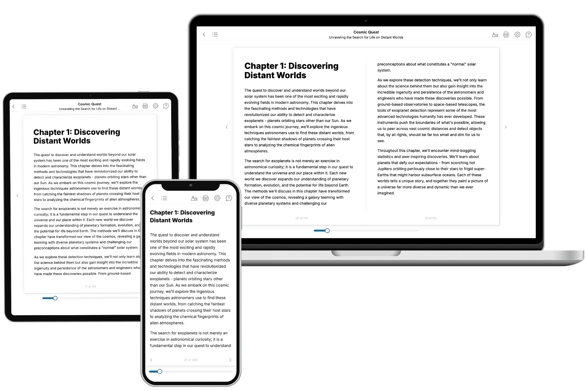
Your book is ready in 10 minutes. Read it online, download as EPUB or PDF, or send to Kindle.

Frequently asked questions
- What is TailoredRead?
TailoredRead creates complete, full-length nonfiction ebooks, written specifically for you. Tell us what you want to learn and why, and TailoredRead writes you a complete ebook on it, with charts, tables, and diagrams throughout. Read it online, on Kindle, or on any e-reader, or download it as a PDF or EPUB. Every book is written fresh the day you order it.
- How is the book tailored to me?
Before anything is written, you answer a few short questions about your goals, your background, and the topics you care about. Your book is then planned around your answers: written at your level, focused on your goals, and covering the topics you chose, instead of the one-size-fits-all book a store sells everyone.
- Will it read like generic AI content?
No. Your book isn't a chat transcript: it's planned chapter by chapter before a single word is written, told in a clear book voice, and explains ideas visually with charts, tables, and diagrams. After writing, every book goes through an editorial pass that polishes chapters so the book reads consistent from start to finish. Judge for yourself: browse thousands of sample books on our Explore Books page.
- How accurate is the content?
Our AI first checks whether your subject needs live research. Niche and fast-moving subjects are researched before writing, and the book plan and chapters are grounded in what it finds. After writing, every book goes through a quality pass that checks its facts and consistency, and researched books list their sources in a Notes section at the back.
- How long is the book?
You choose the length: Comprehensive (250-300 pages), Detailed (150-200 pages), Essential (70-100 pages), or Short (30-50 pages).
- Does the book include visuals?
Yes. Every book includes charts, tables, and diagrams: timelines, checklists, step-by-step guides, decision trees, comparison tables, matrices, and bar charts. A typical book includes dozens of these visual elements.
- Can I read it on Kindle? What formats can I download?
Yes. Every book can be read in our online reader or downloaded as a PDF or EPUB. The EPUB can be opened with Kindle, Apple Books, Kobo, and any e-reader. The Commercial and Author licenses also include an editable Word (DOCX) and Markdown download.
- How much does it cost?
About the price of a regular ebook. The exact price depends on your book's length and complexity, from $2 for the shortest books to $35 for the most comprehensive, and it's shown when you review your book, before you commit to anything. Each book is a one-time purchase, yours to keep, no subscription. Licenses that let you share, teach, or publish your book range from $35.99 to $49.99.
- Can I preview the book before purchasing?
Before you pay, you'll see what your book will cover: the title, a detailed description, the estimated length, and the complete chapter-by-chapter table of contents, so you know what you're getting before you decide. The full text itself is written after you purchase.
- How long does it take to create a book?
About 10 minutes. You can start reading the opening chapters while the rest is still being written.