The Lymphatic System Coloring Book
Learn Anatomy Through Interactive Illustration and Detailed Exploration
Imagine being able to visualize the intricate network of vessels and organs that silently work to protect your body every single day.
222 pages10 chaptersMar 17, 2026Anatomy
About this book
This coloring book transforms the complex anatomy of the lymphatic system into an engaging, interactive learning experience. By combining detailed anatomical illustrations with hands-on coloring activities, you'll develop a deeper understanding of how lymph vessels transport fluid, how lymph nodes filter pathogens, and how specialized organs like the spleen and thymus support your immune defense. Each page guides you through different regions and structures of the lymphatic system, with clear labels and explanations that build your knowledge progressively. Whether you're a student, healthcare professional, or anatomy enthusiast, this book makes learning the lymphatic system accessible and memorable. The act of coloring engages your visual memory, helping you retain complex anatomical relationships far more effectively than passive reading alone. Discover how this often-overlooked system is absolutely essential to your health and immunity.
Table of contents
- IntroductionWhy the Lymphatic System Matters, How to Use This Coloring Book, Coloring Tips for Anatomical Learning, Overview of Lymphatic System FunctionsRead below
- Chapter 1: Lymphatic System BasicsWhat Is the Lymphatic System?, Lymphatic System vs. Circulatory System, Understanding Lymph Fluid
- Chapter 2: Lymph Vessels and TransportLymphatic Capillaries and Vessel Structure, Major Lymphatic Vessel Pathways, The Thoracic Duct and Right Lymphatic Duct, How the One-Way Valve System Works
- Chapter 3: Lymph Nodes and FilteringLymph Node Anatomy and Structure, How Lymph Nodes Filter and Protect, Regional Lymph Node Groups
- Chapter 4: Primary Lymphoid OrgansThe Thymus Gland and T-Cell Development, Bone Marrow and Immune Cell Production, Coloring Primary Lymphoid Organs
- Chapter 5: Secondary Lymphoid OrgansThe Spleen: Structure and Function, Tonsils, Adenoids, and Mucosa-Associated Lymphoid Tissue, Peyer's Patches and Gut-Associated Lymphoid Tissue, Coloring Secondary Lymphoid Organs
- Chapter 6: Lymphatic System in the Head and NeckLymphatic Vessels of the Head and Neck, Cervical Lymph Nodes and Drainage Patterns, Coloring Head and Neck Lymphatics
- Chapter 7: Lymphatic System in the Thorax and AbdomenThoracic Lymphatic Vessels and Nodes, Abdominal and Pelvic Lymphatic Drainage, Mesenteric Nodes and Intestinal Lymphatics, Coloring Thoracic and Abdominal Lymphatics
- Chapter 8: Lymphatic System in the LimbsUpper Limb Lymphatic Drainage, Lower Limb Lymphatic Drainage, Axillary and Inguinal Lymph Node Groups, Coloring Limb Lymphatics
- Chapter 9: Lymphatic System and ImmunityImmune Cells in the Lymphatic System, How Lymphatics Support Immune Response, Lymphocyte Circulation and Activation
- Chapter 10: Clinical Significance and HealthUnderstanding Lymphedema and Dysfunction, Lymph Node Enlargement and What It Means, Maintaining Healthy Lymphatic Function
- ConclusionKey Takeaways About Lymphatic Anatomy, Continuing Your Anatomical Learning Journey
- Glossary of Lymphatic Terms
- Further Reading and Resources
Introduction
There is a vast, silent network running through your body right now. It moves fluid, hunts down pathogens, and keeps your tissues from swelling with excess liquid. It works around the clock without any conscious effort on your part. Yet most people who study anatomy spend far more time on the heart, lungs, and digestive tract than they ever do on this remarkable system. The lymphatic system is one of the most underappreciated networks in the human body, and this book is here to change that.
Learning anatomy is not just about memorizing names and locations. It is about building a mental picture of how structures relate to one another, how they move, and what they do. That kind of spatial understanding is genuinely difficult to develop from text alone. Coloring anatomical illustrations is one of the most effective tools available for building that picture. When you choose a color, apply it to a vessel, and trace its path across a page, your brain encodes that information in a way that passive reading simply cannot match. Research in educational psychology consistently shows that active, hands-on engagement with material leads to stronger long-term retention than reading or listening alone.[1]
This book is designed for learners who already have some foundational knowledge of anatomy and want to go deeper. Whether you are a nursing student preparing for clinical practice, a massage therapist wanting to understand the tissue systems you work with, a medical professional refreshing your knowledge, or simply someone fascinated by how the body works, this coloring book meets you where you are. Each chapter builds on the last, moving from basic concepts to regional anatomy to clinical relevance. By the time you reach the final pages, you will have a thorough, visually grounded understanding of the lymphatic system that will serve you for years to come.
Before diving into the chapters themselves, this introduction covers four essential areas. First, it explains why the lymphatic system deserves serious attention. Second, it walks you through how to get the most out of this coloring book format. Third, it offers practical tips for using color as a learning tool. Finally, it provides a broad overview of the major functions the lymphatic system performs, giving you a conceptual framework to carry into every chapter that follows.
Why the Lymphatic System Matters
The lymphatic system does not get the same attention as the cardiovascular or nervous systems, but its importance to your health is just as significant. Every day, your blood capillaries leak a small amount of fluid into the surrounding tissues. This is a normal part of how nutrients and oxygen are delivered to cells. But that fluid needs to be collected and returned to the bloodstream. Without the lymphatic system performing that job continuously, your tissues would swell with fluid, your blood volume would drop, and your body would quickly face serious problems.[2]
Beyond fluid balance, the lymphatic system is a cornerstone of your immune defense. Lymph nodes are not just passive filters. They are active surveillance stations where immune cells examine everything passing through the lymph fluid. When a pathogen, foreign particle, or abnormal cell is detected, the immune response is triggered right there in the node. This is why lymph nodes swell when you have an infection. They are working hard, multiplying immune cells and mounting a targeted response to whatever threat has been identified.
The system also plays a critical role in fat absorption. In the small intestine, specialized lymphatic vessels called lacteals absorb dietary fats and fat-soluble vitamins that cannot enter the bloodstream directly through the capillary walls. These fats travel through the lymphatic system before eventually entering the bloodstream via the thoracic duct. Without this pathway, your body could not properly absorb essential nutrients from the food you eat.
Understanding the lymphatic system also has direct clinical relevance. Conditions like lymphedema, in which lymphatic drainage is impaired and tissues swell chronically, affect millions of people worldwide. Cancer staging frequently depends on whether cancer cells have spread to nearby lymph nodes. Autoimmune diseases involve dysregulation of the very immune cells that the lymphatic system produces and circulates. A solid grasp of lymphatic anatomy is not just academic knowledge. It is foundational to understanding a wide range of health conditions and medical procedures.
Consider a practical example: a patient undergoes surgery to remove breast cancer, and several axillary lymph nodes are also removed to check for cancer spread. Afterward, the patient develops swelling in the arm on that side. Understanding why this happens requires knowing exactly how lymphatic drainage from the arm passes through the axillary nodes. Without that anatomical knowledge, the connection between the surgery and the swelling is not obvious. With it, the mechanism is immediately clear and the rationale for treatment makes complete sense.
The lymphatic system also connects to the body's hormonal and metabolic functions in ways that are still being actively researched. Recent studies have identified lymphatic vessels in the brain, a discovery that overturned decades of assumptions about the central nervous system being isolated from lymphatic drainage.[3] This finding has opened new lines of research into neurological diseases like Alzheimer's, where the clearance of waste products from brain tissue may depend on these newly identified vessels. The lymphatic system, it turns out, is even more important than previously understood.
In short, the lymphatic system matters because it touches nearly every aspect of your body's normal function. Fluid balance, immune defense, nutrient absorption, waste clearance, and disease progression all involve this network in fundamental ways. Learning its anatomy is not a niche interest. It is essential knowledge for anyone serious about understanding how the human body works.
How to Use This Coloring Book
This book is structured to guide you through the lymphatic system in a logical, progressive sequence. It begins with foundational concepts and gradually moves into more detailed regional anatomy and clinical applications. You do not have to follow the chapters in strict order, but doing so will give you the strongest conceptual foundation. Each chapter builds on vocabulary and concepts introduced in earlier sections, so jumping ahead may occasionally leave you without context for what you are seeing.
Each chapter contains two types of content working together. The written explanations give you the conceptual and functional knowledge you need to understand what you are looking at. The illustrations give you the spatial, visual representation of those same structures. Your job is to bring them together by actively engaging with both. Read the explanation for a structure, locate it in the illustration, and then color it according to the suggested color scheme or your own system. This back-and-forth between text and image is where the real learning happens.
Here is a recommended approach for working through each chapter:
- Read the written section for the subchapter completely before picking up any coloring tools.
- Review the illustration and identify the labeled structures without coloring yet.
- Go back to the text and re-read descriptions of individual structures, then locate each one again in the illustration.
- Begin coloring, working structure by structure rather than filling in large areas at once.
- After coloring, cover the labels and test yourself by naming each structure from memory.
- Review any structures you could not name and repeat the coloring step if needed.
The self-testing step in that process is particularly important. Research on learning consistently shows that retrieval practice, the act of trying to recall information from memory, is one of the most powerful ways to strengthen long-term retention.[4] Covering the labels and naming structures from memory is a simple form of retrieval practice that you can do right on the page. Do not skip this step even when it feels difficult. The difficulty itself is a sign that learning is happening.
You can also use this book as a reference tool after your initial study. Once you have worked through a chapter, the colored illustrations become personalized reference diagrams that reflect your own color choices and annotations. Many learners find it helpful to add their own notes in the margins, write brief function descriptions next to structures, or draw arrows to show the direction of lymph flow. Make this book your own. The more you interact with it, the more useful it becomes.
If you are using this book as part of a course or study group, consider comparing your color choices and annotations with others. Different people notice different details, and discussing why a particular vessel connects to a particular node can deepen everyone's understanding. There is no single correct way to use a coloring book for anatomy. The goal is active engagement, and any approach that keeps you thinking and questioning is the right one.
Finally, do not worry about making your coloring perfect. This is not an art project. Neat, precise coloring is helpful because it forces you to pay attention to boundaries and shapes, but the primary goal is learning, not aesthetics. A slightly messy page that you engaged with deeply is far more valuable than a perfectly colored page you completed on autopilot.
Coloring Tips for Anatomical Learning
Choosing the right tools and developing a consistent color system will make your coloring sessions significantly more productive. The most important principle is consistency. When you assign a color to a specific type of structure, use that same color every time that structure appears throughout the book. This consistency trains your brain to associate the color with the structure automatically, which speeds up recognition and recall.
Here is a suggested color system to get you started. You are welcome to modify it based on the tools you have available:
- Lymphatic vessels: Light green or yellow-green, to distinguish them clearly from blood vessels
- Lymph nodes: Dark green or teal, so they stand out as distinct structures along vessel pathways
- Arteries (for reference): Red, following the standard anatomical convention
- Veins (for reference): Blue, again following standard convention
- The thymus: Purple or violet, to mark it as a primary lymphoid organ
- The spleen: Dark red or maroon, reflecting its blood-filtering role
- Bone marrow regions: Orange or amber
- Tonsils and mucosa-associated lymphoid tissue: Pink or light purple
For tools, colored pencils are generally the best choice for anatomical coloring books. They allow precise control, can be layered for shading, and do not bleed through pages the way markers often do. Fine-tipped colored pencils work especially well for small structures and detailed labels. If you prefer markers, use alcohol-based fine-liners and place a blank sheet behind each page to protect the illustration on the reverse side.
Shading is a technique worth developing even at a basic level. When a structure has depth or three-dimensional form, applying a slightly darker shade of the same color to one side creates a sense of volume. This is particularly useful for lymph nodes, which are roughly bean-shaped, and for organs like the spleen and thymus. Shading helps your brain encode the three-dimensional reality of these structures rather than seeing them as flat shapes on a page.
Another useful technique is color coding by function rather than just by structure type. For example, you might use one shade of green for afferent lymphatic vessels (those carrying lymph into a node) and a slightly different shade for efferent vessels (those carrying lymph out). This functional distinction is anatomically meaningful and coloring it reinforces the concept every time you look at the page. Similarly, you could use different intensities of the same color to indicate the direction of lymph flow, with lighter shades at the origin and darker shades closer to the drainage point.
Take breaks during long coloring sessions. Sustained focus on detailed illustrations is mentally demanding, and fatigue reduces the quality of both your coloring and your learning. A 10-minute break after 30 to 40 minutes of focused work helps maintain concentration and prevents the kind of mechanical, inattentive coloring that does not reinforce learning. Use break time to close the book and mentally review what you just colored. This brief mental rehearsal is another form of retrieval practice that strengthens memory consolidation.
Label as you go. Many illustrations in this book include blank label lines alongside pre-printed ones. Use these to write in additional terms, functional notes, or clinical connections that you want to remember. For example, next to the axillary lymph nodes, you might write breast cancer staging
as a reminder of their clinical significance. These personal annotations transform the illustrations into richer, more meaningful reference tools that reflect your own learning priorities.
Finally, revisit completed pages periodically. Spaced repetition, the practice of reviewing material at increasing intervals over time, is one of the most well-supported strategies in learning science for building durable long-term memory.[5] Flipping back through earlier chapters every few days, covering labels, and testing yourself on structures you have already colored will dramatically improve how much you retain over the long term.
Overview of Lymphatic System Functions
Before exploring the detailed anatomy in the chapters ahead, it helps to have a clear picture of what the lymphatic system actually does. The system performs several distinct but interconnected functions, and understanding all of them gives you a richer context for interpreting the structures you will be coloring. Think of this overview as the conceptual map you will carry with you through the rest of the book.
The first and most fundamental function is fluid balance. Blood plasma constantly leaks out of capillaries into the surrounding interstitial space, the fluid-filled gaps between cells. Most of this fluid is reabsorbed directly back into the capillaries, but roughly 10 to 20 percent is not.[6] That remaining fluid, along with proteins and other large molecules that cannot re-enter blood capillaries, is collected by lymphatic capillaries and transported back to the bloodstream. Without this drainage, the interstitial space would accumulate fluid continuously, causing the tissue swelling known as edema.
The second major function is immune surveillance and defense. As lymph fluid travels through the lymphatic vessels, it passes through lymph nodes distributed along the pathway. Inside these nodes, immune cells called lymphocytes and macrophages examine the fluid for pathogens, foreign particles, and abnormal cells. When a threat is detected, the immune response is activated locally. This is why swollen lymph nodes are a reliable sign of infection or immune activity. The nodes are doing exactly what they are designed to do.
The third function is fat and nutrient transport. In the lining of the small intestine, specialized lymphatic capillaries called lacteals absorb dietary fats, cholesterol, and fat-soluble vitamins (A, D, E, and K) after digestion. These nutrients form a milky fluid called chyle, which travels through the intestinal lymphatics and eventually enters the bloodstream through the thoracic duct. This pathway is essential for proper nutrition, and it is one reason why the lymphatic system is closely connected to digestive health.
The fourth function involves immune cell production and maturation. The lymphatic system includes specialized organs where immune cells are born and trained. Bone marrow produces all blood and immune cells, including the B cells and T cells that are central to adaptive immunity. The thymus gland is where T cells mature and learn to distinguish the body's own cells from foreign invaders. Without these primary lymphoid organs functioning correctly, the entire immune system would be compromised.
Here is a summary of the key functions at a glance:
- Fluid balance: Collects excess interstitial fluid and returns it to the bloodstream
- Immune defense: Filters lymph through nodes where pathogens are identified and destroyed
- Fat absorption: Transports dietary fats and fat-soluble vitamins from the intestine via lacteals
- Immune cell development: Provides sites for the production and maturation of B cells and T cells
- Waste clearance: Removes cellular debris, dead cells, and large protein molecules from tissues
- Hormone and signaling molecule transport: Carries certain hormones and cytokines through the body
It is worth noting how these functions interact with one another. The fluid balance function and the immune function are not separate processes. The same fluid that is being drained from tissues is also being screened for pathogens as it passes through lymph nodes. The fat transport function and the immune function overlap in the gut, where the intestinal lymphoid tissue monitors the contents of the digestive tract for harmful microorganisms while simultaneously absorbing nutrients. The lymphatic system is not a collection of separate mechanisms. It is an integrated network where structure and function are deeply intertwined.
As you work through this book, keep these functions in mind as an interpretive lens. When you color a lymph node, think about what it is filtering. When you trace a lymphatic vessel, think about where the fluid it carries came from and where it is going. When you color the thymus, think about the T cells being trained inside it. Connecting structure to function at every step is what transforms coloring from a mechanical activity into genuine anatomical understanding.
The chapters ahead will take you through each component of this system in detail, from the microscopic structure of lymphatic capillaries to the regional drainage patterns of the limbs, from the internal architecture of lymph nodes to the clinical consequences of lymphatic dysfunction. Each chapter is designed to be both informative and interactive, giving you the knowledge and the visual tools to build a complete, lasting picture of one of the body's most essential systems. The journey starts now, and it begins with the basics.
Notes
Introduction
- 1. S. M. Kosslyn and R. S. Rosenberg, Psychology: The Brain, the Person, the World, Pearson Education, 2004
- 2. E. N. Marieb and K. Hoehn, Human Anatomy and Physiology (11th ed.), Pearson, 2019
- 3. A. Louveau and others, "Structural and functional features of central nervous system lymphatic vessels," Nature, 2015
- 4. H. L. Roediger and J. D. Karpicke, "Test-enhanced learning," Psychological Science, 2006
- 5. H. Ebbinghaus, Über das Gedächtnis, Duncker & Humblot. (Translated as Memory: A Contribution to Experimental Psychology, 1885
- 6. A. C. Guyton and J. E. Hall, Textbook of Medical Physiology (13th ed.), Elsevier, 2016
This is the end of the free sample. The full book has 10 chapters.
Want this book written for you?
Tell us your goals and background, and get your own version of this book, tailored by AI to you.


Benefits of AI-tailored books
- Read one book, not ten:
- all the Anatomy knowledge you need, gathered into a single book.
- Save days of learning:
- tell us your goals, choose your topics, and the whole book is built around them.
- Learn effortlessly:
- Anatomy book written for your specific background and expertise.
- Understand at a glance:
- includes charts, tables, timelines, checklists, decision trees, and more.
- Stay current:
- written the day you order it, not years ago.
- Trust what you read:
- every book is checked for accuracy, with real research when your subject needs it.
Create your unique book in 3 steps
- 1. Select your focus
Select the focus of your Anatomy book and share your background
- 2. Personalize your book
Specify your goals and choose sub-topics to include
- 3. Get your tailored book
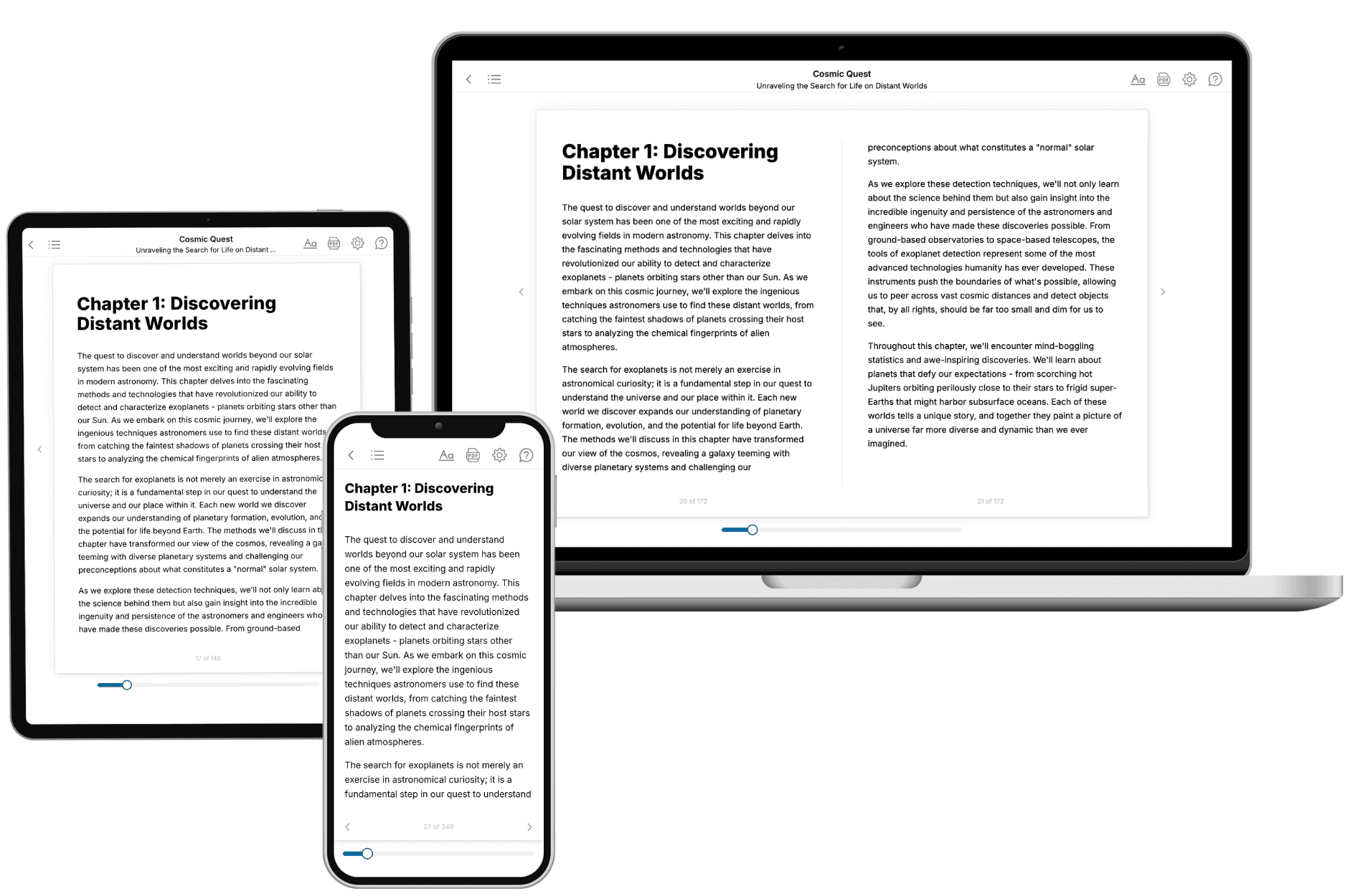
Your book is ready in 10 minutes. Read it online, download as EPUB or PDF, or send to Kindle.

Frequently asked questions
- What is TailoredRead?
TailoredRead creates complete, full-length nonfiction ebooks, written specifically for you. Tell us what you want to learn and why, and TailoredRead writes you a complete ebook on it, with charts, tables, and diagrams throughout. Read it online, on Kindle, or on any e-reader, or download it as a PDF or EPUB. Every book is written fresh the day you order it.
- How is the book tailored to me?
Before anything is written, you answer a few short questions about your goals, your background, and the topics you care about. Your book is then planned around your answers: written at your level, focused on your goals, and covering the topics you chose, instead of the one-size-fits-all book a store sells everyone.
- Will it read like generic AI content?
No. Your book isn't a chat transcript: it's planned chapter by chapter before a single word is written, told in a clear book voice, and explains ideas visually with charts, tables, and diagrams. After writing, every book goes through an editorial pass that polishes chapters so the book reads consistent from start to finish. Judge for yourself: browse thousands of sample books on our Explore Books page.
- How accurate is the content?
Our AI first checks whether your subject needs live research. Niche and fast-moving subjects are researched before writing, and the book plan and chapters are grounded in what it finds. After writing, every book goes through a quality pass that checks its facts and consistency, and researched books list their sources in a Notes section at the back.
- How long is the book?
You choose the length: Comprehensive (250-300 pages), Detailed (150-200 pages), Essential (70-100 pages), or Short (30-50 pages).
- Does the book include visuals?
Yes. Every book includes charts, tables, and diagrams: timelines, checklists, step-by-step guides, decision trees, comparison tables, matrices, and bar charts. A typical book includes dozens of these visual elements.
- Can I read it on Kindle? What formats can I download?
Yes. Every book can be read in our online reader or downloaded as a PDF or EPUB. The EPUB can be opened with Kindle, Apple Books, Kobo, and any e-reader. The Commercial and Author licenses also include an editable Word (DOCX) and Markdown download.
- How much does it cost?
About the price of a regular ebook. The exact price depends on your book's length and complexity, from $2 for the shortest books to $35 for the most comprehensive, and it's shown when you review your book, before you commit to anything. Each book is a one-time purchase, yours to keep, no subscription. Licenses that let you share, teach, or publish your book range from $35.99 to $49.99.
- Can I preview the book before purchasing?
Before you pay, you'll see what your book will cover: the title, a detailed description, the estimated length, and the complete chapter-by-chapter table of contents, so you know what you're getting before you decide. The full text itself is written after you purchase.
- How long does it take to create a book?
About 10 minutes. You can start reading the opening chapters while the rest is still being written.