The Lymphatic System Coloring Guide
Master Anatomy Through Interactive Illustration and Deep Learning
Have you ever wondered how your body manages the fluid that constantly bathes your cells, or how your immune system knows where to send its defenders?
207 pages9 chaptersMay 26, 2026Anatomy
About this book
The lymphatic system is one of your body's most vital yet underappreciated networks, and understanding its anatomy is key to grasping how your body maintains health and fights disease. This coloring guide transforms complex anatomical concepts into engaging, interactive learning experiences. By combining detailed illustrations with scientifically accurate explanations, you'll discover how lymphatic vessels work in concert with your circulatory system, how lymph nodes filter threats, and why this network matters for your overall wellness. Whether you're a student preparing for exams, a healthcare professional seeking deeper knowledge, or simply curious about how your body works, this guide makes lymphatic anatomy accessible and memorable. The act of coloring engages your brain differently than reading alone, helping you retain information longer and understand spatial relationships more intuitively. Explore the pathways of lymph, the architecture of vessels, and the strategic placement of nodes throughout your body.
Table of contents
- IntroductionWhy the Lymphatic System Matters, How to Use This Coloring Guide, The Science of Learning Through ColorRead below
- Chapter 1: Foundations of the Lymphatic SystemOverview and Primary Functions, Comparison with the Circulatory System, What Is Lymph? Composition and Origin
- Chapter 2: Lymphatic Capillaries and Vessel StructureLymphatic Capillary Anatomy and Function, Classification of Lymphatic Vessels, One-Way Valves: The Key to Lymph Movement
- Chapter 3: Major Lymphatic Vessels and DuctsThe Thoracic Duct: The Body's Main Highway, The Right Lymphatic Duct and Upper Body Drainage, Regional Lymphatic Vessels and Pathways
- Chapter 4: Lymph Nodes: Structure and Strategic PlacementInternal Structure of Lymph Nodes, Major Lymph Node Groups Throughout the Body, How Lymph Nodes Filter and Protect
- Chapter 5: Lymphatic Organs and Immune IntegrationThe Spleen: Structure and Immune Role, The Thymus Gland and T-Cell Development, Tonsils, Adenoids, and Mucosa-Associated Lymphoid Tissue
- Chapter 6: Lymphatic Drainage Patterns and RegionsHead and Neck Lymphatic Drainage, Thoracic and Abdominal Drainage Patterns, Upper and Lower Limb Lymphatic Pathways
- Chapter 7: Mechanisms of Lymph MovementThe Skeletal Muscle Pump, The Respiratory Pump and Breathing, Pressure Gradients and Passive Movement
- Chapter 8: Clinical Significance and Common ConditionsLymphedema: Causes, Types, and Manifestations, Lymphatic Infections and Inflammatory Conditions, The Lymphatic System and Cancer Spread
- Chapter 9: Coloring Exercises and Anatomical MapsFull-Body Lymphatic System Map, Regional Anatomy Coloring Exercises, Cellular and Microscopic Structure Illustrations
- ConclusionIntegrating Your Knowledge, Next Steps in Your Learning Journey
- Glossary of Terms
- Further Reading and Resources
Introduction
Your body is running an extraordinary operation right now, and most people never think about it. While your heart pumps blood through arteries and veins in a closed loop, a parallel network of vessels is quietly collecting fluid, filtering threats, and returning essential proteins back to your bloodstream. This is the lymphatic system, and it is one of the most underappreciated networks in the entire human body. Without it, your tissues would swell, your immune defenses would collapse, and the fats you absorb from food would have no way to reach your blood. It is that fundamental.
Despite its importance, the lymphatic system rarely gets the attention it deserves in anatomy education. Students often spend far more time on the cardiovascular system, the nervous system, or the musculoskeletal system, leaving lymphatics as an afterthought. This is a missed opportunity. Understanding how lymph moves, where nodes are positioned, and how vessels are structured gives you a much richer picture of how the body maintains balance and defends itself. It also has real clinical value, from understanding why cancer spreads the way it does to recognizing the early signs of lymphedema.
This guide was designed to change that. Rather than presenting lymphatic anatomy as a dry list of structures to memorize, it invites you to engage with the material actively, through coloring, labeling, and exploring the spatial relationships between vessels, nodes, and organs. Each chapter builds on the last, moving from foundational concepts to regional drainage patterns to clinical applications. By the time you finish, you will not just know the lymphatic system. You will understand it in a way that sticks.
Before diving into the anatomy itself, this introduction covers three things that will make your experience with this guide more effective. First, it explains why the lymphatic system deserves your full attention. Second, it walks you through how to use this coloring guide to get the most out of every page. Third, it explores the science behind why coloring and active illustration are such powerful tools for learning anatomy. These three sections set the stage for everything that follows.
Why the Lymphatic System Matters
The lymphatic system performs three core jobs that no other system in the body can fully replace. It maintains fluid balance by collecting the excess fluid that leaks out of blood capillaries into surrounding tissues. It supports immune function by transporting pathogens, cellular debris, and foreign particles to lymph nodes where immune cells can neutralize them. And it absorbs dietary fats and fat-soluble vitamins from the small intestine, delivering them into the bloodstream through specialized vessels called lacteals. Each of these roles is essential to survival.
Consider the fluid balance function alone. Every day, your blood capillaries leak roughly three to four liters of fluid into the interstitial spaces between your cells.[1] This fluid carries proteins, hormones, and other large molecules that cannot easily re-enter the bloodstream directly. The lymphatic system collects this fluid, now called lymph, and returns it to the venous circulation. If this process were to stop for even a few hours, tissues would begin to swell noticeably. Over days, the swelling would become severe and life-threatening. This condition, when it occurs chronically, is known as lymphedema.
The immune function of the lymphatic system is equally critical. Lymph nodes are not just passive filters. They are active surveillance stations packed with lymphocytes, macrophages, and dendritic cells. When lymph flows through a node, these immune cells scan for anything that should not be there, including bacteria, viruses, cancer cells, and foreign particles. If a threat is detected, the immune response is activated locally and then communicated to the broader immune network. This is why lymph nodes swell when you have an infection. They are working hard.
The digestive role of the lymphatic system is less well known but equally fascinating. In the lining of the small intestine, specialized lymphatic capillaries called lacteals absorb the products of fat digestion. These fats, packaged into particles called chylomicrons, are too large to enter blood capillaries directly. Instead, they enter the lacteals and travel through the lymphatic system before eventually draining into the bloodstream near the heart. This is why lymph from the gut, called chyle, appears milky white after a fatty meal rather than the clear or pale yellow of lymph from other regions.
From a clinical perspective, the lymphatic system is relevant to an enormous range of conditions. Surgeons mapping lymph node involvement in cancer use a technique called sentinel node biopsy to determine whether cancer has begun to spread. Physical therapists use manual lymphatic drainage to treat patients with chronic swelling. Infectious disease specialists track how pathogens travel through lymphatic channels to understand disease progression. Dermatologists consider lymphatic drainage patterns when treating skin conditions. In short, knowledge of lymphatic anatomy is not just academic. It has direct, practical applications across many fields of healthcare.
There is also a growing body of research revealing new dimensions of the lymphatic system that were not well understood even a decade ago. In 2015, researchers discovered a network of lymphatic vessels in the brain's meningeal lining, a finding that overturned the long-held belief that the brain lacked a conventional lymphatic drainage system.[2] This discovery has opened new lines of research into neurodegenerative diseases like Alzheimer's, where the clearance of waste products from the brain may be impaired. The lymphatic system, it turns out, is still revealing its secrets.
Understanding this system also deepens your appreciation for how interconnected the body's systems truly are. The lymphatic system does not operate in isolation. It works alongside the cardiovascular system, the immune system, the digestive system, and even the nervous system. Grasping these connections transforms your understanding of human physiology from a collection of separate systems into a unified, integrated picture. That is the kind of understanding this guide is designed to build.
How to Use This Coloring Guide
This guide is structured to take you from broad concepts to fine anatomical detail in a logical, progressive sequence. Each chapter introduces new structures and functions, then reinforces them through coloring exercises and labeled diagrams. The final chapter brings everything together with full-body maps and regional exercises that let you apply everything you have learned. You can work through the guide from start to finish, or you can jump to specific chapters if you are focusing on a particular region or topic.
Before you begin any coloring exercise, read the accompanying text for that section first. This is important. The explanations provide the context you need to understand what you are coloring and why each structure looks and functions the way it does. Coloring without understanding is just art. Coloring with understanding is learning. Take a few minutes to read, then pick up your pencils or markers and engage with the illustration actively.
When it comes to choosing your coloring tools, a few practical tips will help. Colored pencils give you the most control and allow for shading, which is useful for showing depth in three-dimensional structures. Felt-tip markers produce vivid, saturated colors that make structures easy to distinguish at a glance, but they can bleed through thinner paper. Watercolor pencils offer a middle ground. Whatever you choose, the most important principle is consistency. Use the same color for the same type of structure throughout the guide. For example, if you color lymphatic capillaries in light green, use light green every time they appear. This builds a visual vocabulary that your brain will start to recognize automatically.
Here is a suggested color-coding system to get you started, though you are free to adapt it to your own preferences:
- Light green or yellow-green: Lymphatic capillaries
- Medium green: Collecting lymphatic vessels
- Dark green: Lymphatic ducts (thoracic duct, right lymphatic duct)
- Blue: Lymph nodes
- Purple: Lymphatic organs (spleen, thymus, tonsils)
- Red: Arteries (for reference and comparison)
- Dark blue: Veins (for reference and comparison)
Each coloring exercise includes a set of labels and a corresponding legend. As you color each structure, write the name of the structure next to its label line if it is not already filled in. This act of writing reinforces the name-to-structure connection in your memory. After completing an exercise, try covering the labels and testing yourself. Can you identify each structure by color and position alone? If not, go back and review the text before moving on. Self-testing is one of the most effective study strategies available, and it works especially well when combined with visual learning.[3]
At the end of each chapter, you will find a short review section with key terms and concept questions. These are not just filler. They are designed to prompt you to think about what you have just learned in a slightly different way, connecting new information to what you already know. Spend a few minutes on these before moving to the next chapter. Even brief reflection dramatically improves how well information transfers to long-term memory.
If you are using this guide as part of a course or study group, consider working through the coloring exercises with a partner. Explaining what you are coloring and why to another person forces you to articulate your understanding, which reveals gaps you might not notice when studying alone. Teaching is one of the most powerful forms of learning, and even informal explanation to a study partner counts.
Finally, do not worry about making your illustrations look perfect. The goal is not artistic accuracy. It is anatomical understanding. A slightly uneven line or an imperfect shading job does not matter. What matters is that you are actively engaging with the structures, thinking about their relationships, and building a mental map of the lymphatic system that you can recall and apply. Give yourself permission to be a learner, not an artist.
The Science of Learning Through Color
There is a reason anatomy coloring books have been used in medical and health education for decades. It is not just because they are more engaging than plain diagrams, though they certainly are. It is because the act of coloring activates multiple cognitive processes simultaneously in ways that passive reading simply cannot match. When you color an anatomical illustration, you are not just looking at a structure. You are making decisions about it, moving your hand in response to its shape, and encoding it through both visual and motor memory pathways at the same time.
This multi-channel encoding is the key to why coloring works so well for anatomy. Cognitive scientists refer to this as dual coding theory, first proposed by Allan Paivio in the 1970s. The theory holds that information is better retained when it is encoded through both verbal and visual channels rather than just one.[4] When you read a description of the thoracic duct and then color it on a diagram, you are creating two separate but linked memory traces. When you try to recall the information later, you have two pathways to access it instead of one, which makes retrieval significantly more reliable.
Research specifically on anatomy coloring books supports this effect. A study published in the journal Anatomical Sciences Education found that students who used coloring-based learning activities showed significantly better performance on spatial anatomy tasks compared to students who studied using traditional text and diagram methods alone.[5] Spatial understanding is particularly important in anatomy, where knowing the three-dimensional relationships between structures is often just as important as knowing their names and functions. Coloring helps build that spatial intuition in a way that reading descriptions rarely does.
Color itself also plays a functional role in memory encoding. The brain's visual cortex is highly sensitive to color contrast, and using distinct colors for different structures helps the brain categorize and separate them more efficiently. This is why the color-coding system described in the previous section is worth following consistently. When you always see lymph nodes in blue and lymphatic ducts in dark green, your brain begins to associate those colors with those structures automatically. Over time, this association becomes a retrieval cue. Seeing the color triggers the memory of the structure, even before you consciously read the label.
There is also a motor learning component to coloring that is easy to overlook. The act of tracing the path of a vessel with a colored pencil engages your hand and arm in a way that mirrors the path of the vessel itself. This is not trivial. Motor memory is stored differently from declarative memory, and engaging it adds another layer of encoding. Students who have colored the path of the thoracic duct from the cisterna chyli up through the chest to the left subclavian vein often report that they can mentally trace that path during an exam, almost as if their hand remembers the movement. This is motor memory at work.
Attention is another factor. One of the biggest challenges in studying anatomy from a textbook is maintaining focused attention on complex diagrams. It is easy for your eyes to glaze over a detailed illustration without really processing what you are seeing. Coloring forces sustained, focused attention on each part of the illustration in sequence. You cannot color the afferent lymphatic vessels entering a lymph node without first finding them, identifying them, and distinguishing them from the efferent vessels leaving the node. That process of active discrimination is itself a form of deep learning.
The concept of desirable difficulty in learning science is also relevant here. Research by Robert Bjork and others has shown that learning conditions that feel slightly harder in the moment often produce better long-term retention than conditions that feel easy.[6] Coloring an anatomy diagram is more effortful than simply reading a labeled diagram. That extra effort is not a drawback. It is a feature. The cognitive work required to identify, color, and label each structure is exactly what drives deeper encoding and longer retention.
To get the maximum benefit from the coloring exercises in this guide, approach each one with intention. Before you start coloring, scan the entire illustration and try to identify as many structures as you can from memory. Then read the labels and the accompanying text. Then color, starting with the structures you are most confident about and working toward the ones that are less familiar. After you finish, close the guide and try to sketch the basic layout of what you just colored from memory. This sequence, preview, read, color, recall, is a powerful learning cycle that will accelerate your mastery of lymphatic anatomy considerably.
A practical note on sessions: Research on spaced practice suggests that shorter, more frequent study sessions produce better long-term retention than long, infrequent ones. Rather than working through three chapters in a single sitting, consider completing one chapter per session and returning to review previous material at the start of each new session. Even five minutes of review before starting something new can significantly strengthen memory consolidation.
The combination of visual engagement, motor activity, color-based categorization, and active recall makes coloring one of the most effective self-study tools available for anatomy learners at any level. This guide is built around that principle. Every illustration, every label, and every exercise has been designed to maximize the learning value of your time and effort. The lymphatic system is complex, but with the right approach, it is entirely within your reach to understand it deeply and retain that understanding long after you close this book.
Notes
Introduction
- 1. A. C. Guyton and J. E. Hall, Textbook of Medical Physiology, 13th ed. Elsevier, Chapter 16: The Lymphatic System, 2016
- 2. A. Louveau and others, "Structural and functional features of central nervous system lymphatic vessels," Nature, 2015
- 3. H. L. Roediger and J. D. Karpicke, "Test-enhanced learning," Psychological Science, 2006
- 4. A. Paivio, Imagery and Verbal Processes, Holt, Rinehart, and Winston. New York, 1971
- 5. D. Venes, "The role of active illustration in anatomical learning," Anatomical Sciences Education, 2013
- 6. R. A. Bjork, "Memory and metamemory considerations in the training of human beings," J. Metcalfe & A. Shimamura, Metacognition: Knowing about knowing. MIT Press, 1994
This is the end of the free sample. The full book has 9 chapters.
Want this book written for you?
Tell us your goals and background, and get your own version of this book, tailored by AI to you.


Benefits of AI-tailored books
- Read one book, not ten:
- all the Anatomy knowledge you need, gathered into a single book.
- Save days of learning:
- tell us your goals, choose your topics, and the whole book is built around them.
- Learn effortlessly:
- Anatomy book written for your specific background and expertise.
- Understand at a glance:
- includes charts, tables, timelines, checklists, decision trees, and more.
- Stay current:
- written the day you order it, not years ago.
- Trust what you read:
- every book is checked for accuracy, with real research when your subject needs it.
Create your unique book in 3 steps
- 1. Select your focus
Select the focus of your Anatomy book and share your background
- 2. Personalize your book
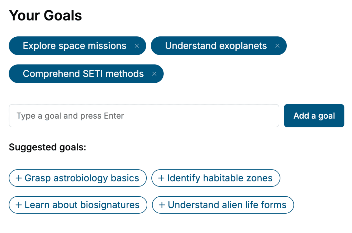
Specify your goals and choose sub-topics to include
- 3. Get your tailored book
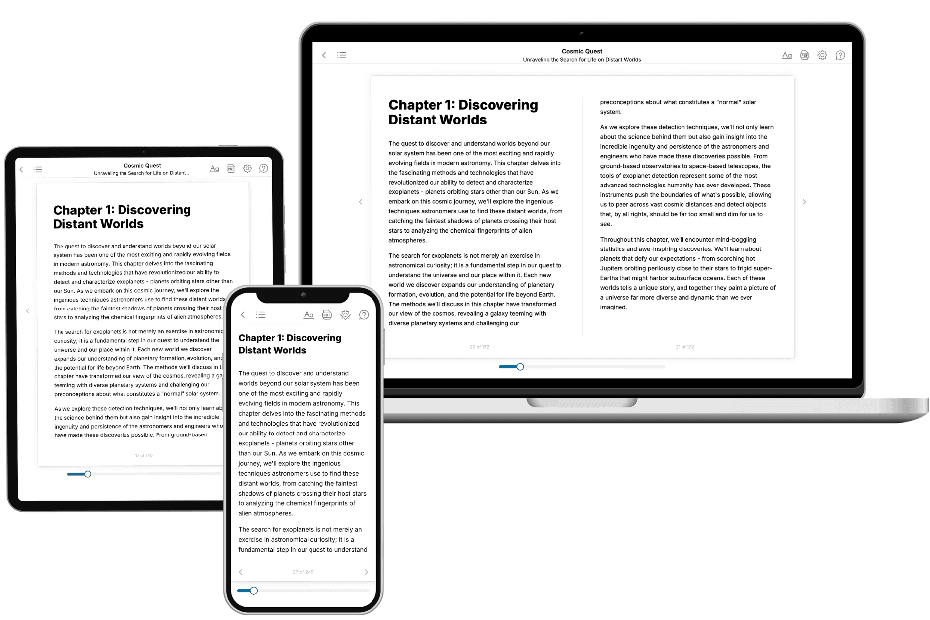
Your book is ready in 10 minutes. Read it online, download as EPUB or PDF, or send to Kindle.

Frequently asked questions
- What is TailoredRead?
TailoredRead creates complete, full-length nonfiction ebooks, written specifically for you. Tell us what you want to learn and why, and TailoredRead writes you a complete ebook on it, with charts, tables, and diagrams throughout. Read it online, on Kindle, or on any e-reader, or download it as a PDF or EPUB. Every book is written fresh the day you order it.
- How is the book tailored to me?
Before anything is written, you answer a few short questions about your goals, your background, and the topics you care about. Your book is then planned around your answers: written at your level, focused on your goals, and covering the topics you chose, instead of the one-size-fits-all book a store sells everyone.
- Will it read like generic AI content?
No. Your book isn't a chat transcript: it's planned chapter by chapter before a single word is written, told in a clear book voice, and explains ideas visually with charts, tables, and diagrams. After writing, every book goes through an editorial pass that polishes chapters so the book reads consistent from start to finish. Judge for yourself: browse thousands of sample books on our Explore Books page.
- How accurate is the content?
Our AI first checks whether your subject needs live research. Niche and fast-moving subjects are researched before writing, and the book plan and chapters are grounded in what it finds. After writing, every book goes through a quality pass that checks its facts and consistency, and researched books list their sources in a Notes section at the back.
- How long is the book?
You choose the length: Comprehensive (250-300 pages), Detailed (150-200 pages), Essential (70-100 pages), or Short (30-50 pages).
- Does the book include visuals?
Yes. Every book includes charts, tables, and diagrams: timelines, checklists, step-by-step guides, decision trees, comparison tables, matrices, and bar charts. A typical book includes dozens of these visual elements.
- Can I read it on Kindle? What formats can I download?
Yes. Every book can be read in our online reader or downloaded as a PDF or EPUB. The EPUB can be opened with Kindle, Apple Books, Kobo, and any e-reader. The Commercial and Author licenses also include an editable Word (DOCX) and Markdown download.
- How much does it cost?
About the price of a regular ebook. The exact price depends on your book's length and complexity, from $2 for the shortest books to $35 for the most comprehensive, and it's shown when you review your book, before you commit to anything. Each book is a one-time purchase, yours to keep, no subscription. Licenses that let you share, teach, or publish your book range from $35.99 to $49.99.
- Can I preview the book before purchasing?
Before you pay, you'll see what your book will cover: the title, a detailed description, the estimated length, and the complete chapter-by-chapter table of contents, so you know what you're getting before you decide. The full text itself is written after you purchase.
- How long does it take to create a book?
About 10 minutes. You can start reading the opening chapters while the rest is still being written.