Mastering Transmission Electron Microscopy
A Practical Guide to Advanced Imaging and Analysis in Modern Laboratories
Transmission electron microscopy remains one of the most powerful tools for visualizing atomic and nanoscale structures, yet many researchers struggle to move beyond basic operation to truly master this complex technology.
222 pages10 chaptersMar 16, 2026Microscopy
About this book
The challenge lies not just in understanding the equipment, but in knowing how to prepare samples correctly, interpret images accurately, and extract meaningful scientific conclusions from your data. This practical guide bridges the gap between theoretical knowledge and real-world laboratory practice. You'll learn the fundamental physics underlying TEM operation, master essential sample preparation techniques that determine success or failure, navigate the full range of imaging modes available on modern instruments, and develop systematic approaches to troubleshooting and maintenance. Whether you're optimizing existing protocols, training new team members, or pushing the boundaries of what's possible with your microscope, this book provides the actionable knowledge and proven strategies you need to achieve publication-quality results consistently.
Table of contents
- IntroductionWhy TEM Matters in Modern Science, What This Book Covers and How to Use It, Your Learning Path to TEM MasteryRead below
- Chapter 1: Fundamentals of Electron MicroscopyLight Microscopy vs. Electron Microscopy: Key Differences, Understanding Electron Wavelength and Resolution, Unique Advantages and Limitations of TEM
- Chapter 2: TEM Instrument Architecture and ComponentsThe Electron Gun: Generation and Acceleration, Magnetic Lens Systems and Beam Control, Specimen Holders and Stage Mechanics, Detectors and Image Acquisition Systems
- Chapter 3: Sample Preparation: The Foundation of SuccessWhy Sample Preparation Determines Your Results, Mechanical Thinning and Polishing Techniques, Ion Milling and Final Thinning, Recognizing and Avoiding Preparation Artifacts
- Chapter 4: Electron-Matter Interactions and Image FormationElastic Scattering and Diffraction Contrast, Inelastic Scattering and Energy Loss, Contrast Mechanisms in Bright-Field and Dark-Field Imaging
- Chapter 5: Imaging Modes and Operational TechniquesBright-Field Imaging: Fundamentals and Applications, Dark-Field Imaging for Enhanced Contrast, High-Resolution TEM (HRTEM) and Lattice Imaging, Scanning Transmission Electron Microscopy (STEM)
- Chapter 6: Analytical Techniques and SpectroscopyEnergy Dispersive X-ray Spectroscopy (EDS), Electron Energy Loss Spectroscopy (EELS), Selected Area Electron Diffraction (SAED) and Analysis, Combining Multiple Analytical Techniques
- Chapter 7: Image Processing and Quantitative AnalysisDigital Image Fundamentals and File Formats, Noise Reduction and Image Enhancement, Quantitative Measurement and Analysis Techniques, Essential Software Tools and Workflows
- Chapter 8: Troubleshooting and Problem-SolvingCommon Image Quality Problems and Solutions, Alignment and Calibration Challenges, Systematic Troubleshooting Methodology
- Chapter 9: Maintenance, Safety, and Best PracticesRoutine Maintenance and Equipment Care, Safety Protocols and Radiation Protection, Laboratory Best Practices and Documentation
- Chapter 10: Advanced Applications and Emerging TechniquesIn-Situ TEM and Dynamic Observations, Electron Tomography and 3D Reconstruction, Emerging Techniques and Future Directions
- ConclusionKey Takeaways for TEM Excellence, Continuing Your Professional Development
- Glossary of Terms
- Further Reading and Resources
Introduction
There is a moment that nearly every electron microscopist remembers. You're sitting at the console, the column is under vacuum, the beam is aligned, and suddenly an image resolves on the screen that shows you something no human eye has ever directly seen before: individual atomic columns arranged in a crystal lattice, a defect threading through a material at the nanometer scale, or a nanoparticle with a structure that explains everything about how it behaves. That moment is why transmission electron microscopy exists, and it's why mastering this technology matters so deeply to modern science.
Transmission electron microscopy, or TEM, is not simply a more powerful version of the light microscope sitting in a biology classroom. It operates on entirely different physical principles, demands a completely different approach to sample preparation, and requires a distinct way of thinking about how images form and what they actually represent. Many researchers come to TEM with strong backgrounds in their own scientific disciplines but find themselves struggling to bridge the gap between knowing what they want to see and knowing how to actually see it. This book exists to close that gap.
The challenge with TEM is that it rewards depth of understanding. You can learn to press the right buttons and produce images relatively quickly. But producing images that are scientifically meaningful, reproducible, and defensible under peer review is a different matter entirely. That requires understanding why each step in your workflow matters, what can go wrong and why, and how to make deliberate choices about imaging modes, sample preparation strategies, and analytical techniques based on the specific questions your research is asking. This guide is built around that deeper level of understanding, grounded in practical, real-world laboratory experience.
Whether you are optimizing protocols for a new class of materials, training junior researchers in your group, troubleshooting persistent image quality problems, or simply trying to get more out of an instrument you've been using for years, the chapters ahead are designed to give you concrete, actionable knowledge. The goal is not to replace the instrument manuals or the foundational physics textbooks. The goal is to give you the practical framework that connects theory to practice in the way that only hands-on experience usually can.
Why TEM Matters in Modern Science
The ability to directly visualize matter at the atomic scale is one of the most transformative capabilities in the history of science. Before electron microscopy, researchers had to infer atomic structure indirectly through techniques like X-ray diffraction, which provides statistical information about crystal structures averaged across millions of unit cells. TEM changed that fundamentally. It allows direct, spatially resolved imaging of individual atoms, grain boundaries, interfaces, defects, and nanostructures in ways that no other technique can match.[1]
The scientific impact of this capability is difficult to overstate. Consider just a few domains where TEM has been decisive. In semiconductor research, TEM imaging of transistor gate oxides at the atomic scale has guided the development of every generation of microprocessor since the 1990s. In catalysis, the ability to image individual nanoparticles and their surface structures has transformed the rational design of catalysts for energy conversion and chemical synthesis. In materials science, TEM characterization of grain boundaries and dislocations has underpinned advances in structural alloys used in aerospace and energy applications.
In the life sciences, TEM has been equally transformative. Cryo-electron microscopy, a specialized form of TEM applied to biological samples preserved in vitreous ice, has revolutionized structural biology. The 2017 Nobel Prize in Chemistry was awarded to Jacques Dubochet, Joachim Frank, and Richard Henderson for developing cryo-EM methods that now allow researchers to determine protein structures at near-atomic resolution without the need for crystallization.[2] This has opened entire new frontiers in drug discovery and our understanding of molecular machines.
Modern TEM instruments are not single-purpose tools. A well-equipped TEM laboratory can perform imaging across a resolution range from micrometers down to sub-angstrom scales, collect chemical composition data through X-ray spectroscopy, probe electronic structure through electron energy loss spectroscopy, determine crystal structure through diffraction, and even observe dynamic processes in real time through in-situ experiments. This versatility makes TEM one of the most information-rich characterization platforms available to researchers today.
The demand for TEM expertise is growing, not shrinking. As nanotechnology, advanced materials, and structural biology continue to expand, the need for researchers who can not only operate TEM instruments but truly master them is increasing across academia, government laboratories, and industry. Pharmaceutical companies need TEM to characterize drug delivery nanoparticles. Battery manufacturers need it to understand electrode degradation. Semiconductor foundries need it for process control at the atomic scale. The skills covered in this book are directly relevant to all of these contexts.
At the same time, TEM remains genuinely difficult. The instruments are complex, expensive, and sensitive. Sample preparation is technically demanding and often the single biggest source of failure. Image interpretation requires understanding physics that is not always intuitive. These challenges are real, and this book does not minimize them. Instead, it gives you the tools to meet them systematically and confidently.
TEM is not just a microscope. It is a complete analytical platform that, when mastered, gives you direct access to the atomic-scale world that governs the properties of every material and biological structure you study.
What This Book Covers and How to Use It
This book is organized to take you from foundational principles through to advanced applications in a logical, building-block sequence. Each chapter is designed to stand on its own as a reference, but the chapters also build on each other. If you are new to TEM or returning after a gap, reading from the beginning will give you the strongest foundation. If you are an experienced operator looking for specific guidance, each chapter is structured so you can navigate directly to the topic you need.
Here is a brief map of what each part of the book covers:
- Chapters 1 and 2 cover the physics and instrument architecture that underpin everything else. You'll learn how electrons behave differently from light, why that matters for resolution, and how the major components of a TEM column work together to form an image.
- Chapter 3 is dedicated entirely to sample preparation, which is treated here as the most critical practical skill in TEM work. Specific techniques for mechanical thinning, ion milling, and artifact recognition are covered in detail.
- Chapters 4 and 5 address electron-matter interactions and the full range of imaging modes available on modern instruments, from basic bright-field imaging through high-resolution TEM and STEM.
- Chapter 6 covers analytical techniques including EDS, EELS, and electron diffraction, with guidance on how to combine them effectively for comprehensive characterization.
- Chapter 7 addresses image processing and quantitative analysis, including software tools and workflows for turning raw data into publication-quality results.
- Chapters 8 and 9 focus on troubleshooting, maintenance, and safety, giving you the systematic frameworks needed to keep your instrument performing reliably and your laboratory operating safely.
- Chapter 10 surveys advanced and emerging techniques including in-situ TEM, electron tomography, and methods that are reshaping what is possible with this technology.
Throughout the book, practical examples, step-by-step procedures, and specific parameter recommendations are provided wherever possible. The goal is always to connect the underlying principle to the practical action you need to take at the instrument or in the sample preparation laboratory. Where trade-offs exist between different approaches, those trade-offs are explained clearly so you can make informed decisions based on your specific situation.
This book assumes you have some prior exposure to microscopy concepts and a working scientific background, but it does not assume deep expertise in electron optics or solid-state physics. Key concepts are explained from the ground up where they are needed, with enough depth to support real understanding without becoming a physics textbook. References and further reading suggestions are provided throughout for those who want to go deeper into any particular area.
One important note about how to use this book in practice: TEM is a hands-on skill. Reading about alignment procedures, sample preparation techniques, or imaging modes is valuable, but it is not a substitute for time at the instrument. Use this book as a companion to your practical work. Read a chapter, then apply what you've learned at the microscope. Return to the relevant sections when you encounter problems or want to try a new technique. The knowledge here is most powerful when it is actively connected to your real laboratory experience.
The book also includes a comprehensive glossary at the end, covering the key terms and abbreviations used throughout. TEM has its own vocabulary, and having a reliable reference for terminology will help you navigate both this book and the broader scientific literature in electron microscopy. The further reading section points you toward the most important textbooks, review articles, and online resources for continuing your development beyond what any single book can cover.
Finally, this book is written to be useful across different instrument platforms and manufacturers. While specific instruments vary in their interfaces and software, the underlying principles and best practices described here apply broadly. Where instrument-specific details matter, general guidance is given that you can adapt to your particular system with reference to your instrument's documentation.
Your Learning Path to TEM Mastery
Mastery in TEM is not a single destination. It is a progression through distinct levels of competence, each building on the last. Understanding where you are in that progression, and what the next level requires, is one of the most useful things you can do to accelerate your development. Most TEM practitioners move through roughly four stages, and recognizing them helps you focus your learning efforts where they will have the most impact.
The four stages look like this:
- Basic operation: You can load a sample, navigate the instrument controls, acquire images in standard modes, and shut down safely. You follow established protocols reliably but depend heavily on those protocols and on more experienced colleagues when something unexpected happens.
- Informed operation: You understand why each step in your protocol exists. You can make deliberate adjustments to imaging parameters, recognize when something is wrong before it becomes a serious problem, and begin to adapt standard procedures to new sample types or research questions.
- Independent expertise: You can design sample preparation strategies from scratch for unfamiliar materials, select and optimize imaging modes based on specific scientific questions, interpret complex image features correctly, and troubleshoot systematically when problems arise.
- Advanced mastery: You push the boundaries of what the instrument can do, combine multiple analytical techniques in sophisticated ways, contribute to method development, and can train and mentor others effectively.
Most researchers reading this book are somewhere between stages one and three. The book is designed to move you forward through those stages by giving you the conceptual frameworks and practical knowledge that distinguish each level. The jump from basic to informed operation is largely about understanding the physics and instrument architecture covered in Chapters 1 and 2. The jump from informed to independent expertise is largely about sample preparation mastery (Chapter 3), deep understanding of contrast mechanisms (Chapter 4), and systematic troubleshooting skills (Chapter 8).
A common mistake at the early stages is to focus almost entirely on the instrument itself and neglect sample preparation. This is understandable because the instrument is impressive and the controls are engaging, but it leads to a ceiling on your results. The single most impactful investment you can make in your TEM skills, regardless of your current level, is to deepen your understanding of sample preparation. A mediocre sample prepared on an excellent instrument will always produce worse results than an excellent sample prepared on a mediocre instrument. Chapter 3 of this book treats sample preparation with the seriousness it deserves.
Another common pattern is to stay within a narrow set of imaging modes and techniques that feel comfortable, without exploring the full analytical capability of the instrument. Many TEM users spend years imaging in bright-field mode without ever seriously exploring dark-field imaging, STEM, or spectroscopic techniques that could answer their research questions far more directly. Part of the learning path this book supports is broadening your technical repertoire deliberately, so that you are choosing the right tool for each question rather than defaulting to the familiar one.
Developing good documentation habits is also part of the learning path, and it is one that is easy to undervalue early on. Recording your imaging conditions, sample preparation parameters, alignment states, and observations systematically does more than satisfy institutional requirements. It creates the database of your own experience that allows you to reproduce successful results, diagnose recurring problems, and build on your work over time. Chapter 9 covers documentation practices in detail, but the habit should start from your very first session at the instrument.
Peer learning is another accelerator that is worth mentioning explicitly. TEM communities within institutions, at conferences, and through online forums are generally generous with practical knowledge. The Microscopy Society of America, the European Microscopy Society, and similar organizations host meetings where practitioners share techniques, troubleshooting solutions, and methodological advances.[3] Engaging with these communities, asking questions, and sharing your own experience will accelerate your development in ways that no book alone can replicate.
As you work through this book and apply its content in your laboratory, keep returning to the question of what you are actually trying to learn from your samples. TEM is a means to a scientific end, not an end in itself. The best TEM practitioners are always thinking about the scientific question first and then working backward to determine what imaging mode, sample preparation strategy, and analytical technique will answer that question most directly and reliably. That scientific-question-first mindset is the thread that runs through every chapter of this book, and it is the orientation that will serve you best throughout your career in electron microscopy.
The path to TEM mastery is built one experiment at a time, but it is navigated most efficiently by understanding the principles that connect each experiment to the next. That is exactly what this book is designed to give you.
Notes
Introduction
This is the end of the free sample. The full book has 10 chapters.
Want this book written for you?
Tell us your goals and background, and get your own version of this book, tailored by AI to you.


Benefits of AI-tailored books
- Read one book, not ten:
- all the Microscopy knowledge you need, gathered into a single book.
- Save days of learning:
- tell us your goals, choose your topics, and the whole book is built around them.
- Learn effortlessly:
- Microscopy book written for your specific background and expertise.
- Understand at a glance:
- includes charts, tables, timelines, checklists, decision trees, and more.
- Stay current:
- written the day you order it, not years ago.
- Trust what you read:
- every book is checked for accuracy, with real research when your subject needs it.
Create your unique book in 3 steps
- 1. Select your focus
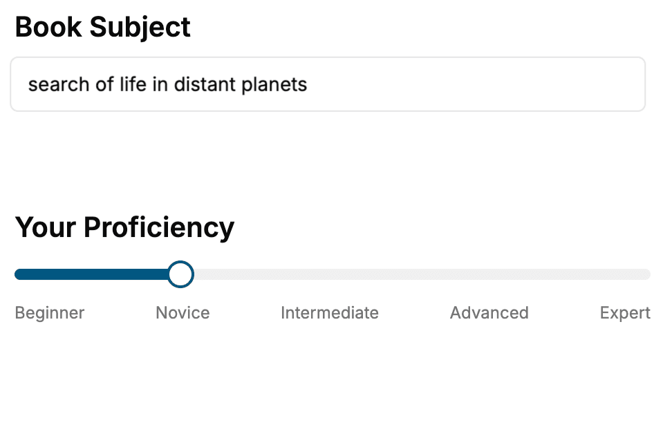
Select the focus of your Microscopy book and share your background
- 2. Personalize your book
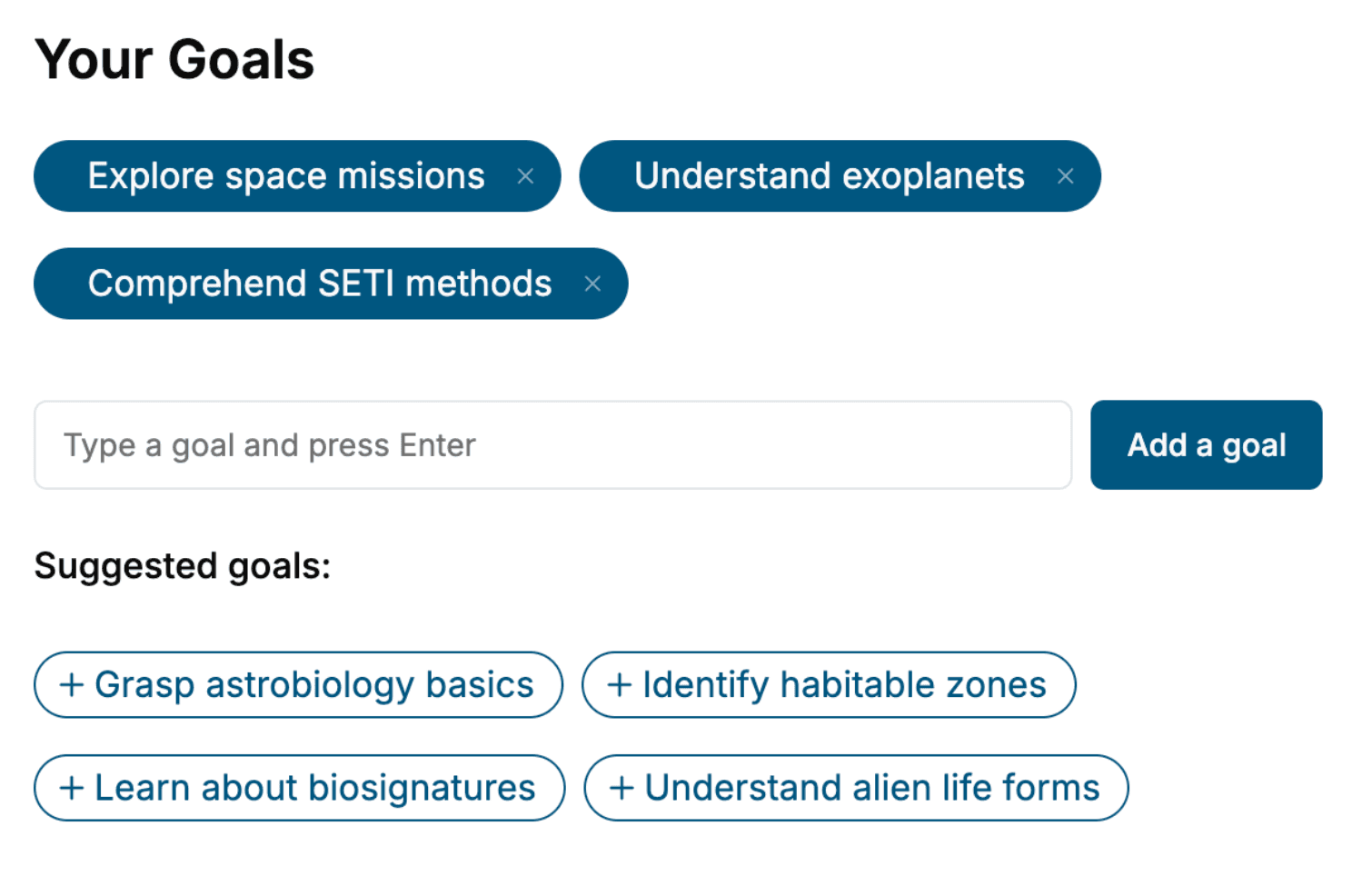
Specify your goals and choose sub-topics to include
- 3. Get your tailored book
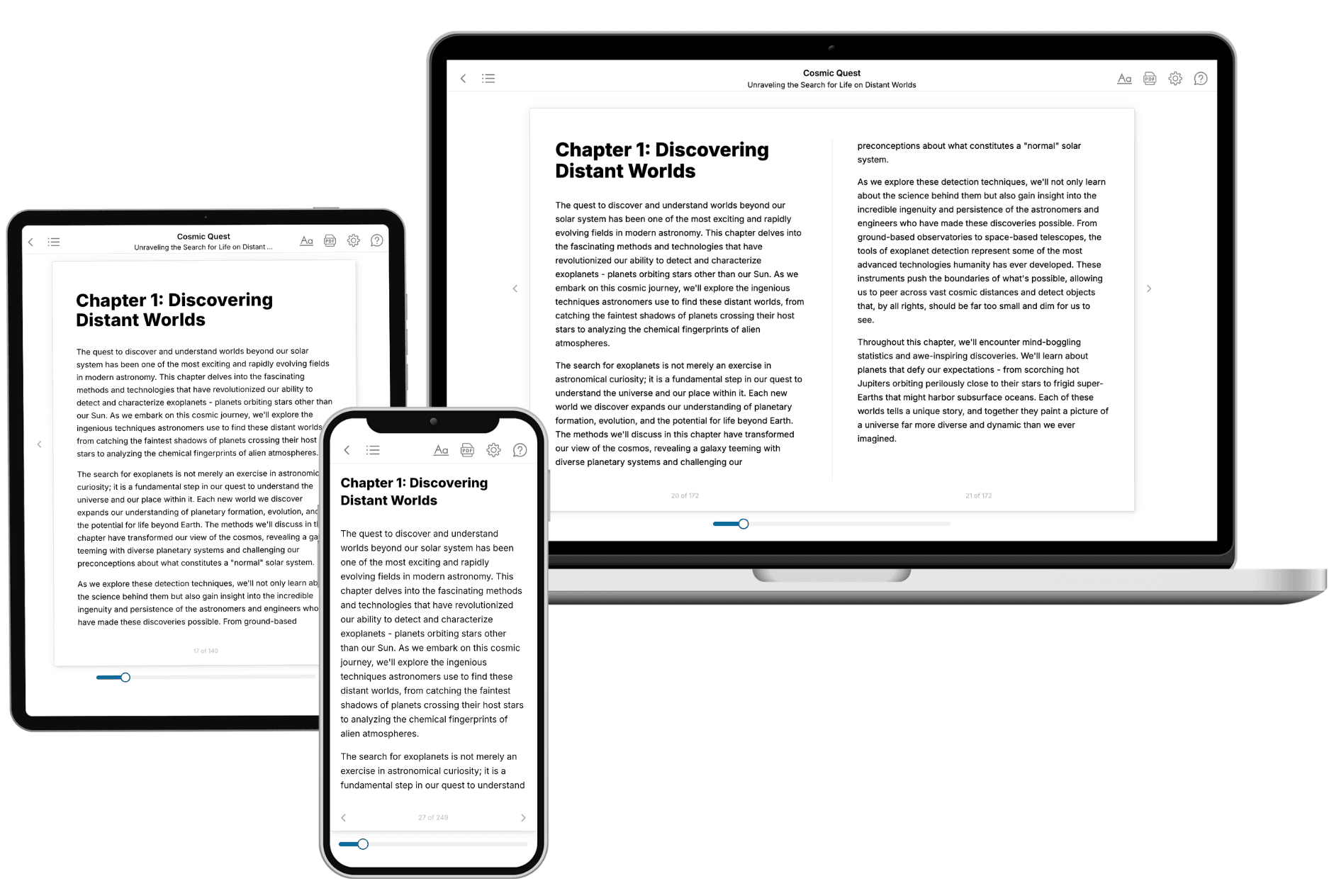
Your book is ready in 10 minutes. Read it online, download as EPUB or PDF, or send to Kindle.
Frequently asked questions
- What is TailoredRead?
TailoredRead creates complete, full-length nonfiction ebooks, written specifically for you. Tell us what you want to learn and why, and TailoredRead writes you a complete ebook on it, with charts, tables, and diagrams throughout. Read it online, on Kindle, or on any e-reader, or download it as a PDF or EPUB. Every book is written fresh the day you order it.
- How is the book tailored to me?
Before anything is written, you answer a few short questions about your goals, your background, and the topics you care about. Your book is then planned around your answers: written at your level, focused on your goals, and covering the topics you chose, instead of the one-size-fits-all book a store sells everyone.
- Will it read like generic AI content?
No. Your book isn't a chat transcript: it's planned chapter by chapter before a single word is written, told in a clear book voice, and explains ideas visually with charts, tables, and diagrams. After writing, every book goes through an editorial pass that polishes chapters so the book reads consistent from start to finish. Judge for yourself: browse thousands of sample books on our Explore Books page.
- How accurate is the content?
Our AI first checks whether your subject needs live research. Niche and fast-moving subjects are researched before writing, and the book plan and chapters are grounded in what it finds. After writing, every book goes through a quality pass that checks its facts and consistency, and researched books list their sources in a Notes section at the back.
- How long is the book?
You choose the length: Comprehensive (250-300 pages), Detailed (150-200 pages), Essential (70-100 pages), or Short (30-50 pages).
- Does the book include visuals?
Yes. Every book includes charts, tables, and diagrams: timelines, checklists, step-by-step guides, decision trees, comparison tables, matrices, and bar charts. A typical book includes dozens of these visual elements.
- Can I read it on Kindle? What formats can I download?
Yes. Every book can be read in our online reader or downloaded as a PDF or EPUB. The EPUB can be opened with Kindle, Apple Books, Kobo, and any e-reader. The Commercial and Author licenses also include an editable Word (DOCX) and Markdown download.
- How much does it cost?
About the price of a regular ebook. The exact price depends on your book's length and complexity, from $2 for the shortest books to $35 for the most comprehensive, and it's shown when you review your book, before you commit to anything. Each book is a one-time purchase, yours to keep, no subscription. Licenses that let you share, teach, or publish your book range from $35.99 to $49.99.
- Can I preview the book before purchasing?
Before you pay, you'll see what your book will cover: the title, a detailed description, the estimated length, and the complete chapter-by-chapter table of contents, so you know what you're getting before you decide. The full text itself is written after you purchase.
- How long does it take to create a book?
About 10 minutes. You can start reading the opening chapters while the rest is still being written.