MRI Essentials
Understanding Magnetic Resonance Imaging from Fundamentals to Clinical Application
Discover how magnetic resonance imaging transforms invisible atomic behavior into detailed medical images that save lives.
238 pages11 chaptersApr 21, 2026Medical Imaging
About this book
This comprehensive guide takes you from the quantum mechanical foundations of nuclear magnetism through the sophisticated engineering that makes modern MRI possible. You'll learn why certain nuclei respond to magnetic fields, how radiofrequency pulses excite tissue, and what determines image quality and contrast. Whether you're a student entering the field, a healthcare professional seeking deeper knowledge, or a technical specialist optimizing imaging protocols, this book provides the conceptual framework and practical insights you need. Each chapter builds systematically on previous concepts, using clear explanations and real-world examples to make complex physics accessible. By the end, you'll understand not just how MRI works, but why specific design choices matter for clinical outcomes.
Table of contents
- IntroductionWhy MRI Matters in Modern Medicine, What You'll Learn in This Book, How to Use This GuideRead below
- Chapter 1: Nuclear Magnetism and SpinUnderstanding Atomic Nuclei and Nuclear Spin, Magnetic Moments and Angular Momentum, Energy States and Spin Populations
- Chapter 2: Magnetic Fields and PrecessionHow Nuclei Interact with Static Magnetic Fields, The Larmor Frequency and Resonance Condition, Precession: The Dance of Spinning Nuclei
- Chapter 3: Radiofrequency Pulses and ExcitationRadiofrequency Pulses and Resonance, Flip Angles and Magnetization Manipulation, The Rotating Frame of Reference
- Chapter 4: Relaxation and Signal RecoveryT1 Relaxation: Spin-Lattice Recovery, T2 Relaxation: Spin-Spin Decay, How Relaxation Times Create Tissue Contrast
- Chapter 5: Spatial Encoding and Image FormationGradient Magnetic Fields and Spatial Localization, Frequency Encoding and Readout Gradients, Phase Encoding and Slice Selection
- Chapter 6: Pulse Sequences and Image AcquisitionSpin Echo Sequences and Signal Recovery, Gradient Echo Sequences and Speed, Inversion Recovery and T1 Weighting
- Chapter 7: Signal, Noise, and Image QualitySignal-to-Noise Ratio and Detection Sensitivity, Spatial Resolution and Acquisition Parameters, Optimizing Image Quality and Acquisition Time
- Chapter 8: Artifacts and Image CorrectionsChemical Shift and Susceptibility Artifacts, Motion Artifacts and Mitigation Techniques, Correction Methods and Post-Processing
- Chapter 9: MRI Safety and Biological EffectsBiological Effects of Static Magnetic Fields, Radiofrequency Energy and Tissue Heating, Gradient Fields and Safety Protocols
- Chapter 10: Advanced MRI ApplicationsFunctional MRI and Brain Mapping, Diffusion Imaging and Tissue Characterization, MR Spectroscopy and Molecular Imaging
- Chapter 11: Clinical Implementation and Best PracticesSelecting Appropriate Imaging Protocols, Patient Preparation and Contraindications, Quality Assurance and Performance Monitoring
- ConclusionKey Takeaways and Core Principles, The Future of MRI Technology
- Glossary of Terms
- Further Reading and Resources
Introduction
Every year, millions of patients lie inside a large cylindrical machine, listening to a rhythmic knocking sound, while invisible forces probe the atomic structure of their bodies. No radiation passes through them. No incision is made. Yet within minutes, clinicians can examine soft tissue in extraordinary detail, spotting tumors, mapping brain activity, or tracing the path of a stroke. Magnetic resonance imaging is one of the most remarkable diagnostic tools ever developed, and its power comes entirely from physics that most people never encounter in daily life. Understanding that physics is what this book is about.
MRI sits at a fascinating crossroads between quantum mechanics, electromagnetism, signal processing, and clinical medicine. Each of those fields contributes something essential to how the technology works. The quantum behavior of atomic nuclei determines which tissues respond to magnetic fields. The principles of electromagnetism govern how those fields are generated and controlled. Signal processing mathematics transforms raw data into recognizable images. And clinical knowledge guides how those images are interpreted and applied. This book walks through each of those layers in a logical, connected sequence, so that by the end, the whole picture comes together clearly.
This guide is written for people who already have some grounding in chemistry and physics and want to go deeper. Whether you are a radiography student preparing for clinical practice, a nurse or physician who wants to understand the tool you rely on every day, or an engineer working on imaging hardware, the explanations here are designed to be rigorous without being impenetrable. Technical terms are introduced carefully, with context and examples. Concepts build on each other chapter by chapter, so skipping ahead is possible but reading in order will give you the strongest foundation. The goal throughout is not just to describe what MRI does, but to explain why it works the way it does.
Why MRI Matters in Modern Medicine
Before MRI became widely available in the 1980s, clinicians had limited options for examining soft tissue inside the body without surgery. X-rays and CT scans excel at imaging bone and dense structures, but they struggle to distinguish between different types of soft tissue. A tumor embedded in brain tissue, a torn ligament in a knee, or early inflammation in a spinal disc were all difficult to visualize clearly. MRI changed that situation dramatically by offering exceptional contrast between different soft tissues, all without exposing patients to ionizing radiation.[1]
The clinical impact of MRI has been enormous across virtually every medical specialty. In neurology, it is the gold standard for diagnosing multiple sclerosis, detecting strokes, and evaluating brain tumors. In orthopedics, it reveals cartilage damage, ligament tears, and bone marrow abnormalities that no other imaging modality can show as clearly. In oncology, MRI guides treatment planning by mapping tumor boundaries and tracking response to therapy. In cardiology, specialized cardiac MRI sequences can assess heart muscle function and detect scarring from previous heart attacks. The breadth of its applications is genuinely remarkable.
Beyond diagnosis, MRI has opened entirely new windows into human biology. Functional MRI (fMRI) measures changes in blood oxygenation to map brain activity in real time, enabling neuroscientists to study cognition, emotion, and sensory processing in living subjects. Diffusion-weighted imaging tracks the movement of water molecules through tissue, revealing the microscopic architecture of white matter tracts in the brain and detecting early signs of stroke within minutes of onset. MR spectroscopy can identify specific chemical compounds within tissue, offering a kind of non-invasive biopsy. These advanced techniques have transformed both clinical medicine and basic research.
The scale of MRI use today reflects its clinical value. In the United States alone, more than 40 million MRI scans are performed each year, a number that has grown steadily as scanner availability has increased and clinical indications have expanded.[2] Globally, MRI is now considered an essential component of modern healthcare infrastructure. Yet despite its widespread use, the underlying physics remains poorly understood by many of the professionals who rely on it daily. That gap between use and understanding matters, because knowing how MRI works helps clinicians choose better protocols, recognize artifacts, troubleshoot image quality problems, and communicate more effectively with physicists and engineers.
MRI also carries important safety considerations that make understanding the technology more than just an academic exercise. The powerful magnetic fields involved can interact dangerously with metallic implants and devices. The radiofrequency energy deposited in tissue generates heat that must be carefully managed. The rapidly switching gradient fields produce acoustic noise and can induce electrical currents in the body. None of these effects are inherently harmful when properly controlled, but they require informed management. Healthcare professionals who understand the physics behind these effects are far better equipped to protect their patients and themselves.
Finally, MRI technology continues to evolve rapidly. Higher field strength scanners, faster acquisition techniques, artificial intelligence-assisted reconstruction, and new contrast mechanisms are all active areas of development. Keeping pace with these advances requires a solid conceptual foundation. The professional who understands the fundamentals can evaluate new techniques critically, adapt to new equipment, and contribute meaningfully to decisions about protocol design and clinical implementation. That kind of informed engagement is what separates a practitioner who uses MRI from one who truly understands it.
What You'll Learn in This Book
This book covers MRI from its deepest physical foundations to its most sophisticated clinical applications. The journey begins at the atomic level, with the quantum mechanical properties of atomic nuclei that make magnetic resonance possible in the first place. From there, it moves through the physics of magnetic fields, radiofrequency excitation, and relaxation processes, before addressing how spatial information is encoded and how images are reconstructed. Later chapters tackle pulse sequences, image quality, artifacts, safety, and advanced applications. Each topic is treated with enough depth to be genuinely useful, while remaining accessible to readers without a graduate-level physics background.
Here is a summary of what each major section covers:
- Chapters 1 and 2 (Nuclear Magnetism and Magnetic Fields): These chapters establish the quantum mechanical foundation of MRI. You will learn why hydrogen nuclei respond to magnetic fields, what nuclear spin means physically, how the Larmor frequency is determined, and what precession looks like at the atomic scale.
- Chapter 3 (Radiofrequency Excitation): This chapter explains how radiofrequency pulses interact with precessing nuclei, what flip angles mean, and how the rotating frame of reference simplifies the mathematics of excitation.
- Chapter 4 (Relaxation): Here you will learn about T1 and T2 relaxation, the two fundamental tissue properties that drive image contrast in MRI. The chapter explains the physical mechanisms behind each relaxation process and shows how differences between tissues create the contrast that makes MRI diagnostically useful.
- Chapter 5 (Spatial Encoding): This chapter addresses one of the most conceptually challenging aspects of MRI: how gradient magnetic fields encode spatial information into the signal, enabling image reconstruction from what is essentially a single detector.
- Chapters 6 and 7 (Pulse Sequences and Image Quality): These chapters cover the practical tools of MRI acquisition, including spin echo, gradient echo, and inversion recovery sequences, as well as the factors that determine signal-to-noise ratio, spatial resolution, and overall image quality.
- Chapter 8 (Artifacts): This chapter identifies the most common image artifacts in MRI, explains their physical origins, and describes strategies for minimizing or correcting them.
- Chapter 9 (Safety): A thorough treatment of MRI safety, covering the biological effects of static fields, radiofrequency heating, and gradient-induced effects, along with the protocols used to manage these risks.
- Chapters 10 and 11 (Advanced Applications and Clinical Practice): The final chapters explore functional MRI, diffusion imaging, MR spectroscopy, protocol selection, patient preparation, and quality assurance, connecting the physics to real-world clinical decision-making.
Throughout the book, the emphasis is on building genuine understanding rather than memorizing facts. Each concept is explained in terms of the underlying physics, and connections between topics are highlighted explicitly. When you understand why T1 relaxation depends on molecular motion, for example, you can predict how different tissues will behave under different conditions, rather than simply recalling a table of values. That kind of transferable understanding is what makes the difference between knowing MRI and truly comprehending it.
Concrete examples appear throughout every chapter. Some are drawn from clinical scenarios, such as why a particular sequence is preferred for liver imaging or how a specific artifact appears on a brain scan. Others are more physical in nature, using analogies and thought experiments to make abstract concepts tangible. Where relevant, specific numbers and quantitative relationships are provided, because understanding the scale of physical quantities matters for developing good intuition about how MRI behaves in practice.
The book also addresses common misconceptions directly. Many people who work with MRI carry mental models that are partially correct but contain subtle errors that lead to confusion later. For example, the idea that MRI "detects hydrogen atoms" is not wrong exactly, but it misses important nuance about which hydrogen atoms contribute to the signal and why. Correcting these partial understandings as they arise helps build a more accurate and durable conceptual framework.
By the time you reach the conclusion, you should be able to explain how an MRI image is formed from first principles, predict how changing acquisition parameters will affect image quality, recognize common artifacts and understand their causes, evaluate the safety implications of different scanning scenarios, and appreciate how advanced techniques like fMRI and diffusion imaging extend the basic principles into new domains. That is a substantial body of knowledge, and this book is designed to deliver it in a way that feels logical and achievable rather than overwhelming.
How to Use This Guide
This book is designed to be read sequentially, at least on a first pass. The chapters are arranged so that each one builds on concepts introduced in the previous ones. Nuclear spin, introduced in Chapter 1, becomes essential for understanding precession in Chapter 2. Precession is necessary for understanding radiofrequency excitation in Chapter 3. Excitation sets up the discussion of relaxation in Chapter 4. And so on through the book. Jumping directly to a later chapter is possible if you already have background in the earlier material, but readers who are new to MRI physics will get the most out of a linear reading.
That said, the book is also designed to serve as a reference after an initial reading. Each chapter is relatively self-contained, with key terms defined within the chapter and cross-references to related material elsewhere in the book. If you are working through a clinical problem and need to refresh your understanding of, say, chemical shift artifacts, you can turn directly to Chapter 8 and find a complete treatment of that topic without needing to re-read everything that came before. The glossary at the back of the book provides quick definitions of all major terms, and the further reading section points toward more advanced resources for topics you want to explore in greater depth.
Different readers will naturally approach this material with different goals. Here are some suggested approaches based on your background and purpose:
- If you are a student new to MRI physics: Read the book from beginning to end, taking time to work through the examples in each chapter before moving on. Pay particular attention to Chapters 1 through 5, which establish the conceptual foundation for everything that follows. Do not rush past material that feels unfamiliar; the later chapters will make much more sense if the early foundations are solid.
- If you are a radiographer or technologist seeking deeper understanding: You likely already have practical familiarity with pulse sequences and acquisition parameters. Chapters 3 through 7 will connect that practical knowledge to the underlying physics. Chapters 8 and 9 on artifacts and safety are particularly relevant to daily clinical work.
- If you are a physician or radiologist: Chapters 4, 6, 10, and 11 are most directly relevant to clinical decision-making, covering tissue contrast, pulse sequences, advanced applications, and protocol selection. However, reading Chapters 1 through 3 first will give you a much richer understanding of why the clinical tools work the way they do.
- If you are an engineer or physicist: The early chapters may cover familiar ground, but Chapters 5 through 8 address the engineering challenges of spatial encoding, image quality optimization, and artifact correction in ways that connect physics to practical implementation.
Throughout the book, certain conventions are used consistently to help you navigate the material. Key terms are bolded when they are first introduced and defined. Italics are used for emphasis and for introducing related concepts that are explained nearby. Blockquotes highlight important examples, clinical scenarios, and notable statements that deserve particular attention. Footnotes provide citations for specific claims and point toward primary sources for readers who want to go deeper on a particular point.
One practical suggestion: do not skip the examples. It can be tempting, especially when reading technical material, to skim past worked examples and scenarios in favor of the explanatory text. In this book, the examples are where many of the most important insights live. A clinical scenario showing how T1 and T2 differences affect the appearance of a brain lesion will do more to cement your understanding of relaxation than any amount of abstract description. The examples are chosen carefully to illustrate principles that are genuinely important, not just to fill space.
It is also worth setting realistic expectations about the learning curve. MRI physics is genuinely complex, and some concepts require time to settle into place. The rotating frame of reference, k-space, and the Bloch equations are all topics that most people find confusing on first encounter. That is completely normal. If a section does not click immediately, move on and come back to it after reading the next chapter. Often, seeing how a concept is used in a new context makes the original explanation suddenly clear. Learning is rarely linear, and this material rewards patience and revisiting.
Finally, consider connecting what you read here to the MRI systems you encounter in practice or in your studies. When you understand that the loud knocking sound during a scan comes from the rapid switching of gradient coils, or that the bore diameter of the magnet is related to the homogeneity of the static field, the abstract physics becomes concrete and memorable. Every time you observe something about an MRI system or an image and ask yourself "why does it work that way?", you are doing exactly the kind of active learning that makes this material stick. This book is designed to give you the tools to answer those questions, and to keep asking better ones.
A note on mathematics: This book uses mathematical expressions where they add clarity, but the focus is always on conceptual understanding rather than derivation. Equations are explained in plain language alongside their symbolic form, and you do not need to be able to solve differential equations to follow the reasoning. Where a formula appears, the goal is to show you the relationship between quantities, not to test your algebra. Readers who want the full mathematical treatment will find references to more advanced texts in the further reading section.
MRI is a technology that rewards curiosity. The more questions you ask about how and why it works, the richer your understanding becomes. This book is an invitation to that kind of engaged, questioning approach to one of medicine's most powerful and fascinating tools. The physics is beautiful, the engineering is impressive, and the clinical impact is profound. All of it starts with a single hydrogen nucleus, spinning in a magnetic field, and the remarkable story of how scientists learned to listen to what it has to say.
This is the end of the free sample. The full book has 11 chapters.
Want this book written for you?
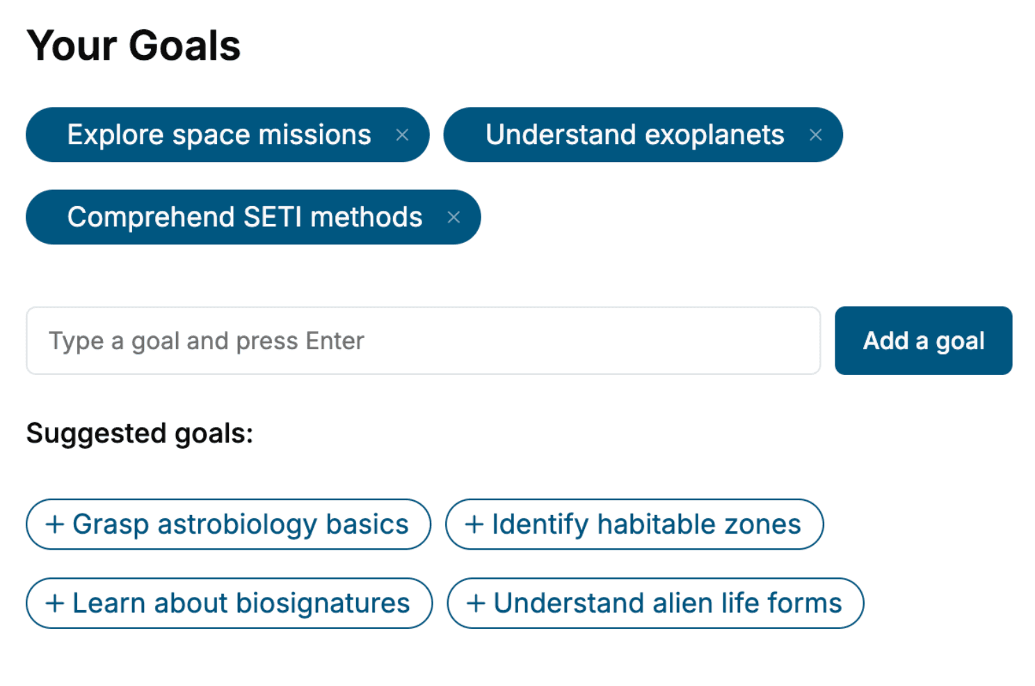
Tell us your goals and background, and get your own version of this book, tailored by AI to you.


Benefits of AI-tailored books
- Read one book, not ten:
- all the Medical Imaging knowledge you need, gathered into a single book.
- Save days of learning:
- tell us your goals, choose your topics, and the whole book is built around them.
- Learn effortlessly:
- Medical Imaging book written for your specific background and expertise.
- Understand at a glance:
- includes charts, tables, timelines, checklists, decision trees, and more.
- Stay current:
- written the day you order it, not years ago.
- Trust what you read:
- every book is checked for accuracy, with real research when your subject needs it.
Create your unique book in 3 steps
- 1. Select your focus
Select the focus of your Medical Imaging book and share your background
- 2. Personalize your book
Specify your goals and choose sub-topics to include
- 3. Get your tailored book
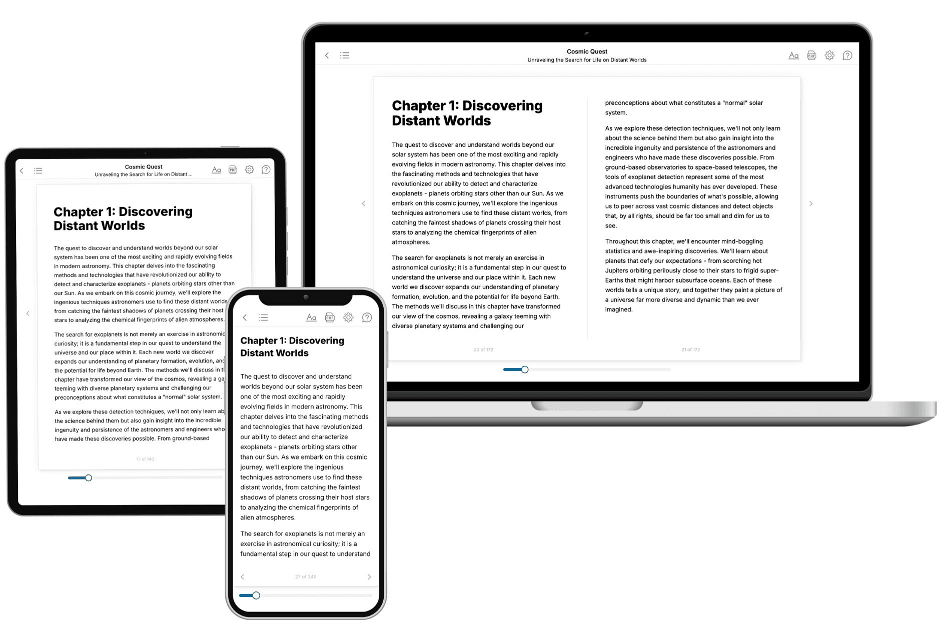
Your book is ready in 10 minutes. Read it online, download as EPUB or PDF, or send to Kindle.

Frequently asked questions
- What is TailoredRead?
TailoredRead creates complete, full-length nonfiction ebooks, written specifically for you. Tell us what you want to learn and why, and TailoredRead writes you a complete ebook on it, with charts, tables, and diagrams throughout. Read it online, on Kindle, or on any e-reader, or download it as a PDF or EPUB. Every book is written fresh the day you order it.
- How is the book tailored to me?
Before anything is written, you answer a few short questions about your goals, your background, and the topics you care about. Your book is then planned around your answers: written at your level, focused on your goals, and covering the topics you chose, instead of the one-size-fits-all book a store sells everyone.
- Will it read like generic AI content?
No. Your book isn't a chat transcript: it's planned chapter by chapter before a single word is written, told in a clear book voice, and explains ideas visually with charts, tables, and diagrams. After writing, every book goes through an editorial pass that polishes chapters so the book reads consistent from start to finish. Judge for yourself: browse thousands of sample books on our Explore Books page.
- How accurate is the content?
Our AI first checks whether your subject needs live research. Niche and fast-moving subjects are researched before writing, and the book plan and chapters are grounded in what it finds. After writing, every book goes through a quality pass that checks its facts and consistency, and researched books list their sources in a Notes section at the back.
- How long is the book?
You choose the length: Comprehensive (250-300 pages), Detailed (150-200 pages), Essential (70-100 pages), or Short (30-50 pages).
- Does the book include visuals?
Yes. Every book includes charts, tables, and diagrams: timelines, checklists, step-by-step guides, decision trees, comparison tables, matrices, and bar charts. A typical book includes dozens of these visual elements.
- Can I read it on Kindle? What formats can I download?
Yes. Every book can be read in our online reader or downloaded as a PDF or EPUB. The EPUB can be opened with Kindle, Apple Books, Kobo, and any e-reader. The Commercial and Author licenses also include an editable Word (DOCX) and Markdown download.
- How much does it cost?
About the price of a regular ebook. The exact price depends on your book's length and complexity, from $2 for the shortest books to $35 for the most comprehensive, and it's shown when you review your book, before you commit to anything. Each book is a one-time purchase, yours to keep, no subscription. Licenses that let you share, teach, or publish your book range from $35.99 to $49.99.
- Can I preview the book before purchasing?
Before you pay, you'll see what your book will cover: the title, a detailed description, the estimated length, and the complete chapter-by-chapter table of contents, so you know what you're getting before you decide. The full text itself is written after you purchase.
- How long does it take to create a book?
About 10 minutes. You can start reading the opening chapters while the rest is still being written.