The Nervous System Coloring Guide
Master Anatomy Through Interactive Illustration and Hands-On Learning
Elevate your understanding of nervous system anatomy with this interactive coloring guide designed for visual learners.
207 pages9 chaptersJul 7, 2026Neuroanatomy
About this book
Rather than passively reading dense textbooks, you'll actively engage with detailed anatomical illustrations, color-coding structures to cement them in your memory. Each page combines scientifically accurate diagrams with guided coloring activities that transform abstract concepts into tangible knowledge. You'll explore the brain's intricate regions, trace neural pathways, and discover how billions of neurons orchestrate every sensation, thought, and movement. Perfect for students, healthcare professionals, educators, and anyone fascinated by how the body's communication network functions, this book makes complex neuroanatomy accessible and genuinely enjoyable. The combination of visual learning and hands-on coloring creates multiple neural pathways in your own brain, dramatically improving retention compared to traditional study methods. Whether you're preparing for exams, deepening professional knowledge, or simply curious about your own biology, this guide transforms learning into an engaging, creative experience.
Table of contents
- IntroductionWhy Coloring Enhances Anatomical Learning, How to Use This Guide Effectively, A Quick Overview of the Nervous SystemRead below
- Chapter 1: Nervous System Organization and DivisionsCentral Nervous System vs. Peripheral Nervous System, Somatic and Autonomic Divisions, Functional Organization and Communication Pathways
- Chapter 2: Neurons: The Building Blocks of the Nervous SystemNeuron Structure and Components, Types of Neurons and Their Functions, Glial Cells and Supporting Structures, Synaptic Transmission and Neurotransmitters
- Chapter 3: The Brain: Structure and Regional FunctionsThe Cerebrum and Cerebral Cortex, Brain Lobes and Their Specialized Functions, Deep Brain Structures: Thalamus, Hypothalamus, and Limbic System, The Cerebellum and Brainstem
- Chapter 4: The Spinal Cord: Central Information HighwaySpinal Cord Structure and Organization, Gray Matter and White Matter Pathways, Spinal Nerves and Segmental Organization
- Chapter 5: Peripheral Nerves and Nerve PathwaysCranial Nerves and Their Functions, Spinal Nerve Branches and Plexuses, Sensory and Motor Pathways Throughout the Body
- Chapter 6: The Autonomic Nervous System: Automatic ControlThe Sympathetic Division: Fight or Flight, The Parasympathetic Division: Rest and Digest, Autonomic Balance and Homeostasis
- Chapter 7: Sensory Systems and Neural ProcessingSensory Receptors and Signal Detection, Sensory Pathways to the Brain, Sensory Processing in the Cerebral Cortex
- Chapter 8: Motor Control and MovementMotor Cortex and Movement Planning, Motor Pathways and Descending Control, Reflex Arcs and Automatic Responses
- Chapter 9: Integrating Knowledge: Systems in ActionIntegrated Nervous System Responses, Clinical Applications and Common Conditions, Comprehensive Coloring Review and Self-Assessment
- ConclusionYour Nervous System Mastery, Next Steps in Your Anatomical Learning Journey
- Glossary of Nervous System Terms
- Further Learning Resources
Introduction
The nervous system is one of the most extraordinary structures in the known universe. It processes billions of signals every second, coordinates every movement you make, generates every thought you have, and keeps your body running without you ever having to think about it. Yet for many students and healthcare learners, studying neuroanatomy feels like trying to memorize a foreign city by reading a phone book. The names are unfamiliar, the structures are layered and complex, and the connections between them can feel impossibly abstract when you're staring at a flat page of text.
This guide takes a different approach. Instead of asking you to passively absorb information, it invites you to actively participate in your own learning. By picking up colored pencils and working through each illustration, you engage your hands, your eyes, and your memory simultaneously. That combination is powerful. Research consistently shows that active, multi-sensory learning produces stronger, longer-lasting retention than passive reading alone.[1] Coloring anatomical diagrams is not a shortcut or a simplification. It is a genuinely effective study strategy used in medical and nursing education worldwide.
Whether you are a student preparing for an anatomy exam, a healthcare professional refreshing your neuroanatomy knowledge, an educator looking for engaging teaching tools, or simply someone curious about how your own brain and nervous system work, this guide is built for you. The chapters move from broad organization down to fine detail, building your understanding layer by layer. Each section pairs clear explanations with structured coloring activities designed to reinforce exactly what you just read. By the time you reach the final chapter, you will not just know the nervous system. You will have drawn it, colored it, and made it your own.
Before diving into the first chapter, this introduction covers three essential foundations. First, it explains the science behind why coloring works as a learning tool. Second, it gives you practical guidance on how to use this book most effectively. Third, it provides a concise overview of the nervous system as a whole, so you have a mental map of the territory before you begin exploring it in detail. Think of this introduction as your orientation session before a fascinating journey through one of biology's greatest achievements.
Why Coloring Enhances Anatomical Learning
At first glance, coloring might seem like something reserved for children's classrooms. In reality, it is a sophisticated cognitive activity that engages multiple brain regions at once. When you color an anatomical diagram, you are not just filling in shapes. You are making decisions about spatial relationships, reinforcing the names of structures, and creating visual associations that your brain can retrieve later. This kind of active engagement is fundamentally different from reading a label on a diagram and moving on.
The concept behind this approach is called dual coding theory, developed by psychologist Allan Paivio in the 1970s. The theory proposes that the brain stores information in two separate but connected systems: one for verbal information (words and language) and one for visual information (images and spatial relationships).[2] When you learn something using both systems at the same time, you create two independent memory traces instead of one. That redundancy makes the information far easier to retrieve later, especially under exam pressure or in clinical situations where you need to recall details quickly.
Color itself plays a specific role in this process. The brain is highly sensitive to color as a distinguishing feature. When you consistently assign one color to motor neurons and a different color to sensory neurons, for example, you are building a visual coding system that your memory can use as a retrieval cue. Studies in educational psychology have found that color-coded study materials improve recall accuracy compared to black-and-white materials, particularly for complex spatial information like anatomical diagrams.[3]
There is also a motor learning component to consider. The physical act of drawing and coloring engages fine motor pathways and creates what researchers call embodied cognition, the idea that physical actions can reinforce conceptual understanding. When your hand traces the path of a nerve from the spinal cord to a muscle, your body is participating in learning the route, not just your eyes. This is the same principle that makes writing notes by hand more effective for retention than typing them.[4]
Coloring also forces a kind of focused attention that passive reading rarely achieves. When you are deciding which structure to color next, checking its label, choosing your color, and carefully staying within the boundaries of a complex diagram, your attention is fully committed to the material. There is no skimming, no glossing over unfamiliar terms, and no drifting. That sustained, focused engagement is exactly the condition under which deep learning happens.
Finally, the process of completing a coloring page creates a tangible artifact of your learning. Looking back at a finished, color-coded diagram of the brain or spinal cord gives you a sense of accomplishment and provides a powerful visual review tool. Many learners find that flipping through their completed pages before an exam is one of the most efficient review strategies available, because each image instantly reactivates the memory network built during the original coloring session.
A practical note on color selection: Throughout this guide, suggested color assignments are provided for each diagram. These suggestions are designed to create meaningful contrast between related structures. You are encouraged to follow them, especially on your first pass through the material. Consistency in color coding across chapters will help your brain build connections between related structures as you progress.
The bottom line is straightforward. Coloring anatomical diagrams is not a passive or trivial activity. It is a research-supported learning strategy that engages visual memory, motor learning, focused attention, and dual coding simultaneously. Used consistently throughout this guide, it will make your understanding of nervous system anatomy deeper, more durable, and more accessible when you need it most.
How to Use This Guide Effectively
Getting the most out of this guide requires a small amount of preparation and a consistent approach. The good news is that neither takes much time. A few simple habits, applied consistently, will dramatically improve what you take away from each session. Before you begin Chapter 1, take a few minutes to set yourself up for success.
Gather your materials before you start. The quality of your coloring tools matters more than you might expect. Here is what works best:
- Colored pencils: The preferred tool for most anatomical coloring. They allow precise control, can be layered for shading, and do not bleed through pages. A set of at least 24 colors gives you enough variety to distinguish structures clearly.
- Fine-tip markers: Useful for bold outlines or labeling, but use them carefully since they can bleed through paper.
- Highlighters: Good for emphasizing key labels or pathways, but not ideal as a primary coloring tool since they obscure printed text.
- A pencil and eraser: Helpful for writing notes directly on diagrams before committing to ink or color.
Follow the chapter sequence on your first pass. This guide is structured deliberately. Each chapter builds on the concepts introduced in the previous one. Jumping ahead to the brain chapter before understanding neuron structure, for example, will leave gaps in your mental model that make the later material harder to absorb. On subsequent reviews, feel free to jump to any chapter you need. But for your initial study, work through the chapters in order.
Read before you color. Each section begins with explanatory text that introduces the structures you are about to illustrate. Read this text fully before picking up your pencils. Understanding what a structure does and how it connects to others gives your coloring session purpose and context. Coloring without reading first reduces the activity to a mechanical exercise rather than a learning one.
Here is a simple session structure that works well for most learners:
- Read the section text once through without stopping to color.
- Review the diagram and its labels before adding any color.
- Color one structure at a time, saying its name aloud as you work.
- After completing the diagram, cover the labels and try to name each structure from memory.
- Write a one-sentence summary of each structure's function in the margin.
Use a consistent color system across chapters. This is one of the most important habits you can build. If you color all motor pathways in red throughout the book, your brain will begin to associate that color with motor function automatically. When you encounter a new diagram in a later chapter, seeing red will immediately cue the right category of information. Suggested color assignments are provided throughout, but the key is consistency, not which specific colors you choose.
Do not rush. A single coloring session of 30 to 45 minutes with full attention will produce better results than two hours of distracted, hurried work. If you find your focus drifting, take a short break rather than pushing through. The goal is deep encoding, not page completion. It is far better to finish one diagram thoroughly than to rush through three.
Use the self-assessment prompts. At the end of each chapter, review questions and self-assessment activities are provided. These are not optional extras. They are a critical part of the learning process. Retrieval practice, the act of pulling information out of memory rather than just reviewing it, is one of the most powerful learning strategies known to cognitive science.[5] Every time you successfully recall a structure's name or function without looking at the label, you strengthen that memory trace significantly.
Return to completed diagrams regularly. Spaced repetition, reviewing material at increasing intervals over time, is the most efficient way to move information into long-term memory. After completing a chapter, revisit its diagrams after one day, then after three days, then after a week. Each review session should take only a few minutes, but the cumulative effect on retention is substantial. Your completed, color-coded diagrams are ideal tools for these quick review sessions.
A note on mistakes: Do not worry about coloring outside the lines or choosing the "wrong" color. The learning value comes from the process of engaging with the material, not from producing a perfect illustration. If you make a mistake, note it, correct your understanding, and move on. Imperfect diagrams that you actually completed are far more valuable than perfect ones you never started.
A Quick Overview of the Nervous System
Before exploring the nervous system in detail, it helps to have a clear picture of the whole. Think of this overview as a map legend. It will not tell you everything about the territory, but it will help you understand what you are looking at as you explore each region in the chapters ahead. The nervous system is the body's master communication and control network, responsible for everything from breathing and heartbeat to language, memory, and emotion.
At the broadest level, the nervous system is divided into two major parts:
- The Central Nervous System (CNS): This includes the brain and the spinal cord. The CNS is the processing center of the entire system. It receives information, interprets it, makes decisions, and sends out instructions. The brain alone contains roughly 86 billion neurons, each capable of forming thousands of connections with other neurons.[6]
- The Peripheral Nervous System (PNS): This includes all the nerves that branch out from the brain and spinal cord to reach every part of the body. The PNS carries sensory information inward to the CNS and motor commands outward to muscles and organs.
Within the peripheral nervous system, there is a further division based on function. The somatic nervous system handles voluntary actions and conscious sensations. When you decide to pick up a pencil, the somatic nervous system executes that command. When you feel the texture of the paper, the somatic nervous system carries that sensation to your brain. The autonomic nervous system, by contrast, operates largely below the level of conscious awareness. It regulates heart rate, digestion, breathing rate, pupil dilation, and dozens of other vital functions automatically, without requiring any deliberate input from you.
The autonomic nervous system itself has two subdivisions that work in opposition to maintain balance. The sympathetic division activates the body's stress response, sometimes called "fight or flight." It accelerates heart rate, redirects blood to muscles, and prepares the body for intense physical activity. The parasympathetic division does the opposite, promoting rest, digestion, and recovery. These two divisions are constantly active, adjusting their relative influence to keep the body in a state of dynamic equilibrium called homeostasis.
At the cellular level, the nervous system is built from two main types of cells. Neurons are the electrically active cells that transmit signals. Each neuron has a cell body (called the soma), branching input fibers (dendrites), and a long output fiber (the axon) that carries signals to the next cell. Glial cells are the supporting cast. They outnumber neurons significantly and perform essential functions including insulating axons, clearing waste, providing structural support, and regulating the chemical environment around neurons. Neither type can function without the other.
Communication between neurons happens at specialized junctions called synapses. When an electrical signal reaches the end of an axon, it triggers the release of chemical messengers called neurotransmitters into the synaptic gap. These chemicals bind to receptors on the next neuron, either exciting it to fire its own signal or inhibiting it from doing so. This process, repeated trillions of times per second across the entire nervous system, underlies every thought, sensation, and movement you experience.
The brain itself is organized into distinct regions, each with specialized roles. The large, wrinkled outer layer called the cerebral cortex handles higher functions like reasoning, language, and voluntary movement. Beneath it, deeper structures including the thalamus, hypothalamus, and limbic system manage sensory relay, hormonal control, and emotional processing. The cerebellum, tucked at the back of the brain, coordinates movement and balance. The brainstem, connecting the brain to the spinal cord, controls the most fundamental life-sustaining functions including breathing, heart rate, and consciousness.
The spinal cord runs from the base of the brain down through the vertebral column, protected by the surrounding bones. It serves as the main communication highway between the brain and the body. Thirty-one pairs of spinal nerves branch off from the cord at regular intervals, each serving a specific region of the body. The cord also processes some information locally, enabling rapid reflex responses that do not require input from the brain at all. When you pull your hand away from a hot surface before you consciously register pain, that is a spinal reflex at work.
Here is a quick reference summary of the major divisions covered in this guide:
- Central Nervous System: Brain and spinal cord
- Peripheral Nervous System: Cranial nerves, spinal nerves, and their branches
- Somatic Division: Voluntary movement and conscious sensation
- Autonomic Division: Involuntary regulation of internal organs
- Sympathetic Division: Stress and activation responses
- Parasympathetic Division: Rest and recovery responses
- Neurons: Signal-transmitting cells with dendrites, soma, and axons
- Glial Cells: Supporting cells that maintain the neural environment
- Synapses: Chemical communication junctions between neurons
This overview is intentionally brief. Each of these components will be explored in much greater depth in the chapters that follow, with detailed diagrams and coloring activities to reinforce every concept. For now, the goal is simply to give you a sense of the overall architecture before you begin filling in the details. The nervous system is complex, but it is not chaotic. It has a clear logic and a beautiful organization that becomes more apparent the more closely you examine it. That examination begins in Chapter 1.
Before you turn the page: Take a moment to gather your coloring materials, find a comfortable, well-lit workspace, and set aside at least 30 minutes for your first session. The nervous system has been waiting a long time to be understood. You are about to give it the attention it deserves.
Notes
Introduction
- 1. S. M. Kosslyn and R. S. Rosenberg, Psychology: The Brain, the Person, the World, Pearson Education, 2004
- 2. A. Paivio, Imagery and Verbal Processes, Holt, Rinehart, and Winston, 1971
- 3. F. A. Wichmann and others, "The contributions of color to recognition memory for natural scenes," Journal of Experimental Psychology: Learning, Memory, and Cognition, 2002
- 4. P. A. Mueller and D. M. Oppenheimer, "The pen is mightier than the keyboard," Psychological Science, 2014
- 5. H. L. Roediger and J. D. Karpicke, "Test-enhanced learning," Psychological Science, 2006
- 6. F. A. C. Azevedo and others, "Equal numbers of neuronal and nonneuronal cells make the human brain an isometrically scaled-up primate brain," Journal of Comparative Neurology, 2009
This is the end of the free sample. The full book has 9 chapters.
Want this book written for you?
Tell us your goals and background, and get your own version of this book, tailored by AI to you.


Benefits of AI-tailored books
- Read one book, not ten:
- all the Neuroanatomy knowledge you need, gathered into a single book.
- Save days of learning:
- tell us your goals, choose your topics, and the whole book is built around them.
- Learn effortlessly:
- Neuroanatomy book written for your specific background and expertise.
- Understand at a glance:
- includes charts, tables, timelines, checklists, decision trees, and more.
- Stay current:
- written the day you order it, not years ago.
- Trust what you read:
- every book is checked for accuracy, with real research when your subject needs it.
Create your unique book in 3 steps
- 1. Select your focus
Select the focus of your Neuroanatomy book and share your background
- 2. Personalize your book
Specify your goals and choose sub-topics to include
- 3. Get your tailored book
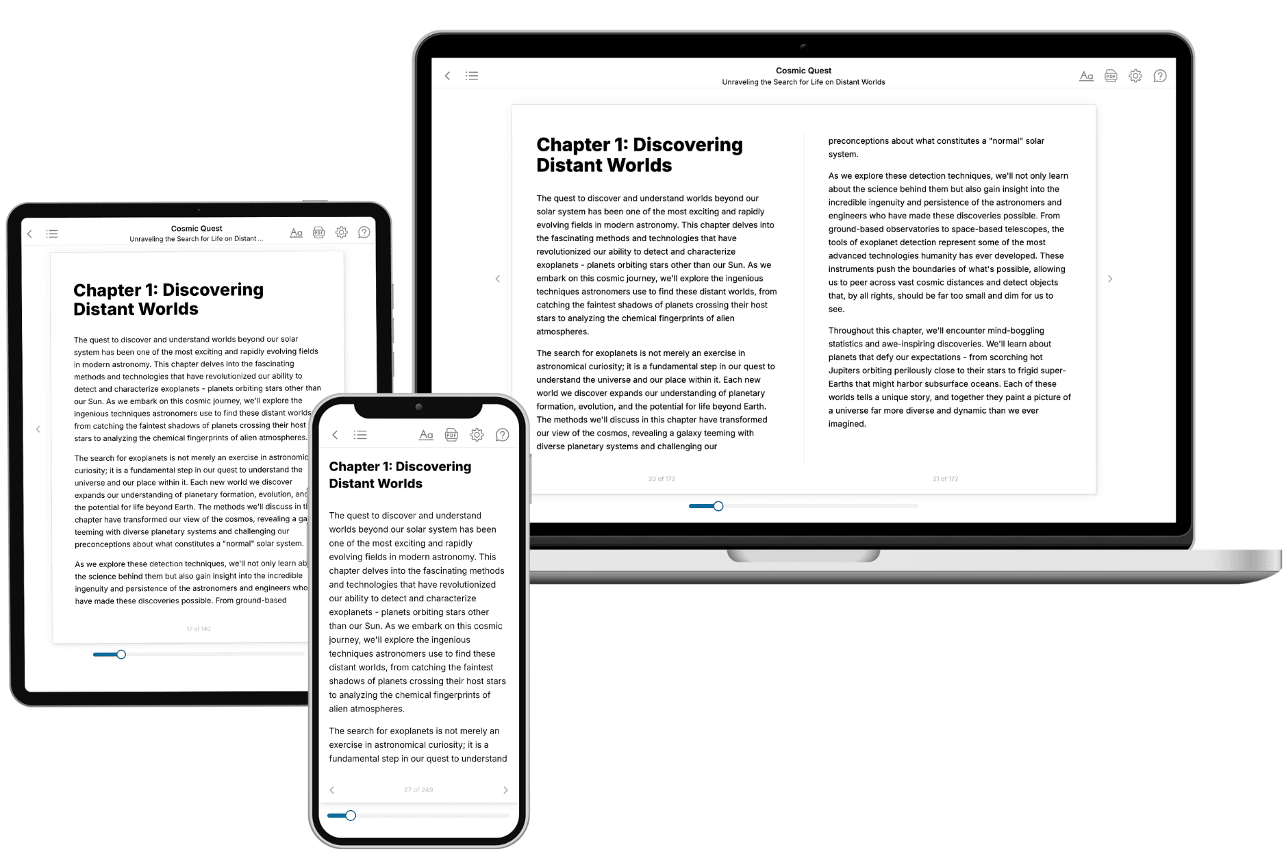
Your book is ready in 10 minutes. Read it online, download as EPUB or PDF, or send to Kindle.

Frequently asked questions
- What is TailoredRead?
TailoredRead creates complete, full-length nonfiction ebooks, written specifically for you. Tell us what you want to learn and why, and TailoredRead writes you a complete ebook on it, with charts, tables, and diagrams throughout. Read it online, on Kindle, or on any e-reader, or download it as a PDF or EPUB. Every book is written fresh the day you order it.
- How is the book tailored to me?
Before anything is written, you answer a few short questions about your goals, your background, and the topics you care about. Your book is then planned around your answers: written at your level, focused on your goals, and covering the topics you chose, instead of the one-size-fits-all book a store sells everyone.
- Will it read like generic AI content?
No. Your book isn't a chat transcript: it's planned chapter by chapter before a single word is written, told in a clear book voice, and explains ideas visually with charts, tables, and diagrams. After writing, every book goes through an editorial pass that polishes chapters so the book reads consistent from start to finish. Judge for yourself: browse thousands of sample books on our Explore Books page.
- How accurate is the content?
Our AI first checks whether your subject needs live research. Niche and fast-moving subjects are researched before writing, and the book plan and chapters are grounded in what it finds. After writing, every book goes through a quality pass that checks its facts and consistency, and researched books list their sources in a Notes section at the back.
- How long is the book?
You choose the length: Comprehensive (250-300 pages), Detailed (150-200 pages), Essential (70-100 pages), or Short (30-50 pages).
- Does the book include visuals?
Yes. Every book includes charts, tables, and diagrams: timelines, checklists, step-by-step guides, decision trees, comparison tables, matrices, and bar charts. A typical book includes dozens of these visual elements.
- Can I read it on Kindle? What formats can I download?
Yes. Every book can be read in our online reader or downloaded as a PDF or EPUB. The EPUB can be opened with Kindle, Apple Books, Kobo, and any e-reader. The Commercial and Author licenses also include an editable Word (DOCX) and Markdown download.
- How much does it cost?
About the price of a regular ebook. The exact price depends on your book's length and complexity, from $2 for the shortest books to $35 for the most comprehensive, and it's shown when you review your book, before you commit to anything. Each book is a one-time purchase, yours to keep, no subscription. Licenses that let you share, teach, or publish your book range from $35.99 to $49.99.
- Can I preview the book before purchasing?
Before you pay, you'll see what your book will cover: the title, a detailed description, the estimated length, and the complete chapter-by-chapter table of contents, so you know what you're getting before you decide. The full text itself is written after you purchase.
- How long does it take to create a book?
About 10 minutes. You can start reading the opening chapters while the rest is still being written.