Neuroanatomy Decoded
A Statistician's Guide to Understanding the Brain and Nervous System
As a statistician in the field of neuroscience, you face a daunting challenge: making sense of complex neurological data without a solid foundation in neuroanatomy.
67 pages3 chaptersJul 14, 2025Neuroanatomy
About this book
This knowledge gap can lead to misinterpretations and missed insights in your analyses. Neuroanatomy Decoded: A Statistician's Guide to Understanding the Brain and Nervous System bridges this divide, offering you a clear, accessible pathway to mastering key neuroanatomical concepts. You'll gain the ability to visualize intricate nervous system structures, comprehend neurologic localization, and grasp the fundamentals of clinical neurology—all through the lens of a data professional. This book equips you with practical knowledge to enhance your neuro exam skills and create more insightful Tableau dashboards. You'll explore the intricate world of neurotransmitters, delve into the mysteries of neurodegenerative diseases, and unravel the complexities of proteins like TDP-43. By the final page, you'll possess a robust understanding of neuroanatomy that will elevate your data analysis and interpretation to new heights.
Table of contents
- Chapter 1: Foundations of NeuroanatomyOverview of the Brain and Central Nervous System, The Spinal Cord: Structure and Function, Neurons and Glial Cells: The Building BlocksRead below
- Chapter 2: Major Neurological SystemsThe Visual System: From Retina to Cortex, The Autonomic Nervous System, Motor Control: Cerebellum and Basal Ganglia
- Chapter 3: Clinical Applications for StatisticiansPrinciples of Neurologic Localization, Neurotransmitters and Signaling Pathways, Neurodegenerative Diseases and TDP-43, Data Visualization Techniques for Neuroanatomy, Statistical Analysis in Neuroscience Research
- Conclusion
Chapter 1: Foundations of Neuroanatomy
As you embark on your journey to understand the intricate world of neuroanatomy, it's essential to establish a solid foundation. This chapter will introduce you to the fundamental concepts and structures that form the basis of the nervous system. By grasping these core principles, you'll be better equipped to analyze and interpret neurological data in your role as a Senior Data Manager. The knowledge gained here will serve as a springboard for more advanced topics and applications in subsequent chapters.
Overview of the Brain and Central Nervous System
The central nervous system (CNS) is the command center of the human body, orchestrating a vast array of functions from basic survival mechanisms to complex cognitive processes. At its core, the CNS consists of two main components: the brain and the spinal cord. These structures work in harmony to process information, control bodily functions, and shape our experiences of the world around us.
The human brain, weighing approximately 1.4 kilograms (3 pounds), is a marvel of biological engineering. It contains an estimated 86 billion neurons, forming trillions of connections known as synapses[1]. This intricate network allows for the rapid transmission of electrical and chemical signals, enabling everything from reflexes to abstract thought. The brain's complexity is reflected in its structure, which can be divided into several major regions, each with specialized functions.
The cerebrum, the largest part of the brain, is divided into two hemispheres connected by a bundle of nerve fibers called the corpus callosum. The outer layer of the cerebrum, known as the cerebral cortex, is responsible for higher-order functions such as perception, decision-making, and language processing. It's characterized by its folded appearance, which increases its surface area and computational power. The cerebral cortex is further divided into four lobes: frontal, parietal, temporal, and occipital, each associated with specific functions.
Beneath the cerebrum lies the brainstem, which connects the brain to the spinal cord. This region is crucial for maintaining vital functions such as breathing, heart rate, and blood pressure. The brainstem consists of three main structures: the midbrain, pons, and medulla oblongata. These components play essential roles in relaying information between the brain and the rest of the body, as well as regulating sleep-wake cycles and arousal.
Adjacent to the brainstem is the cerebellum, often referred to as the "little brain." Despite its small size, the cerebellum contains more neurons than the rest of the brain combined[2]. It's primarily responsible for motor coordination, balance, and fine-tuning of movement. Recent research has also implicated the cerebellum in cognitive functions, highlighting its broader role in neural processing.
The limbic system, a group of structures including the hippocampus, amygdala, and hypothalamus, forms a ring around the brainstem. This system is integral to emotion, memory formation, and motivation. The hippocampus, in particular, plays a crucial role in converting short-term memories to long-term storage, while the amygdala is central to processing fear and other emotions.
At the base of the brain lies the hypothalamus, a small but powerful structure that acts as a link between the nervous system and the endocrine system. It regulates homeostasis, controlling functions such as body temperature, hunger, thirst, and circadian rhythms. The hypothalamus also produces hormones that influence the pituitary gland, often called the "master gland" of the endocrine system.
The thalamus, often described as the brain's relay station, sits near the center of the brain. It processes and filters sensory information before sending it to the appropriate areas of the cerebral cortex. This structure plays a crucial role in attention, consciousness, and sleep regulation.
Understanding the organization and function of these brain regions is essential for interpreting neurological data. As a Senior Data Manager, you'll often encounter datasets related to specific brain areas or neural pathways. By visualizing these structures and their interconnections, you can develop more insightful analyses and create more meaningful data visualizations for your team and stakeholders.
The central nervous system's complexity extends beyond its structural organization. The brain and spinal cord are protected by three layers of membranes called meninges: the dura mater (outermost), arachnoid mater (middle), and pia mater (innermost). These layers, along with cerebrospinal fluid (CSF), provide crucial protection against physical trauma and infection. CSF, produced in the brain's ventricles, circulates through the central nervous system, delivering nutrients and removing waste products.
As you delve deeper into neuroanatomy, you'll discover that the brain's organization follows certain principles that can aid in data analysis. For instance, many functions are lateralized, meaning they are predominantly associated with one hemisphere. Language processing, for example, is typically lateralized to the left hemisphere in most right-handed individuals. This lateralization can be crucial when interpreting data from neuroimaging studies or analyzing the effects of localized brain injuries.
Moreover, the brain exhibits both functional segregation and integration. Specific regions are specialized for particular functions (segregation), but these areas also work together in complex networks to produce higher-order cognition and behavior (integration). This concept is fundamental to modern neuroimaging techniques like functional magnetic resonance imaging (fMRI), which measures brain activity by detecting changes in blood oxygenation and flow. As a data manager, understanding these principles will help you design more effective analyses and interpret results in the context of broader brain function.
In conclusion, this overview of the brain and central nervous system provides a foundation for understanding the complex organ that governs all aspects of human behavior and physiology. As you progress through this book, you'll build upon this knowledge, exploring how these structures and their functions relate to specific neurological processes, disorders, and data analysis techniques. By mastering these fundamentals, you'll be better equipped to tackle the challenges of neurological data management and contribute valuable insights to the field of neuroscience.
The Spinal Cord: Structure and Function
The spinal cord, often overshadowed by the brain in discussions of the central nervous system, is a crucial component that deserves thorough understanding. As a cylindrical bundle of nervous tissue extending from the medulla oblongata of the brainstem to the lumbar region of the vertebral column, the spinal cord serves as the primary conduit for information traveling between the brain and the rest of the body. Its importance in both sensory and motor functions cannot be overstated, and a grasp of its structure and function is essential for any comprehensive analysis of neurological data.
Structurally, the spinal cord is divided into 31 segments, each giving rise to a pair of spinal nerves. These segments are grouped into four regions: 8 cervical, 12 thoracic, 5 lumbar, 5 sacral, and 1 coccygeal. The spinal cord is not uniform throughout its length; it has two enlargements - the cervical enlargement (associated with the arms) and the lumbar enlargement (associated with the legs). These enlargements contain more neurons to handle the increased neural traffic for limb control. Understanding this segmental organization is crucial when interpreting data related to specific body regions or when analyzing the effects of localized spinal cord injuries.
A cross-section of the spinal cord reveals its internal structure, which is organized into gray and white matter. The gray matter, shaped like a butterfly or the letter "H," contains neuronal cell bodies and is the site of synaptic connections. It's divided into dorsal (posterior) and ventral (anterior) horns. The dorsal horn primarily receives sensory information, while the ventral horn contains motor neurons that control muscle movement. The white matter surrounds the gray matter and consists of myelinated axon tracts that carry information up and down the spinal cord. These tracts are organized into specific pathways, each responsible for transmitting different types of information.
The ascending tracts in the white matter carry sensory information from the body to the brain. Key ascending pathways include the dorsal column-medial lemniscus pathway (transmitting fine touch, vibration, and proprioception), the spinothalamic tract (conveying pain, temperature, and crude touch), and the spinocerebellar tracts (relaying proprioceptive information to the cerebellum). Conversely, descending tracts carry motor commands from the brain to the body. The corticospinal tract, also known as the pyramidal tract, is the primary pathway for voluntary motor control. Other descending tracts, such as the rubrospinal and reticulospinal tracts, are involved in more automatic aspects of movement and posture.
The spinal cord's function extends beyond mere signal transmission. It also contains neural circuits that can process information and generate responses without direct input from the brain. These circuits, known as central pattern generators, are responsible for rhythmic activities like walking or swimming. Additionally, the spinal cord is the site of numerous reflex arcs, which allow for rapid, automatic responses to stimuli. The simplest of these, the monosynaptic reflex (e.g., the knee-jerk reflex), involves only one synapse between the sensory and motor neurons. More complex polysynaptic reflexes involve interneurons and can produce coordinated responses across multiple muscle groups.
From a data analysis perspective, understanding the organization and function of the spinal cord is crucial for several reasons. First, it provides a framework for interpreting data related to sensory and motor functions. For instance, when analyzing electromyography (EMG) data, knowledge of spinal cord segmentation can help localize the source of abnormal signals. Second, in studies of spinal cord injuries or diseases like multiple sclerosis, understanding the specific tracts affected can help predict functional outcomes and guide treatment strategies. Third, the spinal cord's role in reflexes and central pattern generation is important for research into motor control and rehabilitation, areas where statistical analysis of movement data is often crucial.
The spinal cord is also a site of significant plasticity, capable of reorganization following injury or in response to training. This neuroplasticity is of great interest in neurorehabilitation research, where statistical analyses often aim to quantify changes in neural function over time. For example, studies using transcranial magnetic stimulation (TMS) to assess motor evoked potentials can provide data on how spinal cord circuits adapt following injury or intervention. As a data manager, you might encounter datasets from such studies, requiring an understanding of spinal cord function to interpret the results accurately.
Moreover, the spinal cord's role in pain processing is an area of intense research, particularly relevant to the development of new analgesics. The gate control theory of pain, which proposes that the spinal cord can modulate pain signals before they reach the brain, has led to numerous studies generating complex datasets on pain perception and modulation. Analyzing such data requires an understanding of spinal cord circuitry and its interaction with descending pain control systems from the brain.
The spinal cord is also intimately connected with the autonomic nervous system, which regulates involuntary bodily functions. The sympathetic nervous system, responsible for "fight or flight" responses, has its preganglionic neurons located in the thoracic and upper lumbar segments of the spinal cord. The parasympathetic nervous system, which promotes "rest and digest" functions, has preganglionic neurons in the brainstem and sacral spinal cord. This organization is crucial for interpreting data related to autonomic functions, such as heart rate variability or skin conductance, often used as measures of stress or emotional arousal in neuroscientific studies.
In conclusion, the spinal cord is a complex and vital component of the central nervous system, serving as both a conduit for information and a center for neural processing. Its segmental organization, internal structure, and diverse functions provide a rich context for interpreting neurological data. As you encounter datasets related to sensory processing, motor control, pain, or autonomic function, your understanding of spinal cord anatomy and physiology will be invaluable. This knowledge will enable you to design more targeted analyses, interpret results more accurately, and contribute more meaningfully to neuroscientific research and clinical applications.
Neurons and Glial Cells: The Building Blocks
At the microscopic level, the nervous system is composed of two main types of cells: neurons and glial cells. These cellular building blocks form the foundation of all neural function, from simple reflexes to complex cognitive processes. As a Senior Data Manager working with neurological data, understanding the structure, function, and interactions of these cells is crucial for interpreting a wide range of neuroscientific datasets and for developing meaningful analyses.
Neurons, often called nerve cells, are the primary functional units of the nervous system. They are specialized for the rapid transmission of electrical and chemical signals, allowing for the processing and relay of information throughout the body. A typical neuron consists of three main parts: the cell body (soma), dendrites, and axon. The soma contains the nucleus and most of the cell's organelles, serving as the metabolic center. Dendrites are branching extensions that receive signals from other neurons or sensory receptors. The axon is a long, slender projection that conducts electrical impulses away from the cell body, transmitting signals to other neurons or effector organs like muscles or glands.
The unique structure of neurons allows for the directional flow of information. Signals are typically received by dendrites, integrated in the cell body, and then transmitted along the axon. This process involves both electrical and chemical signaling. Electrical signals, known as action potentials, are rapid changes in the electrical potential across the neuron's membrane. These signals propagate along the axon at speeds of up to 120 meters per second, allowing for near-instantaneous communication across long distances within the body[3].
At the end of the axon, the electrical signal triggers the release of chemical messengers called neurotransmitters. These molecules cross the small gap (synapse) between neurons and bind to receptors on the receiving neuron, either exciting or inhibiting it. This chemical transmission allows for the fine-tuning of neural signaling and forms the basis for the complex information processing capabilities of the nervous system. The diversity of neurotransmitters and their receptors contributes to the wide range of functions performed by neural circuits, from regulating mood to controlling muscle contractions.
Neurons can be classified based on their structure or function. Structurally, neurons may be unipolar, bipolar, or multipolar, referring to the number of processes extending from the cell body. Functionally, neurons are often categorized as sensory neurons (carrying information from sensory receptors to the CNS), motor neurons (transmitting commands from the CNS to muscles or glands), or interneurons (forming local circuits within the CNS). This functional diversity is crucial for interpreting data from different neural systems and understanding how information flows through the nervous system.
While neurons are the stars of the nervous system, glial cells, once thought to be mere support cells, are now recognized as essential partners in neural function. Glial cells outnumber neurons by about 10 to 1 in the human brain and come in several types, each with specific roles[4]. In the central nervous system, the main types of glial cells are astrocytes, oligodendrocytes, and microglia.
Astrocytes, star-shaped cells, are the most numerous glial cells in the CNS. They perform a wide range of functions, including maintaining the blood-brain barrier, regulating blood flow, providing nutrients to neurons, and removing excess neurotransmitters from synapses. Recent research has also revealed that astrocytes can modulate synaptic transmission and may even participate in information processing, challenging the traditional neuron-centric view of brain function. From a data analysis perspective, understanding astrocyte function is crucial when interpreting neuroimaging data, as changes in blood flow (often used as a proxy for neural activity) can be influenced by astrocyte activity.
Oligodendrocytes are responsible for producing myelin, the insulating sheath that surrounds axons in the CNS. Myelin is crucial for the rapid conduction of action potentials, allowing for efficient information transmission over long distances. In the peripheral nervous system, Schwann cells perform a similar myelinating function. Disruptions in myelination, as seen in diseases like multiple sclerosis, can have profound effects on neural function. When analyzing data related to conduction velocities or in studies of demyelinating diseases, understanding the role of oligodendrocytes is essential for accurate interpretation.
Microglia are the immune cells of the CNS, constantly surveying the environment for signs of injury or infection. When activated, they can change shape, proliferate, and engulf debris or damaged cells. Microglia also play roles in synaptic pruning during development and in modulating neural plasticity. In recent years, the role of microglia in neurodegenerative diseases has become a major focus of research. As a data manager, you may encounter datasets from studies investigating microglial activation in conditions like Alzheimer's disease or following brain injury. Understanding the functions of microglia can help in designing analyses that capture the complex interactions between the immune system and neural function.
The interactions between neurons and glial cells are complex and multifaceted. For example, astrocytes form part of the "tripartite synapse," where they interact with both the pre- and post-synaptic neurons to modulate synaptic transmission. Oligodendrocytes not only provide myelin but also support axon metabolism and survival. These intricate relationships highlight the importance of considering both neuronal and glial contributions when analyzing neural data.
From a data analysis perspective, understanding the cellular basis of neural function is crucial for several reasons. First, it provides a framework for interpreting electrophysiological data, such as EEG or single-unit recordings. Knowing how neurons generate and propagate electrical signals can help in designing analyses that accurately capture neural dynamics. Second, in neuroimaging studies, understanding the roles of different cell types can inform the interpretation of signals. For instance, fMRI signals reflect not just neuronal activity but also associated changes in blood flow, which are regulated in part by astrocytes.
Moreover, many neurological disorders involve specific cellular or molecular disruptions. For example, Parkinson's disease is characterized by the loss of dopaminergic neurons, while multiple sclerosis involves damage to oligodendrocytes and myelin. When analyzing data related to these conditions, understanding the underlying cellular pathology can guide the choice of analytical approaches and help in identifying relevant biomarkers.
In the realm of neural plasticity and learning, the cellular perspective is equally important. Long-term potentiation (LTP), a key mechanism of synaptic plasticity thought to underlie learning and memory, involves changes in both neuronal and glial function. Studies investigating neural plasticity often generate complex datasets tracking changes in synaptic strength, dendritic spine morphology, or gene expression. Interpreting these data requires an understanding of the cellular processes involved in plasticity.
As neuroscience moves towards increasingly high-resolution and high-throughput techniques, such as single-cell RNA sequencing or large-scale electron microscopy reconstructions, the ability to analyze and interpret data at the cellular level becomes ever more critical. These techniques generate vast datasets that can reveal new insights into neural cell types, their molecular profiles, and their connectivity patterns. As a data manager, familiarity with the basic cellular components of the nervous system will be invaluable in working with these complex datasets and in collaborating with researchers to extract meaningful biological insights.
In conclusion, neurons and glial cells form the fundamental building blocks of the nervous system, each playing crucial and complementary roles in neural function. As you navigate the complex landscape of neurological data, this cellular perspective will provide a solid foundation for understanding the origins of neural signals, the mechanisms of information processing, and the pathological changes associated with neurological disorders. By integrating this knowledge into your data management and analysis strategies, you'll be better equipped to contribute to cutting-edge neuroscience research and to derive meaningful insights from diverse neurological datasets.
Notes
Chapter 1: Foundations of Neuroanatomy
- 1. S. Herculano-Houzel, The human brain in numbers: a linearly scaled-up primate brain, Frontiers in Human Neuroscience, 2009
- 2. I. S. Zagon and others, "Neural populations in the human cerebellum," Brain Research, 1977
- 3. Purves D, Augustine GJ, Fitzpatrick D, et al., editors. Neuroscience. 2nd edition. Sunderland (MA): Sinauer Associates; 2001.
- 4. C. S. von Bartheld and others, "The search for true numbers of neurons and glial cells in the human brain," Journal of Comparative Neurology, 2016
This is the end of the free sample. The full book has 3 chapters.
Want this book written for you?
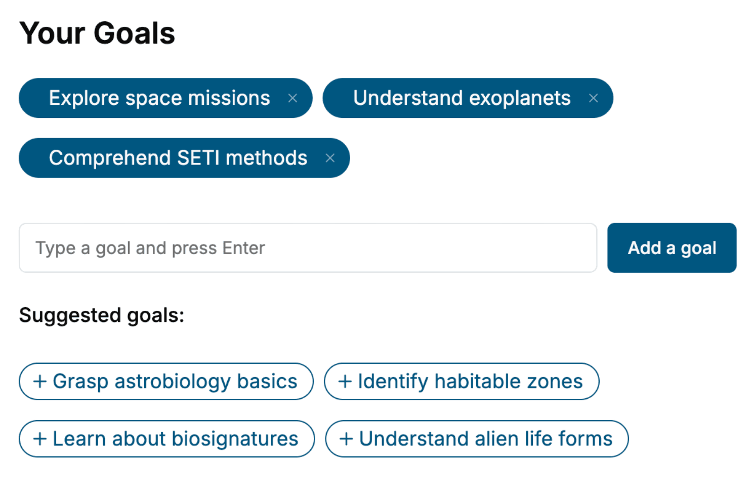
Tell us your goals and background, and get your own version of this book, tailored by AI to you.


Benefits of AI-tailored books
- Read one book, not ten:
- all the Neuroanatomy knowledge you need, gathered into a single book.
- Save days of learning:
- tell us your goals, choose your topics, and the whole book is built around them.
- Learn effortlessly:
- Neuroanatomy book written for your specific background and expertise.
- Understand at a glance:
- includes charts, tables, timelines, checklists, decision trees, and more.
- Stay current:
- written the day you order it, not years ago.
- Trust what you read:
- every book is checked for accuracy, with real research when your subject needs it.
Create your unique book in 3 steps
- 1. Select your focus
Select the focus of your Neuroanatomy book and share your background
- 2. Personalize your book
Specify your goals and choose sub-topics to include
- 3. Get your tailored book
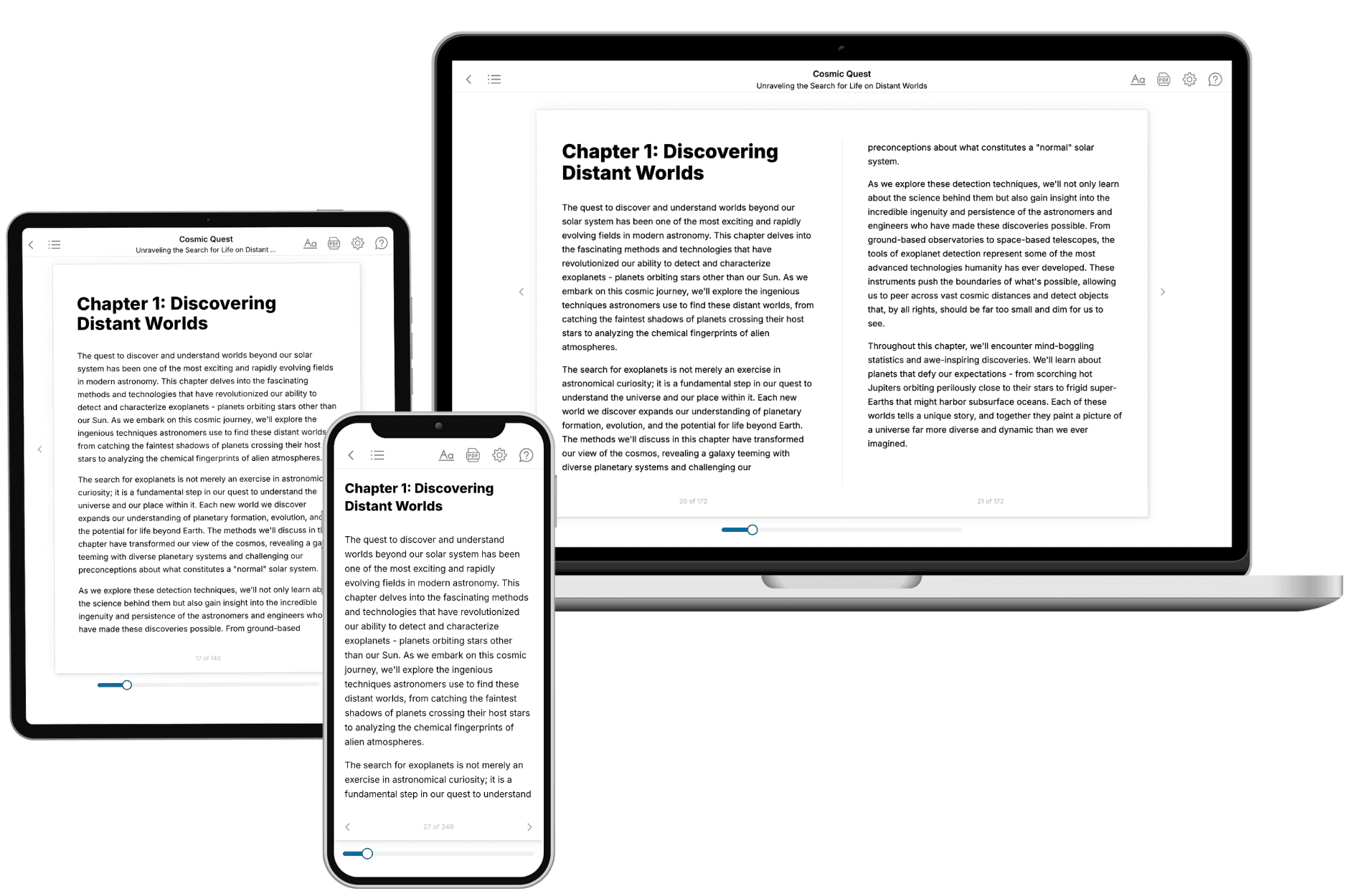
Your book is ready in 10 minutes. Read it online, download as EPUB or PDF, or send to Kindle.

Frequently asked questions
- What is TailoredRead?
TailoredRead creates complete, full-length nonfiction ebooks, written specifically for you. Tell us what you want to learn and why, and TailoredRead writes you a complete ebook on it, with charts, tables, and diagrams throughout. Read it online, on Kindle, or on any e-reader, or download it as a PDF or EPUB. Every book is written fresh the day you order it.
- How is the book tailored to me?
Before anything is written, you answer a few short questions about your goals, your background, and the topics you care about. Your book is then planned around your answers: written at your level, focused on your goals, and covering the topics you chose, instead of the one-size-fits-all book a store sells everyone.
- Will it read like generic AI content?
No. Your book isn't a chat transcript: it's planned chapter by chapter before a single word is written, told in a clear book voice, and explains ideas visually with charts, tables, and diagrams. After writing, every book goes through an editorial pass that polishes chapters so the book reads consistent from start to finish. Judge for yourself: browse thousands of sample books on our Explore Books page.
- How accurate is the content?
Our AI first checks whether your subject needs live research. Niche and fast-moving subjects are researched before writing, and the book plan and chapters are grounded in what it finds. After writing, every book goes through a quality pass that checks its facts and consistency, and researched books list their sources in a Notes section at the back.
- How long is the book?
You choose the length: Comprehensive (250-300 pages), Detailed (150-200 pages), Essential (70-100 pages), or Short (30-50 pages).
- Does the book include visuals?
Yes. Every book includes charts, tables, and diagrams: timelines, checklists, step-by-step guides, decision trees, comparison tables, matrices, and bar charts. A typical book includes dozens of these visual elements.
- Can I read it on Kindle? What formats can I download?
Yes. Every book can be read in our online reader or downloaded as a PDF or EPUB. The EPUB can be opened with Kindle, Apple Books, Kobo, and any e-reader. The Commercial and Author licenses also include an editable Word (DOCX) and Markdown download.
- How much does it cost?
About the price of a regular ebook. The exact price depends on your book's length and complexity, from $2 for the shortest books to $35 for the most comprehensive, and it's shown when you review your book, before you commit to anything. Each book is a one-time purchase, yours to keep, no subscription. Licenses that let you share, teach, or publish your book range from $35.99 to $49.99.
- Can I preview the book before purchasing?
Before you pay, you'll see what your book will cover: the title, a detailed description, the estimated length, and the complete chapter-by-chapter table of contents, so you know what you're getting before you decide. The full text itself is written after you purchase.
- How long does it take to create a book?
About 10 minutes. You can start reading the opening chapters while the rest is still being written.