Optical Coherence Tomography
Principles, Applications, and Innovations in High-Resolution Imaging
Dive into the fascinating world of Optical Coherence Tomography (OCT) with this comprehensive guide.
300 pages15 chaptersNov 22, 2024Medical Imaging
About this book
This book offers an in-depth exploration of OCT, a cutting-edge imaging technology that has revolutionized various scientific and medical fields. You'll discover the fundamental principles behind OCT, understanding how it utilizes light waves to create high-resolution, cross-sectional images of biological tissues and materials. The book covers a wide range of applications, from ophthalmology and cardiology to materials science and art conservation. As you progress through the chapters, you'll gain insights into the latest technological advancements in OCT, including emerging techniques and improved resolution capabilities. Real-world case studies and practical examples will help you grasp the significance of OCT in research and clinical settings. Whether you're a researcher, clinician, or technology enthusiast, this book will equip you with the knowledge to understand, apply, and stay up-to-date with the rapidly evolving field of Optical Coherence Tomography.
Table of contents
- IntroductionOverview of Optical Coherence Tomography, Historical Context and Development, Structure of the BookRead below
- Chapter 1: Fundamentals of Optical Coherence TomographyProperties of Light Relevant to OCT, Principles of Interferometry, Coherence and Its Role in OCT, Axial and Lateral Resolution in OCT
- Chapter 2: OCT System ArchitectureTime-Domain OCT Systems, Fourier-Domain OCT Systems, Swept-Source OCT Systems, Full-Field OCT Systems
- Chapter 3: Light Sources for OCTSuperluminescent Diodes, Tunable Lasers, Femtosecond Lasers, Supercontinuum Sources
- Chapter 4: Detectors and Signal ProcessingPhotodetectors for OCT, Spectrometers in Spectral-Domain OCT, Analog-to-Digital Conversion, Signal Processing Algorithms
- Chapter 5: Image Formation and ReconstructionInterference Patterns and Depth Encoding, Fourier Transform in OCT, Dispersion Compensation Techniques, Common Image Artifacts and Corrections
- Chapter 6: Light-Sample Interaction in OCTScattering and Absorption, Penetration Depth and Tissue Optics, Speckle Formation and Reduction, Polarization Effects in OCT
- Chapter 7: OCT in OphthalmologyRetinal Imaging and Analysis, Anterior Segment Imaging, Glaucoma Assessment, Diagnosis of Ocular Pathologies
- Chapter 8: OCT in CardiologyIntravascular OCT, Atherosclerosis Assessment, Stent Evaluation and Optimization, Plaque Characterization
- Chapter 9: OCT in DermatologyImaging Skin Layers and Structures, Skin Cancer Detection, Burn Depth Assessment, Cosmetic and Aesthetic Applications
- Chapter 10: OCT in OncologyEarly Cancer Detection, Tumor Margin Assessment, Treatment Response Monitoring, OCT-Guided Biopsy
- Chapter 11: OCT in Materials ScienceNon-Destructive Testing of Materials, Polymer Characterization, Applications in Art Conservation, Semiconductor Inspection
- Chapter 12: Functional OCT TechniquesDoppler OCT for Flow Imaging, Polarization-Sensitive OCT, OCT Angiography, OCT Elastography
- Chapter 13: Multimodal Imaging with OCTOCT and Fluorescence Imaging, OCT and Photoacoustic Imaging, OCT and Raman Spectroscopy, OCT and Ultrasound Integration
- Chapter 14: OCT Data Analysis and InterpretationImage Segmentation Techniques, Feature Extraction and Quantification, Machine Learning in OCT Analysis, 3D Visualization and Rendering
- Chapter 15: Emerging OCT TechnologiesQuantum OCT, Advancements in Full-Field OCT, OCT-based Microscopy, Miniaturization and OCT Endoscopy
- ConclusionFuture Directions in OCT, Challenges and Opportunities
- Glossary
- Further Reading
Introduction
Welcome to the fascinating world of Optical Coherence Tomography (OCT), a revolutionary imaging technology that has transformed numerous scientific and medical fields. This introduction will provide you with a comprehensive overview of OCT, its historical context, and the structure of this book. As you embark on this journey, you'll discover how OCT has become an indispensable tool for researchers, clinicians, and technologists across various disciplines.
OCT is a non-invasive imaging technique that utilizes light waves to produce high-resolution, cross-sectional images of biological tissues and materials. Its ability to provide detailed, real-time visualization of structures at the micrometer scale has made it an invaluable asset in fields such as ophthalmology, cardiology, dermatology, and materials science. Throughout this book, you'll explore the principles underlying OCT, its diverse applications, and the latest advancements that continue to expand its capabilities.
As you delve deeper into the world of OCT, you'll gain a thorough understanding of its core principles, including low-coherence interferometry, which allows for depth-resolved imaging. You'll also learn about the various OCT system architectures, light sources, detectors, and signal processing techniques that contribute to the creation of high-quality OCT images. By the end of this book, you'll be well-equipped to appreciate the strengths and limitations of OCT, enabling you to effectively implement this technology in your research or clinical practice.
Overview of Optical Coherence Tomography
Optical Coherence Tomography (OCT) is a powerful imaging modality that has revolutionized the way we visualize and analyze biological tissues and materials. At its core, OCT is an interferometric technique that uses low-coherence light to produce high-resolution, cross-sectional images of samples. The fundamental principle behind OCT is analogous to ultrasound imaging, but instead of using sound waves, OCT employs light waves to create detailed images of structures below the surface of the sample.
The key advantage of OCT lies in its ability to provide non-invasive, real-time, in situ imaging with micrometer-scale resolution. This unique combination of features has made OCT an invaluable tool across a wide range of applications, from medical diagnostics to materials characterization. In the medical field, OCT has become particularly prominent in ophthalmology, where it is used for retinal imaging and the diagnosis of various eye diseases. Its ability to provide detailed cross-sectional images of the retina has revolutionized the early detection and management of conditions such as age-related macular degeneration, diabetic retinopathy, and glaucoma[1].
Beyond ophthalmology, OCT has found applications in numerous other medical specialties. In cardiology, intravascular OCT provides high-resolution images of coronary arteries, aiding in the assessment of atherosclerosis and the optimization of stent placement. Dermatologists use OCT for non-invasive imaging of skin layers, assisting in the diagnosis of skin cancers and the evaluation of treatment efficacy. In oncology, OCT is being explored for early cancer detection and tumor margin assessment during surgery.
The versatility of OCT extends beyond the medical field into various scientific and industrial applications. In materials science, OCT serves as a powerful tool for non-destructive testing and characterization of materials. It allows for the visualization of internal structures in polymers, composites, and ceramics, providing valuable insights into material properties and defects. In the field of art conservation, OCT has been employed to study the layered structure of paintings and other artifacts, aiding in their preservation and restoration.
One of the key strengths of OCT is its ability to provide both structural and functional information about the sample being imaged. While traditional OCT primarily focuses on structural imaging, advanced functional OCT techniques have been developed to extract additional information. These include Doppler OCT for blood flow imaging, polarization-sensitive OCT for assessing tissue birefringence, OCT angiography for visualizing blood vessels without the need for contrast agents, and OCT elastography for measuring tissue elasticity.
The basic principle of OCT involves splitting a low-coherence light beam into two paths: a reference arm and a sample arm. The light reflected from the sample interferes with the light from the reference arm, creating an interference pattern. By analyzing this interference pattern, the OCT system can determine the depth and intensity of reflections from different layers within the sample. This information is then used to construct a cross-sectional image, often referred to as an A-scan. By scanning the beam across the sample, multiple A-scans can be combined to create 2D cross-sectional images (B-scans) or even 3D volumetric datasets.
Over the years, OCT technology has evolved significantly, leading to improvements in imaging speed, resolution, and sensitivity. Early OCT systems were based on time-domain detection, which required mechanical scanning of the reference arm. The introduction of Fourier-domain OCT techniques, including spectral-domain and swept-source OCT, has dramatically increased imaging speeds and sensitivity. These advancements have enabled real-time, high-resolution imaging of dynamic processes and have expanded the range of applications for OCT.
As OCT continues to evolve, new frontiers are being explored. Researchers are working on pushing the boundaries of resolution, with some systems now capable of achieving sub-micrometer resolution. Efforts are also underway to extend the imaging depth of OCT, which is typically limited to a few millimeters in most biological tissues due to light scattering. Novel approaches, such as optical clearing techniques and the use of longer wavelength light sources, are being investigated to overcome this limitation.
The integration of OCT with other imaging modalities is another area of active research. Multimodal imaging systems that combine OCT with techniques such as fluorescence microscopy, photoacoustic imaging, or Raman spectroscopy are being developed to provide complementary information about the sample. These hybrid systems have the potential to offer a more comprehensive understanding of tissue structure and function.
As you progress through this book, you'll gain a deeper understanding of the principles underlying OCT, the various system architectures and components, and the wide range of applications across different fields. You'll also explore the latest advancements in OCT technology and get a glimpse into the future directions of this rapidly evolving field. Whether you're a researcher, clinician, or technology enthusiast, this comprehensive guide to OCT will equip you with the knowledge to appreciate and harness the power of this remarkable imaging technology.
Historical Context and Development
The development of Optical Coherence Tomography (OCT) represents a fascinating journey through the realms of optics, interferometry, and biomedical imaging. To fully appreciate the significance of OCT, it's essential to understand its historical context and the key milestones that led to its emergence as a powerful imaging modality. The roots of OCT can be traced back to the fundamental principles of interferometry, which were established in the 19th century.
The concept of interferometry, which forms the basis of OCT, was first demonstrated by Albert A. Michelson in 1881. Michelson invented the Michelson interferometer, a device that splits a beam of light into two paths, reflects them back, and recombines them to create an interference pattern. This invention laid the groundwork for numerous interferometric techniques, including OCT. Michelson's work was so significant that he was awarded the Nobel Prize in Physics in 1907 for his optical precision instruments and the spectroscopic and metrological investigations carried out with their aid[2].
In the decades that followed, interferometry found applications in various fields, including astronomy, metrology, and spectroscopy. However, it wasn't until the late 20th century that the principles of interferometry were applied to biological imaging in the form of OCT. The development of OCT was driven by the need for high-resolution, non-invasive imaging techniques in medicine, particularly in ophthalmology.
The direct precursor to OCT was optical time-domain reflectometry (OTDR), a technique used in the telecommunications industry to characterize optical fibers. OTDR uses short pulses of light to detect reflections along the length of an optical fiber, allowing for the identification of breaks, splices, and other features. The principles of OTDR, particularly the use of low-coherence interferometry to measure reflections from different depths, would later be adapted for biological imaging in OCT.
The birth of OCT as we know it today can be traced back to the early 1990s. In 1991, David Huang and colleagues at the Massachusetts Institute of Technology (MIT) published a seminal paper in Science titled "Optical Coherence Tomography"[3]. This paper described the first OCT system capable of producing cross-sectional images of biological tissues with micrometer-scale resolution. The authors demonstrated OCT imaging of the human retina and coronary artery, showcasing its potential for medical applications.
The initial OCT systems developed in the early 1990s were based on time-domain detection. In these systems, the reference arm of the interferometer was mechanically scanned to produce depth-resolved images. While groundbreaking, these early systems were relatively slow, limiting their practical applications. Despite this limitation, the potential of OCT for medical imaging, particularly in ophthalmology, was quickly recognized.
The late 1990s and early 2000s saw rapid advancements in OCT technology. One of the most significant developments was the introduction of Fourier-domain OCT techniques. In 1995, Adolf Fercher and colleagues proposed the concept of spectral-domain OCT, which eliminated the need for mechanical scanning of the reference arm by using a spectrometer to detect the interference spectrum[4]. This approach significantly increased the imaging speed and sensitivity of OCT systems.
Around the same time, another Fourier-domain OCT technique called swept-source OCT was developed. Swept-source OCT uses a rapidly tunable laser source and a single photodetector to achieve high-speed, high-sensitivity imaging. These Fourier-domain OCT techniques represented a major leap forward in OCT technology, enabling real-time, high-resolution imaging of dynamic processes.
The early 2000s saw the commercialization of OCT technology, particularly in the field of ophthalmology. The first commercial OCT system for retinal imaging was introduced by Carl Zeiss Meditec in 1996. This marked the beginning of OCT's widespread adoption in clinical practice. Over the next decade, OCT quickly became an indispensable tool for diagnosing and monitoring various retinal diseases, revolutionizing the field of ophthalmology.
As OCT technology matured, its applications expanded beyond ophthalmology. In the mid-2000s, intravascular OCT systems were developed for coronary artery imaging, providing cardiologists with unprecedented views of arterial walls and implanted stents. OCT also found applications in dermatology, dentistry, and various other medical specialties.
The late 2000s and 2010s saw continued advancements in OCT technology. Researchers focused on improving image resolution, increasing imaging speed, and developing new functional OCT techniques. One notable development was OCT angiography, a technique that allows for non-invasive visualization of blood vessels without the need for contrast agents. This technique has had a significant impact on the diagnosis and management of retinal vascular diseases.
Another important trend in OCT development has been the miniaturization of OCT systems. Advances in photonics and microelectronics have enabled the development of compact, portable OCT devices. These handheld and even smartphone-based OCT systems have the potential to bring high-resolution imaging capabilities to point-of-care settings and resource-limited environments.
In recent years, OCT has also made significant inroads in non-medical applications. In materials science, OCT has been used for non-destructive testing of materials, characterization of 3D-printed structures, and quality control in manufacturing processes. In the field of art conservation, OCT has been employed to study the layered structure of paintings and other artifacts, providing valuable insights for restoration efforts.
The integration of artificial intelligence (AI) and machine learning techniques with OCT has been a major focus of research in recent years. AI algorithms are being developed to automatically analyze OCT images, detect abnormalities, and assist in diagnosis. These developments have the potential to enhance the efficiency and accuracy of OCT-based diagnostics.
Looking to the future, OCT continues to evolve. Researchers are exploring new frontiers, such as quantum OCT, which promises to push the boundaries of resolution and sensitivity. Efforts are also underway to extend the imaging depth of OCT and to develop novel functional OCT techniques that can provide even more detailed information about tissue properties and function.
The history of OCT is a testament to the power of interdisciplinary collaboration and the impact of technological innovation on medical practice and scientific research. From its origins in the principles of interferometry to its current status as a versatile, high-resolution imaging modality, OCT has transformed our ability to visualize and understand the microscopic world. As you delve deeper into the principles and applications of OCT in the following chapters, keep in mind the rich history and ongoing evolution of this remarkable technology.
Structure of the Book
This book is designed to provide you with a comprehensive understanding of Optical Coherence Tomography (OCT), from its fundamental principles to its diverse applications and latest advancements. The structure of the book is carefully organized to guide you through the complexities of OCT in a logical and accessible manner. Whether you're a researcher, clinician, or technology enthusiast, this book will equip you with the knowledge to appreciate and harness the power of OCT in your field of interest.
The book begins with this introduction, which provides an overview of OCT, its historical context, and the structure of the book. This foundation will help you understand the significance of OCT and prepare you for the more detailed discussions in the subsequent chapters. Following the introduction, the book is divided into several main sections, each focusing on a specific aspect of OCT technology and its applications.
The first section of the book delves into the fundamentals of OCT. Chapter 1, "Fundamentals of Optical Coherence Tomography," explores the basic principles that underpin OCT technology. You'll learn about the properties of light relevant to OCT, the principles of interferometry, the concept of coherence and its role in OCT, and the factors that determine axial and lateral resolution in OCT imaging. This chapter lays the groundwork for understanding the more advanced concepts discussed later in the book.
The next section focuses on the technical aspects of OCT systems. Chapter 2, "OCT System Architecture," introduces you to the various types of OCT systems, including time-domain, Fourier-domain, swept-source, and full-field OCT. You'll gain insights into the strengths and limitations of each architecture and understand how these different approaches have shaped the evolution of OCT technology.
Chapters 3 and 4 delve deeper into the key components of OCT systems. In Chapter 3, "Light Sources for OCT," you'll explore the various light sources used in OCT, including superluminescent diodes, tunable lasers, femtosecond lasers, and supercontinuum sources. Chapter 4, "Detectors and Signal Processing," covers the detection systems used in OCT, including photodetectors and spectrometers, as well as the signal processing algorithms that transform raw data into meaningful images.
The book then moves on to the process of image formation in OCT. Chapter 5, "Image Formation and Reconstruction," explains how interference patterns are used to encode depth information, the role of Fourier transform in OCT image reconstruction, techniques for dispersion compensation, and common image artifacts and their corrections. Chapter 6, "Light-Sample Interaction in OCT," explores the physics of how light interacts with biological tissues and materials in OCT imaging, covering topics such as scattering, absorption, penetration depth, speckle formation, and polarization effects.
The next section of the book focuses on the applications of OCT across various fields. Chapters 7 through 11 cover the use of OCT in ophthalmology, cardiology, dermatology, oncology, and materials science, respectively. Each of these chapters provides an in-depth look at how OCT is used in these specific fields, the unique challenges and opportunities it presents, and case studies that illustrate its impact on research and clinical practice.
Chapter 12, "Functional OCT Techniques," introduces you to advanced OCT modalities that go beyond structural imaging to provide functional information about tissues. You'll learn about Doppler OCT for flow imaging, polarization-sensitive OCT, OCT angiography, and OCT elastography. These techniques represent the cutting edge of OCT technology and are expanding its capabilities in various applications.
The integration of OCT with other imaging modalities is explored in Chapter 13, "Multimodal Imaging with OCT." This chapter discusses how OCT can be combined with techniques such as fluorescence imaging, photoacoustic imaging, Raman spectroscopy, and ultrasound to provide complementary information and enhance diagnostic capabilities.
Chapter 14, "OCT Data Analysis and Interpretation," focuses on the crucial aspect of extracting meaningful information from OCT images. You'll learn about image segmentation techniques, feature extraction and quantification methods, the application of machine learning in OCT analysis, and advanced 3D visualization and rendering techniques.
The book concludes with a look towards the future of OCT technology. Chapter 15, "Emerging OCT Technologies," explores cutting-edge developments such as quantum OCT, advancements in full-field OCT, OCT-based microscopy, and the miniaturization of OCT systems for endoscopic applications. This chapter will give you a glimpse into the exciting future directions of OCT research and development.
Throughout the book, you'll find numerous illustrations, diagrams, and real-world examples to help clarify complex concepts. Each chapter includes a summary of key points and a list of references for further reading. The book also includes a comprehensive glossary of OCT-related terms and a section on further reading resources for those who wish to delve deeper into specific topics.
By structuring the book in this way, the aim is to provide you with a logical progression from basic principles to advanced applications and future perspectives. Whether you're new to OCT or an experienced practitioner looking to expand your knowledge, this book offers a comprehensive guide to understanding and applying OCT technology. As you work through the chapters, you'll gain a deep appreciation for the power and versatility of OCT and its potential to drive future innovations in imaging science and technology.
Notes
Introduction
- 1. W. Drexler and others, Optical coherence tomography, Springer, 2015
- 2. A. A. Michelson, "The relative motion of the Earth and the luminiferous ether," American Journal of Science, 1881
- 3. D. Huang and others, "Optical coherence tomography," Science, 1991
- 4. A. F. Fercher and others, "Measurement of intraocular distances by backscattering spectral interferometry," Optics Communications, 1995
This is the end of the free sample. The full book has 15 chapters.
Want this book written for you?
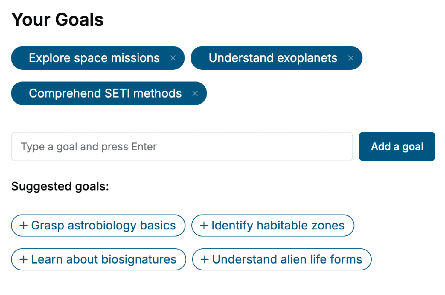
Tell us your goals and background, and get your own version of this book, tailored by AI to you.


Benefits of AI-tailored books
- Read one book, not ten:
- all the Medical Imaging knowledge you need, gathered into a single book.
- Save days of learning:
- tell us your goals, choose your topics, and the whole book is built around them.
- Learn effortlessly:
- Medical Imaging book written for your specific background and expertise.
- Understand at a glance:
- includes charts, tables, timelines, checklists, decision trees, and more.
- Stay current:
- written the day you order it, not years ago.
- Trust what you read:
- every book is checked for accuracy, with real research when your subject needs it.
Create your unique book in 3 steps
- 1. Select your focus
Select the focus of your Medical Imaging book and share your background
- 2. Personalize your book
Specify your goals and choose sub-topics to include
- 3. Get your tailored book
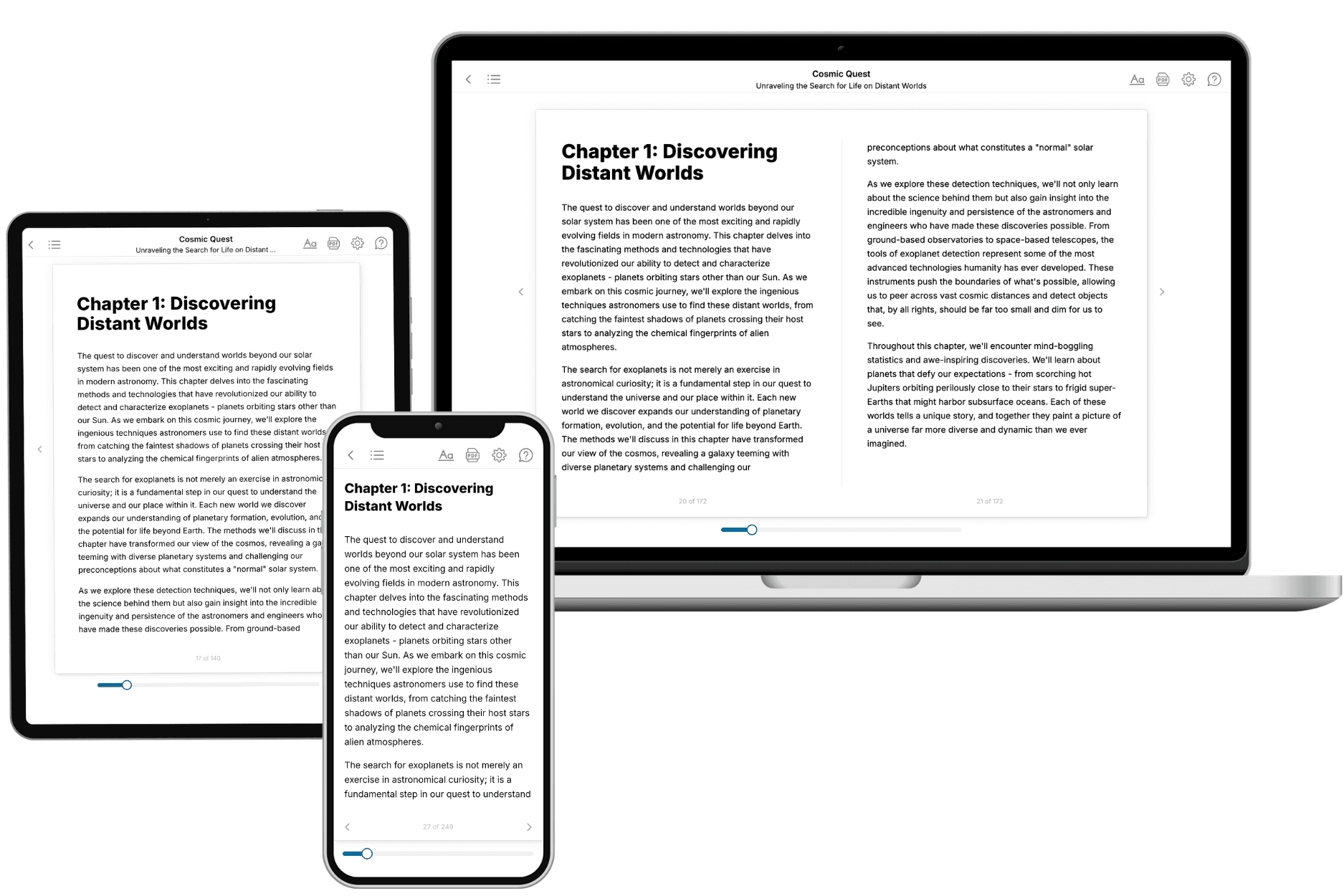
Your book is ready in 10 minutes. Read it online, download as EPUB or PDF, or send to Kindle.

Frequently asked questions
- What is TailoredRead?
TailoredRead creates complete, full-length nonfiction ebooks, written specifically for you. Tell us what you want to learn and why, and TailoredRead writes you a complete ebook on it, with charts, tables, and diagrams throughout. Read it online, on Kindle, or on any e-reader, or download it as a PDF or EPUB. Every book is written fresh the day you order it.
- How is the book tailored to me?
Before anything is written, you answer a few short questions about your goals, your background, and the topics you care about. Your book is then planned around your answers: written at your level, focused on your goals, and covering the topics you chose, instead of the one-size-fits-all book a store sells everyone.
- Will it read like generic AI content?
No. Your book isn't a chat transcript: it's planned chapter by chapter before a single word is written, told in a clear book voice, and explains ideas visually with charts, tables, and diagrams. After writing, every book goes through an editorial pass that polishes chapters so the book reads consistent from start to finish. Judge for yourself: browse thousands of sample books on our Explore Books page.
- How accurate is the content?
Our AI first checks whether your subject needs live research. Niche and fast-moving subjects are researched before writing, and the book plan and chapters are grounded in what it finds. After writing, every book goes through a quality pass that checks its facts and consistency, and researched books list their sources in a Notes section at the back.
- How long is the book?
You choose the length: Comprehensive (250-300 pages), Detailed (150-200 pages), Essential (70-100 pages), or Short (30-50 pages).
- Does the book include visuals?
Yes. Every book includes charts, tables, and diagrams: timelines, checklists, step-by-step guides, decision trees, comparison tables, matrices, and bar charts. A typical book includes dozens of these visual elements.
- Can I read it on Kindle? What formats can I download?
Yes. Every book can be read in our online reader or downloaded as a PDF or EPUB. The EPUB can be opened with Kindle, Apple Books, Kobo, and any e-reader. The Commercial and Author licenses also include an editable Word (DOCX) and Markdown download.
- How much does it cost?
About the price of a regular ebook. The exact price depends on your book's length and complexity, from $2 for the shortest books to $35 for the most comprehensive, and it's shown when you review your book, before you commit to anything. Each book is a one-time purchase, yours to keep, no subscription. Licenses that let you share, teach, or publish your book range from $35.99 to $49.99.
- Can I preview the book before purchasing?
Before you pay, you'll see what your book will cover: the title, a detailed description, the estimated length, and the complete chapter-by-chapter table of contents, so you know what you're getting before you decide. The full text itself is written after you purchase.
- How long does it take to create a book?
About 10 minutes. You can start reading the opening chapters while the rest is still being written.