The Respiratory System Coloring Book
Learn Anatomy Through Interactive Illustrations and Detailed Exploration
Discover how your body breathes through detailed anatomical illustrations designed specifically for interactive learning.
222 pages10 chaptersApr 14, 2026Anatomy
About this book
This coloring book guides you through the complete respiratory system—from the moment air enters your nose to the precise moment oxygen crosses into your bloodstream. Each chapter presents carefully detailed drawings of respiratory structures, accompanied by clear explanations that help you understand not just what you're coloring, but why each component matters. By engaging both your visual and creative faculties, you'll develop a deeper, more intuitive grasp of how your lungs, airways, diaphragm, and supporting muscles work together seamlessly. Whether you're a student, healthcare professional, or simply curious about human anatomy, this interactive approach transforms learning from passive reading into active discovery. The combination of accurate anatomical detail and hands-on coloring creates multiple pathways for your brain to process and retain information, making complex respiratory concepts accessible and memorable.
Table of contents
- IntroductionWhy Understanding Your Respiratory System Matters, How Coloring Enhances Anatomical Learning, How to Use This Book, Your Journey Through the Respiratory SystemRead below
- Chapter 1: The Upper Respiratory TractThe Nasal Cavity and Septum, The Pharynx: Nasopharynx, Oropharynx, and Laryngopharynx, The Larynx: Voice Box and Airway Guardian
- Chapter 2: The Lower Respiratory TractThe Trachea: Windpipe Structure and Function, Primary and Secondary Bronchi: The Branching Airways, Bronchioles and Alveoli: Where Gas Exchange Happens
- Chapter 3: The Lungs and Pleural MembranesRight and Left Lungs: Structure and Lobes, The Pleural Membranes and Pleural Cavity, The Hilum: Gateway for Vessels and Nerves
- Chapter 4: Muscles of RespirationThe Diaphragm: Primary Breathing Muscle, Intercostal Muscles: External and Internal, Accessory Respiratory Muscles
- Chapter 5: The Rib Cage and Thoracic CavityRibs, Sternum, and Vertebral Column, Key Anatomical Landmarks of the Thorax, The Mediastinum and Its Contents
- Chapter 6: Gas Exchange and Alveolar StructureAlveolar Walls and Respiratory Membrane, Pulmonary Capillaries and Blood Flow, The Gas Exchange Process at the Cellular Level
- Chapter 7: Blood Supply and InnervationPulmonary Arteries and Veins, Bronchial Arteries and Systemic Supply, Vagus Nerve and Respiratory Control
- Chapter 8: Breathing Mechanics and VentilationInspiration: How Air Enters the Lungs, Expiration: How Air Leaves the Lungs, Pressure Changes During the Breathing Cycle
- Chapter 9: Protective Mechanisms and DefenseFiltration and Warming in the Upper Airways, Mucus and Cilia: The Respiratory Cleaning System, Immune Cells and Pathogen Defense
- Chapter 10: Regulation of BreathingBrainstem Respiratory Centers, Chemical Regulation: CO2, O2, and pH, Voluntary Control and Reflex Responses
- ConclusionHow It All Works Together, Continuing Your Anatomical Exploration
- Glossary of Respiratory Terms
- Coloring Guide and Color Suggestions
Introduction
Every single moment of your life, without any conscious effort on your part, your body performs one of its most essential acts: breathing. Right now, as you read these words, air is moving through a remarkable network of passages, chambers, and membranes, delivering oxygen to trillions of cells and carrying away the carbon dioxide they produce. This process happens roughly 20,000 times per day, and yet most people never stop to consider the extraordinary anatomical machinery that makes it possible. This book invites you to slow down, look closely, and truly understand the system that keeps you alive with every breath.
The respiratory system is far more than just a pair of lungs. It is an integrated network of structures that spans from the tip of your nose to the microscopic walls of your alveoli, each component precisely designed to filter, warm, humidify, and ultimately exchange gases with your bloodstream. Understanding this system in detail is not just an academic exercise. It builds a foundation for appreciating how the body maintains balance, how diseases disrupt that balance, and how medical interventions work to restore it. Whether you are a student preparing for exams, a healthcare professional refreshing your knowledge, or simply someone fascinated by human biology, this level of understanding has real and lasting value.
What makes this book different from a standard anatomy text is its approach. Rather than asking you to passively absorb diagrams and descriptions, it invites you to actively participate in your own learning. By coloring detailed anatomical illustrations, you engage your hands, your eyes, and your analytical mind simultaneously. You make decisions about which structure is which, you trace pathways with your pencil or marker, and you build a personal, tactile connection to the material. Research consistently shows that this kind of active engagement produces stronger memory formation and deeper conceptual understanding than reading alone. This introduction will walk you through why the respiratory system deserves your focused attention, how the coloring method works in your favor, and how to get the most out of every page that follows.
Why Understanding Your Respiratory System Matters
The respiratory system sits at the center of human physiology in a way that few other systems do. Every other organ in your body depends on the oxygen that your lungs deliver. Your heart pumps blood, but that blood is only useful because your lungs have loaded it with oxygen and cleared it of carbon dioxide. Your brain controls every function in your body, but it consumes roughly 20% of your total oxygen supply and will begin to fail within minutes if that supply is interrupted.[1] Understanding the respiratory system means understanding the foundation on which all other physiological processes rest.
From a clinical perspective, respiratory conditions are among the most common and most serious health challenges worldwide. Asthma affects more than 260 million people globally, while chronic obstructive pulmonary disease (COPD) is the third leading cause of death worldwide.[2] Pneumonia, lung cancer, pulmonary fibrosis, and sleep apnea collectively affect hundreds of millions more. When you understand the anatomy of the respiratory system in detail, you gain the ability to understand what goes wrong in these conditions, why certain symptoms appear, and how treatments target specific structures or mechanisms. That knowledge transforms abstract medical information into something concrete and meaningful.
Beyond disease, understanding your respiratory system has practical everyday applications. Athletes who understand how breathing mechanics affect performance can train more effectively. People who practice meditation or yoga benefit from knowing how controlled breathing influences the nervous system. Parents who understand how a child's airway differs from an adult's are better equipped to respond to emergencies. Even something as simple as understanding why you get out of breath when you climb stairs becomes more interesting and actionable when you know the physiology behind it.
There is also a deeper, more personal reason to study this system carefully. Breathing is one of the few physiological processes that sits at the intersection of the automatic and the voluntary. Your brainstem controls your breathing rate without any conscious input, yet you can override that control at will, holding your breath, slowing your exhale, or deliberately deepening your inhale. This dual nature makes the respiratory system a fascinating window into how the nervous system balances automatic regulation with conscious control. Exploring that balance is one of the most intellectually rewarding aspects of this subject.
For students in nursing, medicine, respiratory therapy, physical therapy, or any allied health field, a thorough grasp of respiratory anatomy is not optional. It is foundational. Clinical assessments, diagnostic reasoning, and treatment planning all depend on knowing exactly where structures are located, how they connect to one another, and what happens when they are compromised. The visual and hands-on approach in this book is specifically designed to build that kind of solid, reliable anatomical knowledge rather than surface-level familiarity.
Finally, there is something genuinely awe-inspiring about the respiratory system when you examine it closely. The fact that your alveoli, if spread flat, would cover an area roughly the size of a tennis court, yet they fit inside your chest, is a remarkable feat of biological engineering.[3] The way your nasal passages can warm incoming air from near-freezing temperatures to body temperature in a fraction of a second is extraordinary. Studying this system is not just useful. It is genuinely fascinating, and this book is designed to help you experience that fascination firsthand.
How Coloring Enhances Anatomical Learning
The idea of using coloring as a serious learning tool might seem unconventional at first, but the science behind it is well established. When you color an anatomical illustration, you are not simply filling in shapes. You are making active decisions about what each structure is, where its boundaries lie, and how it relates to neighboring structures. This process forces a level of engagement that passive reading simply cannot match. You cannot color the trachea without first identifying it, and you cannot identify it without understanding what distinguishes it from the esophagus sitting just behind it.
Cognitive science research supports this approach through the concept of elaborative encoding. This is the process by which new information becomes more memorable when it is connected to existing knowledge through active processing.[4] When you read a description of the bronchi, your brain processes it at a relatively shallow level. When you then trace and color those bronchi on an illustration, connecting the written description to a visual and motor experience, your brain encodes the information at a much deeper level. The result is stronger, longer-lasting memory.
The dual coding theory, developed by psychologist Allan Paivio, offers another explanation for why this method works so well.[5] According to this theory, the brain stores information in two separate but interconnected systems: one for verbal information and one for visual and spatial information. When you learn anatomy through both reading and coloring, you create representations in both systems simultaneously. This means you have two separate pathways for retrieving the information later, making recall significantly more reliable than if you had used only one approach.
There is also a practical benefit that experienced anatomy students know well: color-coding creates instant visual organization. When you consistently use one color for arteries, another for veins, and a third for nerves across multiple illustrations, your brain begins to recognize these patterns automatically. Over time, you develop an intuitive sense of how structures relate to one another spatially, which is exactly the kind of three-dimensional anatomical thinking that clinical practice requires. This is why coloring books have been used in medical and nursing education for decades, not as a novelty, but as a genuinely effective pedagogical tool.
The motor component of coloring also plays a role that is easy to underestimate. The act of physically moving a pencil or marker along the contours of a structure engages your motor cortex and creates what researchers call embodied cognition, a form of learning that is anchored in physical experience.[6] Students who have traced the path of the vagus nerve with their own hand tend to remember its course more reliably than those who have only read about it. The physical act of drawing creates a kind of muscle memory for the information itself.
Beyond the cognitive benefits, coloring also makes the learning process more enjoyable and sustainable. Anatomy is a demanding subject with a large volume of terminology, spatial relationships, and functional details to absorb. Fatigue and frustration are real obstacles. The coloring format breaks the material into manageable, visually engaging segments that feel rewarding to complete. Each finished illustration is a small achievement, a visible record of what you have learned. That sense of progress is motivating, and motivation is one of the most powerful drivers of effective learning.
It is worth noting that the quality of the illustrations matters enormously. The drawings in this book are designed to be anatomically accurate while remaining clear enough to color effectively. Structures are labeled precisely, proportions are carefully maintained, and the level of detail is calibrated to support learning rather than overwhelm it. Each illustration is a teaching tool in its own right, and the coloring activity is designed to deepen your engagement with that tool rather than distract from it.
How to Use This Book
Getting the most out of this book requires a small amount of preparation and a clear approach. The good news is that the format is flexible enough to accommodate different learning styles and schedules. Whether you prefer to work through the chapters in strict order or jump to the sections most relevant to your current studies, the book is designed to support you either way. That said, working sequentially from the upper respiratory tract downward does have a logical advantage: the anatomy builds on itself, and understanding earlier structures makes later ones easier to grasp.
Before you begin coloring any illustration, take a few minutes to read the accompanying text carefully. The written explanations are not just captions. They provide the conceptual framework that makes the coloring activity meaningful. Understanding what the tracheal rings do before you color them transforms the activity from a mechanical exercise into an act of genuine comprehension. Read first, then color, and then read again briefly to reinforce what you have just done visually.
For your coloring materials, the following setup works well for most learners:
- Colored pencils are the most versatile choice. They allow fine detail work, can be layered for shading, and do not bleed through pages.
- Fine-tip markers work well for bold, clear color-coding but require care near page edges to avoid bleed-through.
- Gel pens can add detail and are useful for labeling or annotating structures in the margins.
- Avoid felt-tip markers with broad tips, as they tend to obscure fine anatomical details.
Developing a consistent color-coding system across the entire book will pay significant dividends. A suggested starting framework is provided in the Coloring Guide at the back of the book, but you are encouraged to adapt it to your own preferences. The key principle is consistency: once you assign a color to a structure type, use it throughout. For example, if you use red for oxygenated blood vessels and blue for deoxygenated ones, maintain that convention in every chapter. Your brain will begin to process these color associations automatically, which speeds up both learning and recall.
Each chapter follows a consistent structure that is worth understanding before you begin:
- Introductory text that provides context and explains the significance of the structures covered in that chapter.
- Detailed anatomical illustrations with labeled structures ready for coloring.
- Explanatory subchapters that describe each structure's anatomy, location, and function in detail.
- Key terms highlighted throughout the text, all of which are defined in the Glossary at the back of the book.
Annotation is strongly encouraged. The margins of each page are intentionally generous to give you space to write notes, draw arrows, add your own labels, or jot down questions. Treating this book as a working document rather than a pristine object will make it far more useful as a study resource. Some learners find it helpful to write brief functional summaries next to each structure as they color it, for example, noting that the epiglottis "folds down to cover the larynx during swallowing." These self-generated notes are among the most effective memory aids available.
If you encounter a term or concept that is unfamiliar, the Glossary at the back of the book provides clear, concise definitions for all key respiratory anatomy terms used throughout the text. Cross-referencing the Glossary as you work through each chapter is a good habit to develop. It reinforces terminology in context, which is far more effective than memorizing definitions in isolation. The goal is not just to know what words mean in the abstract, but to understand them in relation to the structures you are actively coloring and studying.
Finally, do not rush. The value of this book comes from the time you spend with each illustration, not from how quickly you complete it. A single chapter studied carefully over two sessions will serve you far better than the entire book skimmed in an afternoon. Anatomy rewards patience and repetition. Return to earlier chapters as you progress through later ones, and notice how your understanding of earlier structures deepens as you learn more about the system as a whole.
Your Journey Through the Respiratory System
This book takes you on a logical, anatomically coherent journey through the entire respiratory system, following the path that air itself travels from the moment it enters your body to the moment gas exchange occurs at the cellular level. Along the way, you will explore the structures that support, protect, and regulate that journey. Here is a brief overview of what each chapter covers and why it matters.
Chapter 1: The Upper Respiratory Tract begins your journey at the nose and mouth, tracing the path of incoming air through the nasal cavity, the pharynx, and the larynx. You will learn how the nasal cavity filters, warms, and humidifies air before it reaches the lungs, and how the larynx serves the dual role of protecting the airway and producing sound. The structures here are the first line of defense against airborne particles and pathogens, and understanding them sets the stage for everything that follows.
Chapter 2: The Lower Respiratory Tract takes you from the trachea through the branching network of bronchi and bronchioles, all the way to the alveoli. This chapter introduces the concept of the bronchial tree, the progressively branching airway system that distributes air throughout both lungs. By the time you finish this chapter, you will understand why the airways branch the way they do and how that branching pattern maximizes the surface area available for gas exchange.
Chapter 3: The Lungs and Pleural Membranes examines the lungs themselves as organs, including their lobar structure, their surface anatomy, and the pleural membranes that encase them. You will learn why the right and left lungs differ in structure, what the pleural cavity is and why it matters for breathing mechanics, and how the hilum serves as the entry and exit point for all the vessels, nerves, and airways that supply each lung.
Chapters 4 and 5 shift focus to the mechanical framework of breathing. Chapter 4 covers the muscles of respiration, with particular attention to the diaphragm and the intercostal muscles, while Chapter 5 explores the rib cage and thoracic cavity that house and protect the lungs. Together, these chapters explain how the body generates the pressure changes that move air in and out of the lungs with every breath.
Chapter 6: Gas Exchange and Alveolar Structure zooms in to the microscopic level, examining the alveolar walls, the respiratory membrane, and the pulmonary capillaries where oxygen and carbon dioxide are exchanged. This is the functional heart of the entire respiratory system, and the illustrations in this chapter are designed to make the microscopic architecture of the alveolus as clear and tangible as possible.
Chapters 7 and 8 address the circulatory and mechanical dimensions of respiration. Chapter 7 covers the blood supply to the lungs, including both the pulmonary circulation that carries blood for gas exchange and the bronchial circulation that nourishes the lung tissue itself. Chapter 8 explains the mechanics of breathing in detail, walking through the pressure changes, muscle actions, and volume shifts that occur during each cycle of inspiration and expiration.
Chapters 9 and 10 round out the book by exploring the respiratory system's protective and regulatory mechanisms. Chapter 9 examines how the airways filter and defend against pathogens and irritants, from the mucus-coated surfaces of the nasal passages to the immune cells that patrol the alveoli. Chapter 10 explains how the brainstem regulates breathing automatically and how chemical signals, particularly carbon dioxide levels and blood pH, fine-tune that regulation moment to moment.
The Conclusion brings all of these elements together, helping you see the respiratory system not as a collection of separate structures but as a single, integrated system in which every component depends on and supports every other. The Glossary and Coloring Guide at the back of the book are resources you will return to throughout your study, providing quick reference for terminology and color-coding suggestions respectively.
By the time you reach the final page, you will have traced the complete journey of a breath from nose to alveolus and back again, colored every major structure along the way, and built a comprehensive, visually grounded understanding of one of the body's most essential systems. That understanding is not just academic. It is a foundation for better clinical reasoning, deeper scientific curiosity, and a genuine appreciation for the extraordinary complexity of the body you live in every day. Take a breath, pick up your pencils, and begin.
Notes
Introduction
- 1. P. J. Magistretti and I. Allaman, "A cellular perspective on brain energy metabolism and functional imaging," Neuron, 2015
- 2. World Health Organization, Chronic respiratory diseases, WHO Global Health Estimates, 2023
- 3. M. Ochs and others, "The number of alveoli in the human lung," American Journal of Respiratory and Critical Care Medicine, 2004
- 4. F. I. M. Craik and R. S. Lockhart, "Levels of processing," Journal of Verbal Learning and Verbal Behavior, 1972
- 5. A. Paivio, "Dual coding theory," Canadian Journal of Psychology, 1991
- 6. M. Wilson, "Six views of embodied cognition," Psychonomic Bulletin & Review, 2002
This is the end of the free sample. The full book has 10 chapters.
Want this book written for you?
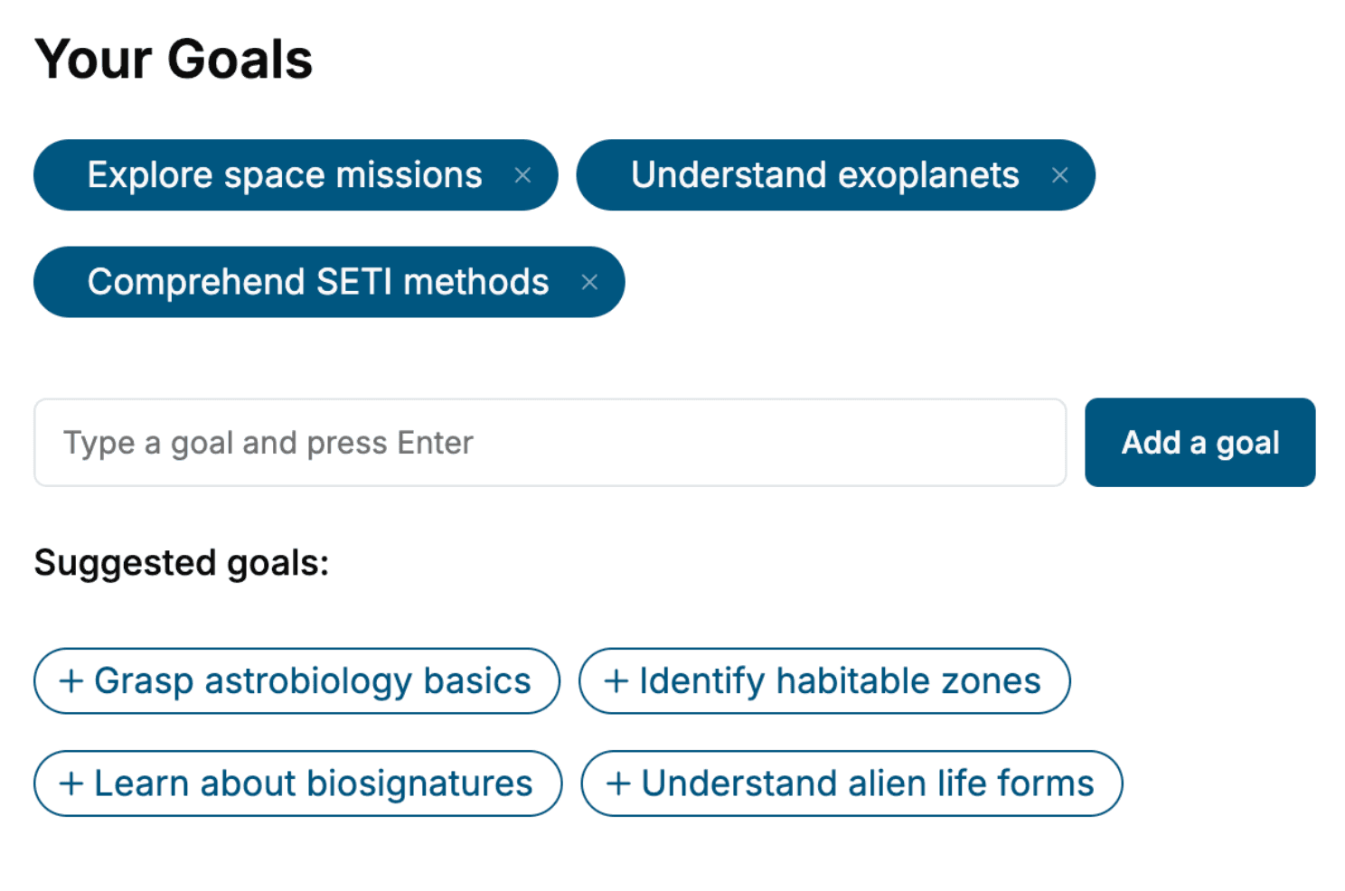
Tell us your goals and background, and get your own version of this book, tailored by AI to you.


Benefits of AI-tailored books
- Read one book, not ten:
- all the Anatomy knowledge you need, gathered into a single book.
- Save days of learning:
- tell us your goals, choose your topics, and the whole book is built around them.
- Learn effortlessly:
- Anatomy book written for your specific background and expertise.
- Understand at a glance:
- includes charts, tables, timelines, checklists, decision trees, and more.
- Stay current:
- written the day you order it, not years ago.
- Trust what you read:
- every book is checked for accuracy, with real research when your subject needs it.
Create your unique book in 3 steps
- 1. Select your focus
Select the focus of your Anatomy book and share your background
- 2. Personalize your book
Specify your goals and choose sub-topics to include
- 3. Get your tailored book
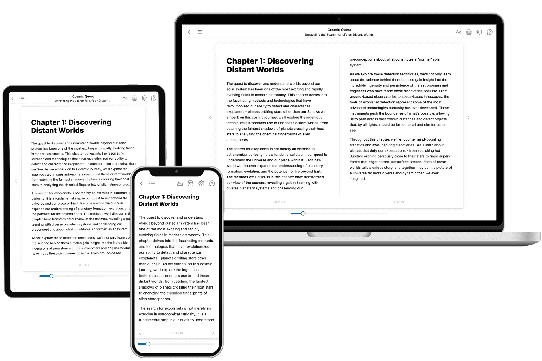
Your book is ready in 10 minutes. Read it online, download as EPUB or PDF, or send to Kindle.

Frequently asked questions
- What is TailoredRead?
TailoredRead creates complete, full-length nonfiction ebooks, written specifically for you. Tell us what you want to learn and why, and TailoredRead writes you a complete ebook on it, with charts, tables, and diagrams throughout. Read it online, on Kindle, or on any e-reader, or download it as a PDF or EPUB. Every book is written fresh the day you order it.
- How is the book tailored to me?
Before anything is written, you answer a few short questions about your goals, your background, and the topics you care about. Your book is then planned around your answers: written at your level, focused on your goals, and covering the topics you chose, instead of the one-size-fits-all book a store sells everyone.
- Will it read like generic AI content?
No. Your book isn't a chat transcript: it's planned chapter by chapter before a single word is written, told in a clear book voice, and explains ideas visually with charts, tables, and diagrams. After writing, every book goes through an editorial pass that polishes chapters so the book reads consistent from start to finish. Judge for yourself: browse thousands of sample books on our Explore Books page.
- How accurate is the content?
Our AI first checks whether your subject needs live research. Niche and fast-moving subjects are researched before writing, and the book plan and chapters are grounded in what it finds. After writing, every book goes through a quality pass that checks its facts and consistency, and researched books list their sources in a Notes section at the back.
- How long is the book?
You choose the length: Comprehensive (250-300 pages), Detailed (150-200 pages), Essential (70-100 pages), or Short (30-50 pages).
- Does the book include visuals?
Yes. Every book includes charts, tables, and diagrams: timelines, checklists, step-by-step guides, decision trees, comparison tables, matrices, and bar charts. A typical book includes dozens of these visual elements.
- Can I read it on Kindle? What formats can I download?
Yes. Every book can be read in our online reader or downloaded as a PDF or EPUB. The EPUB can be opened with Kindle, Apple Books, Kobo, and any e-reader. The Commercial and Author licenses also include an editable Word (DOCX) and Markdown download.
- How much does it cost?
About the price of a regular ebook. The exact price depends on your book's length and complexity, from $2 for the shortest books to $35 for the most comprehensive, and it's shown when you review your book, before you commit to anything. Each book is a one-time purchase, yours to keep, no subscription. Licenses that let you share, teach, or publish your book range from $35.99 to $49.99.
- Can I preview the book before purchasing?
Before you pay, you'll see what your book will cover: the title, a detailed description, the estimated length, and the complete chapter-by-chapter table of contents, so you know what you're getting before you decide. The full text itself is written after you purchase.
- How long does it take to create a book?
About 10 minutes. You can start reading the opening chapters while the rest is still being written.