Scanning the Quantum World
A Practical Guide to Tunneling Microscopy and Atomic-Scale Imaging
Imagine being able to see individual atoms on a material's surface—to visualize the precise arrangement of matter at scales a million times smaller than the width of a human hair.
207 pages9 chaptersAug 21, 2026Microscopy
About this book
Tunneling microscopy makes this possible, revealing the atomic architecture that determines material properties. This book demystifies the quantum mechanics and engineering behind this transformative technology, guiding you from fundamental principles through practical implementation. Whether you're preparing for laboratory work, deepening your understanding of condensed matter physics, or exploring how scientists map the nanoscale world, you'll discover how tunneling microscopy works, what it can reveal, and how to interpret the remarkable images it produces. With clear explanations, real-world examples, and practical insights, this guide equips you to engage confidently with one of modern physics' most powerful investigative tools.
Table of contents
- IntroductionWhy Atomic-Scale Imaging Matters, A Brief History of Tunneling Microscopy, What You'll Learn in This BookRead below
- Chapter 1: Quantum Tunneling: The FoundationClassical Barriers and Quantum Possibilities, Understanding Tunneling Probability, Work Function and Electron Escape, From Tunneling to Measurable Current
- Chapter 2: Scanning Tunneling Microscopy FundamentalsHow the STM Works, The Tip-Sample Interaction, Feedback Control and Constant-Current Imaging, Achieving Atomic Resolution
- Chapter 3: Atomic Force Microscopy and BeyondAFM: Measuring Forces Instead of Currents, Contact and Non-Contact Imaging Modes, When to Use AFM Over STM, Combining STM and AFM Capabilities
- Chapter 4: Building and Optimizing Your MicroscopeEssential Hardware Components, Vibration Isolation and Environmental Control, Electronics and Control Systems, Preparing and Characterizing Probe Tips
- Chapter 5: Spectroscopy and Energy-Resolved ImagingScanning Tunneling Spectroscopy (STS), Mapping Electronic Density of States, Spatially-Resolved Energy Information, Advanced Spectroscopic Techniques
- Chapter 6: Reading and Interpreting ImagesTopography Versus Electronic Structure, Common Artifacts and How to Recognize Them, Tip Convolution and Image Deconvolution, Quantitative Measurements from Images
- Chapter 7: Real-World Applications and Case StudiesSurface Chemistry and Catalysis, Superconductivity and Electronic Properties, Nanomaterials and Nanostructures, Biological and Organic Molecules
- Chapter 8: Troubleshooting and OptimizationDiagnosing Common Problems, Strategies for Improving Image Quality, Sample Preparation Best Practices, Maintenance and Calibration Procedures
- Chapter 9: Emerging Techniques and Future HorizonsHigh-Speed and Time-Resolved Imaging, Multimodal and Correlative Microscopy, Machine Learning and Automated Analysis, Next-Generation Microscopy Technologies
- ConclusionKey Takeaways, Your Next Steps in Tunneling Microscopy
- Glossary of Terms
- Further Reading and Resources
Introduction
A single tungsten needle, sharpened to a point that ends in one atom, hovers less than a nanometer above a crystal surface. No lens sits between the needle and the sample. No light illuminates the scene. Yet within minutes, a computer screen fills with a map so precise that individual atoms appear as neat rows of bumps, each one placed exactly where quantum mechanics says it should be. This is the working reality of a scanning tunneling microscope, an instrument that turned an abstract feature of quantum theory into a everyday laboratory tool.
The gap between the needle and the surface in that scene is not empty in the way a gap between two macroscopic objects would be. Electrons cross it constantly, not by jumping over a barrier but by passing through one that classical physics says they cannot cross at all. That crossing, called quantum tunneling, is the entire basis of the technology this book explores. Understanding it does not require abandoning intuition so much as replacing one intuition with another, one built on wave functions rather than solid billiard balls.
This book exists because tunneling microscopy sits at an unusual intersection. It is simultaneously a piece of applied quantum mechanics, a precision engineering challenge, and a discovery engine for chemistry, materials science, and biology. Few technologies ask a reader to hold all three of these perspectives at once. A physicist trained mostly in theory may find the mechanical engineering unfamiliar. An engineer comfortable with feedback loops and piezoelectric actuators may find the quantum mechanics intimidating. This book is written to bridge that gap, moving between the physics and the hardware without assuming expertise in either beyond a foundation in condensed matter physics.
The chapters ahead build in a deliberate order. The quantum mechanical basis of tunneling comes first, because every other concept in the book rests on it. From there, the discussion moves to the instrument itself, how tips, scanners, and feedback loops turn a tunneling current into an image, and then to atomic force microscopy, spectroscopy, image interpretation, real-world applications, troubleshooting, and finally the frontier of the field today. The three sections that follow in this introduction set the stage: why this kind of imaging matters, how it came to exist, and what to expect from the chapters ahead.
Why Atomic-Scale Imaging Matters
Materials behave the way they do because of arrangements invisible to the naked eye and invisible even to most microscopes. A catalyst that speeds up a chemical reaction does so because of the exact positions of atoms at its surface, not because of its overall shape or color. A superconductor loses electrical resistance because of pairing behavior among electrons that plays out at distances measured in fractions of a nanometer. Without a way to see these arrangements directly, scientists were left inferring atomic structure indirectly, through diffraction patterns, spectroscopic averages, and theoretical models that fit the data reasonably well but never showed the atoms themselves.
Consider the difference between knowing the average composition of an alloy and knowing exactly where each type of atom sits on its surface. Bulk techniques such as X-ray diffraction reveal the repeating unit cell of a crystal with excellent precision, but they average over billions of atoms and cannot show a single defect, step edge, or impurity atom in isolation. Tunneling microscopy fills that gap. It can locate one missing atom in an otherwise perfect lattice, watch a single molecule diffuse across a surface, or catch a chemical reaction happening one bond at a time.
The practical stakes of this capability are large. Modern semiconductor devices, for example, depend on interfaces just a few atomic layers thick, where a single misplaced atom can change electrical performance measurably. Battery researchers need to understand how ions move across electrode surfaces at the atomic scale to design longer lasting, safer batteries. Drug developers studying how molecules bind to protein surfaces benefit from seeing binding sites directly rather than inferring them from crystallographic averages. In each case, atomic-scale imaging turns a hypothesis about structure into a direct observation.
History offers a useful comparison. Optical microscopy let biologists see cells for the first time in the seventeenth century, transforming biology from a science of whole organisms into a science of cellular structure. Electron microscopy pushed resolution down to the nanometer scale in the twentieth century, revealing viruses and macromolecular machines. Tunneling microscopy represents a comparable leap, pushing resolution to the scale of individual atoms and their electronic states. Each leap did not just add detail to existing pictures. It opened entirely new categories of questions that scientists had not been able to ask before, because the objects of study had simply been invisible.
The kind of information available at the atomic scale also differs qualitatively from what larger-scale imaging provides. A microscope image of a surface at the nanometer scale might show a smooth terrace. The same terrace imaged atom by atom might reveal subtle buckling, rows of atoms displaced from ideal lattice positions, or a scattering of adsorbed molecules invisible at lower resolution. These fine details often explain macroscopic behavior that would otherwise remain mysterious, such as why one crystal facet catalyzes a reaction efficiently while another facet, chemically identical in composition, does almost nothing at all.
Atomic-scale imaging also changes how scientists test theoretical predictions. Condensed matter theory generates detailed predictions about electronic structure, magnetic ordering, and defect behavior, but many of these predictions historically could only be tested indirectly through bulk measurements averaged over huge numbers of atoms. With a scanning tunneling microscope, a theorist's prediction about the local electronic structure near a single defect can be checked directly, atom by atom. This tightens the feedback loop between theory and experiment considerably, allowing faster refinement of models describing how electrons behave in real materials.
None of this diminishes the value of bulk characterization techniques, which remain essential for understanding average properties and statistical behavior across large samples. Atomic-scale imaging complements rather than replaces these methods. A materials scientist studying a new alloy might use X-ray diffraction to confirm its crystal structure, then use tunneling microscopy to examine specific surface features that diffraction cannot resolve. The two approaches, one broad and statistical, the other narrow and direct, together give a fuller picture than either could provide alone.
A Brief History of Tunneling Microscopy
The theoretical seeds of tunneling microscopy were planted decades before the instrument existed. Quantum tunneling itself was described mathematically in the late 1920s, as physicists worked out how particles governed by wave functions could have a nonzero probability of appearing on the far side of an energy barrier they classically could not cross[1]. This early work focused on nuclear physics, explaining phenomena such as radioactive decay, and had no obvious connection to surface imaging. Decades passed before anyone recognized that the same tunneling effect could be harnessed to probe surfaces with atomic precision.
The breakthrough came at IBM's research laboratory in Zurich, where Gerd Binnig and Heinrich Rohrer built the first working scanning tunneling microscope in 1981. Their instrument measured the tiny tunneling current flowing between a sharp metal tip and a conductive sample as the tip scanned across the surface, converting variations in that current into a topographic map[2]. Early skepticism within the physics community was considerable, since the tolerances required, positioning a tip with sub-angstrom precision while suppressing vibration, seemed almost unachievable with the technology available at the time.
The moment that silenced most of the skeptics arrived when Binnig and Rohrer's team imaged the surface reconstruction of silicon, a long-standing puzzle in surface science. Silicon atoms at a clean surface rearrange themselves into a complex repeating pattern to minimize energy, a structure that had been debated for years based on indirect diffraction evidence alone. The scanning tunneling microscope image showed the arrangement directly, resolving the debate in a single picture[3]. That single result convinced much of the field that atomic-scale, real-space imaging was not just possible but reliable.
Recognition followed quickly by the standards of scientific prizes. Binnig and Rohrer received the Nobel Prize in Physics in 1986, only five years after their first working instrument, an unusually short interval between discovery and recognition. The same year's prize also honored Ernst Ruska for his earlier work on electron microscopy, a pairing that highlighted how the two techniques, one based on electrons focused by lenses and the other on electrons tunneling through a vacuum gap, had each transformed the limits of visible detail in complementary ways.
The scanning tunneling microscope's central limitation, that it could only image electrically conductive samples, motivated the next major advance. Binnig, together with Christoph Gerber and Calvin Quate, developed the atomic force microscope in 1986, replacing the tunneling current with a measurement of mechanical force between a tip and a sample[4]. This innovation opened atomic-scale imaging to insulators, polymers, and biological samples, categories of material entirely closed off from the original tunneling technique.
Through the 1990s and 2000s, the field diversified rapidly beyond simple topographic imaging. Researchers developed scanning tunneling spectroscopy to extract electronic structure information at each point on a surface, added low-temperature and ultra-high vacuum capabilities to stabilize measurements against thermal noise, and pushed resolution far enough to resolve not just individual atoms but the internal chemical bonds within single molecules. Each advance expanded the range of scientific questions the technique could answer, moving it from a novelty capable of producing striking images to a standard tool found in surface science and condensed matter laboratories worldwide.
The present state of the field reflects four decades of incremental refinement layered onto that original 1981 design. Modern instruments operate at temperatures near absolute zero, inside chambers isolated from vibration by multiple independent systems, and often paired with complementary techniques such as optical spectroscopy or transport measurements performed on the very same sample. The core principle, a sharp tip sensing a surface through quantum tunneling or atomic forces, remains unchanged. What has changed is the precision, stability, and range of information that principle can now deliver.
What You'll Learn in This Book
This book moves through the subject in a sequence designed to build understanding layer by layer, starting with the physics that makes tunneling microscopy possible and ending with the frontier techniques extending it today. The early chapters establish the quantum mechanical groundwork, including tunneling probability, the role of the work function in determining how easily electrons escape a material, and the WKB approximation used to estimate tunneling rates across realistic barriers. These concepts are not presented as abstract theory disconnected from the instrument. Each one connects directly to a measurable quantity that appears on a screen during an actual experiment.
From the quantum foundations, the discussion turns to the scanning tunneling microscope itself: how a piezoelectric scanner positions a tip with sub-angstrom precision, how a feedback loop maintains a constant tip-sample gap while scanning, and how these engineering choices translate directly into atomic resolution. A separate chapter covers atomic force microscopy, explaining how measuring force rather than current opens the technique to insulating and biological samples, and detailing the tradeoffs between contact mode, non-contact mode, and tapping mode. Readers who need to choose between these two families of instruments for a specific sample will find practical guidance rather than abstract comparison.
A substantial portion of the book addresses the practical work of building, operating, and troubleshooting a working microscope. This includes the hardware components required, from vibration isolation platforms to ultra-high vacuum chambers, the electronics behind PID controllers and lock-in amplifier techniques, and the surprisingly delicate art of tip etching and characterization. These chapters are written for readers who may eventually operate or maintain an instrument themselves, not just interpret images produced by someone else. Specific failure modes such as tip crashes, feedback oscillation, and image streaking receive dedicated troubleshooting guidance, since these problems account for a large share of wasted laboratory time in practice.
Spectroscopy and image interpretation form another major thread running through the second half of the book. Scanning tunneling spectroscopy extends the basic imaging technique to extract dI/dV spectra that reveal the local density of states at specific points on a surface, information that goes beyond simple topography to describe the electronic character of a material. Interpreting the resulting images correctly requires distinguishing topographic contrast from electronic contrast, recognizing tip convolution artifacts, and understanding when apparent height on an image reflects real atomic position versus a purely electronic effect. These distinctions matter enormously, since misreading an image can lead to incorrect conclusions about a material's actual structure.
The applications chapter grounds all of this theory and instrumentation in concrete scientific achievements, covering how tunneling microscopy has advanced understanding of surface catalysis, superconductivity, nanomaterials, and biological molecules. Real examples appear throughout, including how quasiparticle interference patterns reveal electronic structure in unconventional superconductors, and how the Kondo resonance and charge density wave phenomena have been mapped atom by atom in various materials. These case studies serve a dual purpose, illustrating both what the technique can achieve scientifically and how the principles from earlier chapters apply in genuine research settings.
The closing chapters look toward where the field is heading, covering high-speed and time-resolved scanning probe microscopy, machine learning image analysis for automating pattern recognition in large datasets, and millikelvin STM systems that push measurements toward the coldest temperatures achievable in a laboratory. These emerging capabilities are extending the reach of tunneling microscopy well past its original scope, into territory that was not imaginable when Binnig and Rohrer built their first instrument. Readers finishing the book should come away with enough grounding in both the quantum mechanics and the instrumentation to read current research papers critically, and with enough practical knowledge to begin planning their own experiments or troubleshooting an instrument already in use.
Throughout the book, technical explanation is paired consistently with concrete examples rather than left as pure abstraction. A formula for tunneling probability appears alongside a worked calculation showing how sensitive that probability is to tip-sample distance, explaining why a change of just one angstrom can shift measured current by an order of magnitude. A description of feedback loop design appears alongside an explanation of what happens when that loop is tuned incorrectly, producing the ringing artifacts that plague many first attempts at STM imaging. This pairing of principle and practice reflects the conviction that tunneling microscopy is best learned not as a set of equations to memorize, but as a discipline where physics, engineering, and careful observation combine to reveal a world too small to see any other way.
Notes
Introduction
- 1. George Gamow, "Zur Quantentheorie des Atomkernes," Zeitschrift fur Physik, 1928
- 2. Gerd Binnig and Heinrich Rohrer, "Scanning Tunneling Microscopy," Helvetica Physica Acta, 1982
- 3. Gerd Binnig and Heinrich Rohrer, "7 times 7 Reconstruction on Si 111 Resolved in Real Space," Physical Review Letters, 1983
- 4. Gerd Binnig and others, "Atomic Force Microscope," Physical Review Letters, 1986
This is the end of the free sample. The full book has 9 chapters.
Want this book written for you?
Tell us your goals and background, and get your own version of this book, tailored by AI to you.


Benefits of AI-tailored books
- Read one book, not ten:
- all the Microscopy knowledge you need, gathered into a single book.
- Save days of learning:
- tell us your goals, choose your topics, and the whole book is built around them.
- Learn effortlessly:
- Microscopy book written for your specific background and expertise.
- Understand at a glance:
- includes charts, tables, timelines, checklists, decision trees, and more.
- Stay current:
- written the day you order it, not years ago.
- Trust what you read:
- every book is checked for accuracy, with real research when your subject needs it.
Create your unique book in 3 steps
- 1. Select your focus
Select the focus of your Microscopy book and share your background
- 2. Personalize your book
Specify your goals and choose sub-topics to include
- 3. Get your tailored book
Your book is ready in 10 minutes. Read it online, download as EPUB or PDF, or send to Kindle.


Frequently asked questions
- What is TailoredRead?
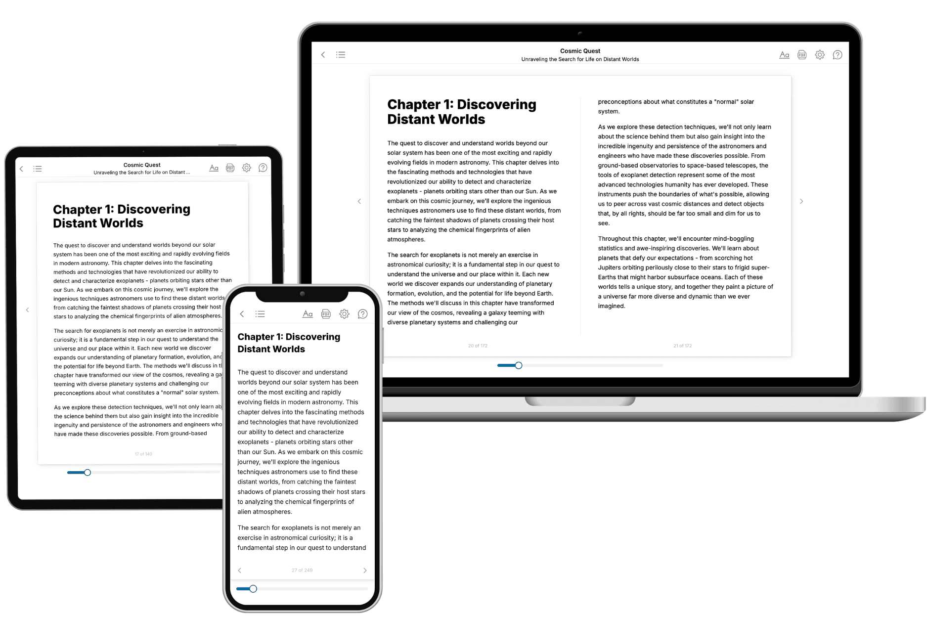
TailoredRead creates complete, full-length nonfiction ebooks, written specifically for you. Tell us what you want to learn and why, and TailoredRead writes you a complete ebook on it, with charts, tables, and diagrams throughout. Read it online, on Kindle, or on any e-reader, or download it as a PDF or EPUB. Every book is written fresh the day you order it.
- How is the book tailored to me?
Before anything is written, you answer a few short questions about your goals, your background, and the topics you care about. Your book is then planned around your answers: written at your level, focused on your goals, and covering the topics you chose, instead of the one-size-fits-all book a store sells everyone.
- Will it read like generic AI content?
No. Your book isn't a chat transcript: it's planned chapter by chapter before a single word is written, told in a clear book voice, and explains ideas visually with charts, tables, and diagrams. After writing, every book goes through an editorial pass that polishes chapters so the book reads consistent from start to finish. Judge for yourself: browse thousands of sample books on our Explore Books page.
- How accurate is the content?
Our AI first checks whether your subject needs live research. Niche and fast-moving subjects are researched before writing, and the book plan and chapters are grounded in what it finds. After writing, every book goes through a quality pass that checks its facts and consistency, and researched books list their sources in a Notes section at the back.
- How long is the book?
You choose the length: Comprehensive (250-300 pages), Detailed (150-200 pages), Essential (70-100 pages), or Short (30-50 pages).
- Does the book include visuals?
Yes. Every book includes charts, tables, and diagrams: timelines, checklists, step-by-step guides, decision trees, comparison tables, matrices, and bar charts. A typical book includes dozens of these visual elements.
- Can I read it on Kindle? What formats can I download?
Yes. Every book can be read in our online reader or downloaded as a PDF or EPUB. The EPUB can be opened with Kindle, Apple Books, Kobo, and any e-reader. The Commercial and Author licenses also include an editable Word (DOCX) and Markdown download.
- How much does it cost?
About the price of a regular ebook. The exact price depends on your book's length and complexity, from $2 for the shortest books to $35 for the most comprehensive, and it's shown when you review your book, before you commit to anything. Each book is a one-time purchase, yours to keep, no subscription. Licenses that let you share, teach, or publish your book range from $35.99 to $49.99.
- Can I preview the book before purchasing?
Before you pay, you'll see what your book will cover: the title, a detailed description, the estimated length, and the complete chapter-by-chapter table of contents, so you know what you're getting before you decide. The full text itself is written after you purchase.
- How long does it take to create a book?
About 10 minutes. You can start reading the opening chapters while the rest is still being written.