The Skin Anatomy Coloring Book
Learn the Structure and Function of Your Body's Largest Organ Through Interactive Illustrations
Imagine being able to see exactly how your skin protects you, regulates your temperature, and senses the world around you.
222 pages10 chaptersNov 24, 2025Human Anatomy
About this book
This coloring book transforms the complex architecture of skin anatomy into an interactive learning experience. By combining detailed, scientifically accurate illustrations with clear explanations, you'll discover how the epidermis, dermis, and hypodermis work together as an integrated system. As you color each structure—from hair follicles to sweat glands to sensory receptors—you'll develop a deeper understanding of how your skin functions and why it matters. Whether you're a student, healthcare professional, or simply curious about your body, this hands-on approach makes anatomy memorable and engaging. Each illustration is designed to highlight specific structures and their relationships, while accompanying text explains their roles and significance. The coloring process itself enhances learning by engaging your visual and motor skills simultaneously, creating stronger neural connections and lasting knowledge.
Table of contents
- IntroductionWhy Understanding Skin Anatomy Matters, How to Use This Coloring Book, The Science Behind Learning Through Coloring, What You'll Discover in These PagesRead below
- Chapter 1: The Big Picture: Skin as a SystemThe Five Critical Functions of Skin, Skin by the Numbers: Size, Weight, and Renewal, Overview of the Three Main Layers
- Chapter 2: The Epidermis: Your Protective BarrierThe Five Layers of the Epidermis, Keratinocytes, Melanocytes, and Langerhans Cells, The 28-Day Renewal Cycle, How Melanin Protects and Colors Your Skin
- Chapter 3: The Dermis: Support and SensationPapillary and Reticular Layers, Collagen and Elastin: The Skin's Framework, Blood Vessels and Nutrient Delivery, Touch, Temperature, and Pain Receptors
- Chapter 4: The Hypodermis: Insulation and StorageAdipose Tissue and Fat Storage, How the Hypodermis Regulates Temperature, Connective Tissue and Structural Support
- Chapter 5: Hair Follicles: Growth and StructureThe Complete Anatomy of a Hair Follicle, The Hair Growth Cycle: Anagen, Catagen, and Telogen, Sebaceous Glands and Oil Production, The Arrector Pili Muscle and Goosebumps
- Chapter 6: Sweat Glands: Cooling and CleansingEccrine Sweat Glands and Thermoregulation, Apocrine Sweat Glands and Body Odor, What Sweat Contains and Why It Matters
- Chapter 7: The Dermal-Epidermal Junction: Where Layers MeetThe Architecture of the Basement Membrane, How the Junction Holds Layers Together, What Happens When the Junction Is Damaged
- Chapter 8: Skin Variations: Across the Body and Between PeopleHow Skin Differs on Different Body Parts, Genetic Influences on Skin Type and Color, How Skin Changes Throughout Life, Understanding Skin Diversity Across Populations
- Chapter 9: Common Skin Conditions: Understanding the Anatomy Behind ThemAcne: When Follicles and Oil Production Go Wrong, Eczema and Psoriasis: Epidermal Dysfunction, How Skin Heals: The Anatomy of Repair
- Chapter 10: Protecting Your Skin: Applying Anatomy to Daily CareUV Damage and Why Sunscreen Matters, Maintaining the Skin Barrier and Hydration, How Common Skincare Ingredients Work at the Anatomical Level, Understanding Aging and Preventive Care
- Conclusion: Your Skin, UnderstoodKey Takeaways from Your Learning Journey, Next Steps in Your Anatomical Education
- Glossary of Skin Anatomy Terms
- Further Reading and Resources
Introduction
Your skin is far more than a simple covering for your body. It's a complex, dynamic organ that performs dozens of critical functions every second of every day. Weighing approximately eight pounds and covering about twenty square feet on an average adult, your skin is the largest organ in your body. Yet despite its constant presence and vital importance, most people know surprisingly little about how it actually works. This coloring book changes that by transforming abstract anatomical concepts into tangible, visual knowledge that you can literally hold in your hands and bring to life with color.
The approach taken in this book is fundamentally different from passive reading or memorization. When you color an anatomical illustration, you engage multiple regions of your brain simultaneously. Your visual cortex processes the shapes and spatial relationships. Your motor cortex controls the precise movements of coloring. Your prefrontal cortex makes decisions about color choices and follows instructions. This multi-sensory engagement creates stronger neural pathways and more durable memories than simply reading text or looking at pre-colored diagrams. Research in educational psychology consistently demonstrates that active learning techniques produce better retention and deeper understanding than passive methods[1].
Throughout these pages, you'll discover the intricate architecture of your skin's three main layers, the specialized structures embedded within them, and the remarkable ways they work together as an integrated system. Each illustration has been carefully designed to highlight specific anatomical features while showing their relationships to surrounding structures. The accompanying text explains not just what each structure is, but why it matters and how it functions in your daily life. By the time you complete this book, you'll have developed a comprehensive understanding of skin anatomy that will serve you whether you're a student, healthcare professional, artist, or simply someone who wants to understand their body better.
This introduction will guide you through the essential information you need to get the most from this learning experience. You'll discover why understanding skin anatomy matters in practical terms, how to use this book effectively, the scientific evidence supporting coloring as a learning tool, and a preview of the fascinating structures and systems you'll explore in the chapters ahead. Think of this introduction as your roadmap to a journey through one of the most remarkable organs in the human body—an organ you carry with you everywhere, yet may never have truly seen until now.
Why Understanding Skin Anatomy Matters
Understanding skin anatomy provides practical benefits that extend far beyond academic knowledge. When you know how your skin is structured, you can make informed decisions about skincare products, understand why certain treatments work, and recognize when something might be going wrong. For example, knowing that the epidermis contains no blood vessels helps you understand why superficial cuts don't bleed, while deeper cuts that penetrate the dermis do. This knowledge transforms your relationship with your body from passive acceptance to active understanding.
Healthcare professionals rely on detailed anatomical knowledge to diagnose and treat skin conditions effectively. A dermatologist examining a rash needs to determine which layer of skin is affected to prescribe the right treatment. Topical medications that work for epidermal conditions may be ineffective for dermal problems, and vice versa. Nurses administering injections must understand the depth and structure of skin layers to choose the correct needle length and injection angle. Physical therapists working with burn patients need to assess damage depth by understanding which structures have been affected. This anatomical foundation is essential for competent clinical practice.
Artists and aestheticians benefit from understanding skin anatomy in different but equally important ways. Portrait artists who understand the underlying structure of skin can render it more realistically, capturing the subtle variations in texture, color, and light reflection that make skin look alive rather than flat. Makeup artists who know where oil glands are concentrated can better address shine and choose appropriate products. Aestheticians performing treatments like microdermabrasion or chemical peels must understand exactly which layers they're affecting to achieve desired results without causing damage. This anatomical knowledge elevates technical skill into true expertise.
Your understanding of skin anatomy also empowers you to evaluate health information critically. The skincare industry generates billions of dollars annually, with countless products making claims about penetrating deep into your skin or targeting specific structures. When you understand that the epidermis is only about 0.1 millimeters thick on your eyelids but up to 1.5 millimeters thick on your palms, you can better evaluate whether a product's claims are realistic. When you know that collagen and elastin are located in the dermis, you can assess whether a topical cream can truly deliver these molecules where they're needed, or whether such claims are primarily marketing.
Understanding skin anatomy also helps you recognize potential problems early. When you know that melanocytes are normally distributed evenly throughout the basal layer of the epidermis, you can better appreciate why a new, irregularly shaped, or rapidly changing mole might warrant medical attention. When you understand that the dermal-epidermal junction is where blisters form, you can better assess the severity of a burn or friction injury. This knowledge doesn't replace professional medical advice, but it does help you make informed decisions about when to seek care and what questions to ask.
The practical applications extend to everyday situations as well. Understanding how sweat glands function helps you choose appropriate antiperspirants or deodorants. Knowing that hair follicles extend deep into the dermis explains why shaving only removes the visible shaft while waxing or plucking removes more of the follicle, leading to longer-lasting results. Recognizing that your skin's barrier function depends on the outermost layer of the epidermis helps you understand why harsh soaps that strip away natural oils can lead to dryness and irritation. These insights transform routine decisions into informed choices based on anatomical reality.
Finally, understanding your skin's anatomy fosters appreciation for the remarkable complexity of your body. Your skin isn't just a passive barrier—it's an active organ that constantly regenerates itself, fights off pathogens, regulates temperature, synthesizes vitamin D, and provides sensory information about your environment. Every square inch contains thousands of cells, dozens of nerve endings, multiple glands, and an intricate network of blood vessels. When you understand this complexity, you develop a deeper respect for your body and a stronger motivation to care for it properly. This shift from ignorance to understanding is one of the most valuable outcomes of studying anatomy.
How to Use This Coloring Book
This coloring book is designed to be used systematically rather than randomly. While you can certainly jump to sections that interest you most, working through the chapters in order provides the most coherent learning experience. The book progresses from broad overview to specific details, with each chapter building on concepts introduced in previous ones. Start with Chapter 1 to establish a foundation of how skin functions as a system, then move through the subsequent chapters that explore each layer and structure in detail. This sequential approach mirrors how anatomy is typically taught in educational settings, moving from general to specific.
Before you begin coloring each illustration, read the accompanying text carefully. The text provides context that makes the coloring process more meaningful and educational. Understanding what you're coloring and why it matters transforms the activity from simple artistic expression into active learning. Pay particular attention to the relationships between structures—how they connect, where they're located relative to each other, and how they interact functionally. These relationships are often more important than memorizing individual structures in isolation.
When selecting colors, use the suggested color schemes provided for each illustration, as these are chosen to maximize clarity and distinguish between different structures. However, don't feel rigidly bound by these suggestions if you find that different colors work better for your learning style. The key is to use consistent colors for similar structures throughout the book. For example, if you color all blood vessels red in one illustration, continue using red for blood vessels in subsequent illustrations. This consistency helps your brain build associations and recognize patterns across different anatomical contexts.
Consider these specific coloring strategies to enhance your learning:
- Layer by layer: Color all structures in one layer before moving to the next, which helps you understand the organization within each layer
- Structure by structure: Color the same type of structure throughout the entire illustration (all nerves, then all blood vessels, etc.), which emphasizes functional systems
- Light to dark: Start with lighter colors and progress to darker ones, which prevents darker colors from bleeding into lighter areas
- Outline first: Trace the outlines of structures before filling them in, which improves accuracy and helps you focus on boundaries
Take your time with each illustration. Rushing through the coloring process defeats the purpose of this learning method. The act of carefully coloring each structure forces you to observe it closely, notice its shape and boundaries, and consider its position relative to other structures. This focused attention is what creates strong memories. Research suggests that spending 15-20 minutes on a single detailed illustration produces better learning outcomes than quickly coloring multiple illustrations[2]. Quality of engagement matters more than quantity of pages completed.
After completing each illustration, test your understanding by covering the labels and trying to identify structures from memory. This retrieval practice is one of the most effective learning techniques available. If you can't remember a structure's name or function, that's valuable feedback indicating you need to review that material. Return to the text, reread the relevant section, and examine the colored illustration again. This cycle of coloring, testing, and reviewing creates robust learning that persists long after you close the book.
Consider keeping a separate notebook alongside this coloring book where you can write additional notes, questions, or observations. When you encounter something particularly interesting or confusing, jot it down. If you notice a pattern or connection that isn't explicitly stated in the text, record it. These personal annotations create additional memory cues and demonstrate active engagement with the material. You might also use this notebook to sketch simplified versions of complex structures from memory, which provides another form of retrieval practice and helps you identify gaps in your understanding.
Finally, don't hesitate to revisit completed sections. Anatomy learning is cumulative, and concepts that seemed confusing initially often become clear after you've learned more advanced material. You might color an illustration of the epidermis early in the book, then return to it after learning about the dermis and suddenly notice details or relationships you missed the first time. This iterative approach, where you repeatedly engage with material at increasing levels of sophistication, is how experts develop their deep understanding of complex subjects.
The Science Behind Learning Through Coloring
The effectiveness of coloring as a learning tool is supported by substantial research in cognitive psychology and neuroscience. When you color an anatomical illustration, you engage in what educators call "active learning"—a process where you manipulate information rather than passively receiving it. Active learning consistently produces better outcomes than passive learning across virtually all subjects and age groups. The physical act of coloring requires you to make decisions, focus attention, and coordinate visual and motor systems, all of which strengthen the neural pathways associated with the information you're learning.
Coloring activates multiple memory systems simultaneously, creating what psychologists call "elaborative encoding." When you color a hair follicle, you're not just reading the word "hair follicle" or looking at a picture of one. You're tracing its shape with your hand, observing its boundaries, noting its position relative to other structures, choosing an appropriate color, and carefully filling in the space it occupies. Each of these actions creates a different type of memory trace in your brain. Later, when you try to recall information about hair follicles, you have multiple retrieval pathways available, making the memory more accessible and durable.
The visual-spatial processing involved in coloring is particularly valuable for learning anatomy. Anatomical knowledge is inherently spatial—it's about where structures are located, how they're shaped, and how they relate to their neighbors. When you color an illustration, you're building a mental map of these spatial relationships. Research using brain imaging shows that people who learn anatomy through hands-on activities like coloring develop stronger activation in the parietal cortex, a brain region critical for spatial reasoning, compared to those who learn through reading alone[3].
Coloring also promotes what psychologists call "desirable difficulty"—a level of challenge that's high enough to require effort but not so high that it becomes frustrating. When you color a complex illustration with many labeled structures, you must constantly refer back and forth between the labels and the structures, which requires sustained attention and working memory. This cognitive effort, while sometimes challenging, is precisely what drives learning. If learning were completely effortless, it would also be ineffective. The mild challenge of accurately coloring detailed anatomical structures hits the sweet spot of desirable difficulty.
The repetitive nature of coloring also contributes to learning through a process called "distributed practice." Rather than trying to memorize all the parts of skin anatomy in one intense study session, coloring spreads the learning over time. You might spend twenty minutes coloring the layers of the epidermis today, then return tomorrow to color the structures in the dermis. This spacing of practice sessions is one of the most robust findings in learning research—distributed practice produces dramatically better long-term retention than massed practice (cramming)[4].
Coloring also reduces cognitive load in helpful ways. When you're learning complex material, your working memory can become overwhelmed by trying to process too much information simultaneously. Coloring provides a structured way to break down complex anatomical illustrations into manageable pieces. As you color each structure individually, you can focus your attention on that specific element without being distracted by everything else in the illustration. This focused attention allows for deeper processing of each component before moving on to the next.
The emotional and motivational benefits of coloring shouldn't be overlooked either. Many people find coloring relaxing and enjoyable, which creates positive associations with the learning material. When learning is associated with positive emotions, the brain releases neurotransmitters like dopamine that enhance memory consolidation. Additionally, completing a colored illustration provides a sense of accomplishment and visible progress, which maintains motivation over time. Unlike reading a textbook where your progress is invisible, a coloring book provides tangible evidence of your learning journey—you can literally see how much you've learned by looking at your completed illustrations.
Finally, colored illustrations become personalized study materials that you can return to repeatedly. Because you created them yourself, they're more meaningful and memorable than pre-colored diagrams in a textbook. When you review your colored illustrations before an exam or when you need to recall anatomical information in a clinical setting, you're not just seeing the structures—you're remembering the experience of coloring them, which provides additional retrieval cues. This personal connection to the material is a powerful but often underestimated component of effective learning.
What You'll Discover in These Pages
Your journey through skin anatomy begins with the big picture in Chapter 1, where you'll explore how skin functions as an integrated system. You'll discover that your skin performs at least five critical functions simultaneously: protection from physical damage and pathogens, regulation of body temperature, sensation of environmental stimuli, synthesis of vitamin D, and excretion of waste products through sweat. Understanding these functions provides context for all the structural details you'll learn in subsequent chapters. You'll also encounter fascinating statistics about your skin—like the fact that you shed approximately 30,000 to 40,000 dead skin cells every minute, or that your skin completely renews itself approximately every 28 days.
Chapters 2 through 4 take you on a detailed tour through each of the three main layers of skin. In Chapter 2, you'll explore the epidermis, the outermost layer that you can see and touch. You'll discover that this seemingly simple surface is actually composed of five distinct sublayers, each with specialized cells performing specific functions. You'll learn about keratinocytes, the cells that make up about 90% of the epidermis and produce the tough protein keratin that makes your skin waterproof. You'll also meet melanocytes, the cells that produce melanin and determine your skin color, and Langerhans cells, which serve as sentinels of your immune system.
Chapter 3 reveals the dermis, the thick middle layer that gives skin its strength and elasticity. Here you'll discover the intricate network of collagen and elastin fibers that form your skin's structural framework. You'll explore the papillary layer with its fingerlike projections that increase surface area for nutrient exchange, and the reticular layer with its dense collagen bundles that resist tearing and stretching. You'll also learn about the rich blood vessel network that nourishes the skin and the diverse array of sensory receptors that allow you to feel everything from a gentle breeze to painful heat. These receptors include:
- Meissner's corpuscles for light touch and texture
- Pacinian corpuscles for deep pressure and vibration
- Ruffini endings for skin stretch and sustained pressure
- Free nerve endings for pain, temperature, and itch
Chapter 4 takes you to the deepest layer, the hypodermis, where you'll discover how adipose tissue does far more than simply store energy. You'll learn how this layer insulates your body, cushions underlying structures from impact, and serves as an endocrine organ that produces hormones affecting metabolism and appetite. You'll also explore how the hypodermis anchors skin to underlying muscles and bones through connective tissue septa, allowing skin to move somewhat independently while remaining securely attached.
Chapters 5 and 6 focus on the specialized structures embedded within the skin. Chapter 5 provides a comprehensive look at hair follicles, revealing them as complex mini-organs with their own blood supply, nerve endings, and associated structures. You'll learn about the three phases of the hair growth cycle and why hair on your scalp can grow much longer than hair on your arms. You'll discover how sebaceous glands produce sebum, the oily substance that waterproofs your skin and hair, and how the tiny arrector pili muscles create goosebumps when they contract. Chapter 6 explores sweat glands, distinguishing between eccrine glands that cover most of your body and produce watery sweat for cooling, and apocrine glands concentrated in your armpits and groin that produce the protein-rich sweat that bacteria metabolize to create body odor.
Chapter 7 examines the dermal-epidermal junction, the critical boundary where the epidermis and dermis meet. You'll discover that this isn't a simple flat interface but rather a complex, undulating structure with fingerlike projections that increase surface area and strengthen the bond between layers. You'll learn about the basement membrane, a specialized structure composed of proteins like collagen IV and laminin that anchors the epidermis to the dermis. Understanding this junction helps explain conditions like epidermolysis bullosa, where genetic defects in junction proteins cause the skin layers to separate easily, forming painful blisters.
Chapter 8 explores the remarkable variations in skin structure across different body regions and between different people. You'll discover why the skin on your eyelids is dramatically thinner than the skin on your palms, and how these regional differences reflect functional demands. You'll learn about the genetic factors that influence skin type, color, and texture, and how skin changes throughout the lifespan from infancy through old age. This chapter also addresses the diversity of skin across different ethnic populations, explaining the anatomical basis for variations in melanin production, skin thickness, and other characteristics.
Chapters 9 and 10 apply your anatomical knowledge to practical concerns. Chapter 9 examines common skin conditions through an anatomical lens, helping you understand why acne develops when hair follicles become clogged, how eczema and psoriasis involve dysfunction of the epidermal barrier, and what happens at the cellular level during wound healing. Chapter 10 connects anatomy to daily skincare, explaining how UV radiation damages DNA in skin cells, why maintaining the skin barrier is crucial for hydration, and how common skincare ingredients like retinoids, alpha-hydroxy acids, and antioxidants work at the anatomical level. You'll gain practical knowledge that helps you make informed decisions about protecting and caring for your skin based on scientific understanding rather than marketing claims.
Notes
Introduction
- 1. E. Dale, Audio-Visual Methods in Teaching, Holt, Rinehart & Winston, 1969
- 2. J. Noh and J. Lee, "Effects of coloring activities on cognitive function and mood in older adults," Journal of Aging Research, 2020
- 3. M. E. Estevez and others, "A novel three-dimensional tool for teaching human neuroanatomy," Anatomical Sciences Education, 2010
- 4. N. J. Cepeda and others, "Distributed practice in verbal recall tasks," Psychological Bulletin, 2006
This is the end of the free sample. The full book has 10 chapters.
Want this book written for you?
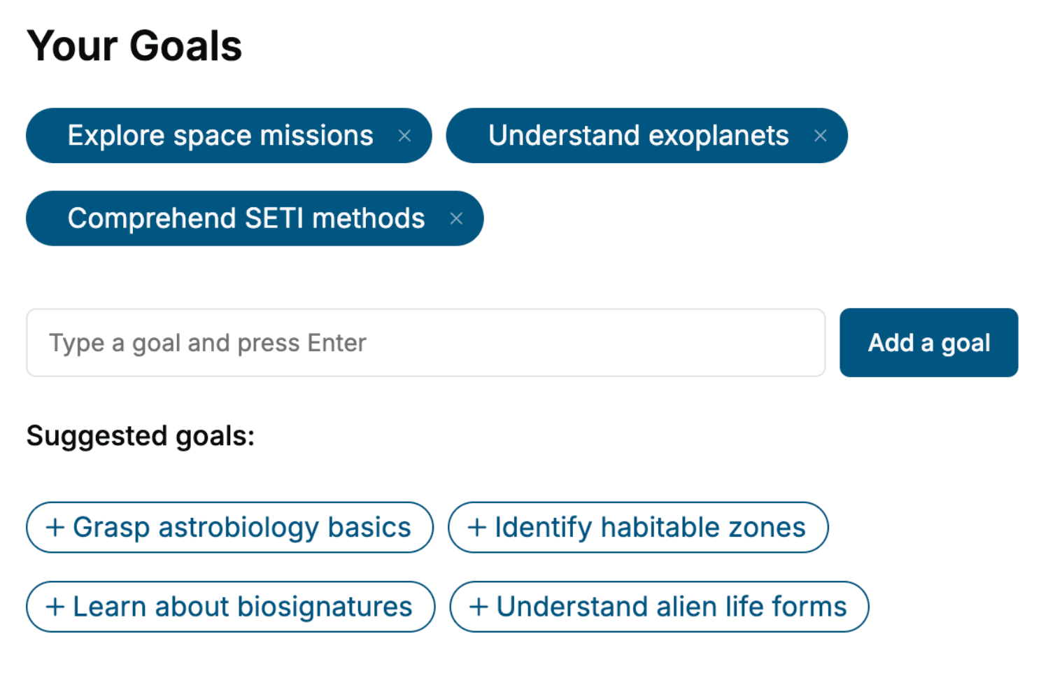
Tell us your goals and background, and get your own version of this book, tailored by AI to you.


Benefits of AI-tailored books
- Read one book, not ten:
- all the Human Anatomy knowledge you need, gathered into a single book.
- Save days of learning:
- tell us your goals, choose your topics, and the whole book is built around them.
- Learn effortlessly:
- Human Anatomy book written for your specific background and expertise.
- Understand at a glance:
- includes charts, tables, timelines, checklists, decision trees, and more.
- Stay current:
- written the day you order it, not years ago.
- Trust what you read:
- every book is checked for accuracy, with real research when your subject needs it.
Create your unique book in 3 steps
- 1. Select your focus
Select the focus of your Human Anatomy book and share your background
- 2. Personalize your book
Specify your goals and choose sub-topics to include
- 3. Get your tailored book
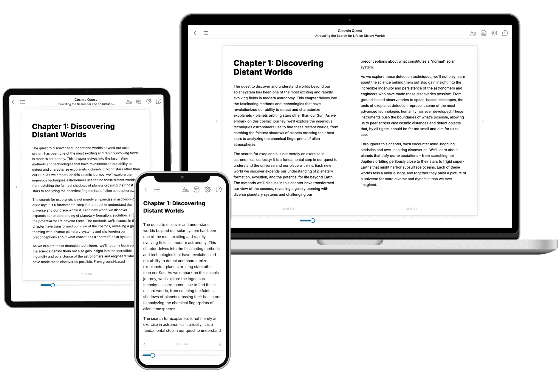
Your book is ready in 10 minutes. Read it online, download as EPUB or PDF, or send to Kindle.

Frequently asked questions
- What is TailoredRead?
TailoredRead creates complete, full-length nonfiction ebooks, written specifically for you. Tell us what you want to learn and why, and TailoredRead writes you a complete ebook on it, with charts, tables, and diagrams throughout. Read it online, on Kindle, or on any e-reader, or download it as a PDF or EPUB. Every book is written fresh the day you order it.
- How is the book tailored to me?
Before anything is written, you answer a few short questions about your goals, your background, and the topics you care about. Your book is then planned around your answers: written at your level, focused on your goals, and covering the topics you chose, instead of the one-size-fits-all book a store sells everyone.
- Will it read like generic AI content?
No. Your book isn't a chat transcript: it's planned chapter by chapter before a single word is written, told in a clear book voice, and explains ideas visually with charts, tables, and diagrams. After writing, every book goes through an editorial pass that polishes chapters so the book reads consistent from start to finish. Judge for yourself: browse thousands of sample books on our Explore Books page.
- How accurate is the content?
Our AI first checks whether your subject needs live research. Niche and fast-moving subjects are researched before writing, and the book plan and chapters are grounded in what it finds. After writing, every book goes through a quality pass that checks its facts and consistency, and researched books list their sources in a Notes section at the back.
- How long is the book?
You choose the length: Comprehensive (250-300 pages), Detailed (150-200 pages), Essential (70-100 pages), or Short (30-50 pages).
- Does the book include visuals?
Yes. Every book includes charts, tables, and diagrams: timelines, checklists, step-by-step guides, decision trees, comparison tables, matrices, and bar charts. A typical book includes dozens of these visual elements.
- Can I read it on Kindle? What formats can I download?
Yes. Every book can be read in our online reader or downloaded as a PDF or EPUB. The EPUB can be opened with Kindle, Apple Books, Kobo, and any e-reader. The Commercial and Author licenses also include an editable Word (DOCX) and Markdown download.
- How much does it cost?
About the price of a regular ebook. The exact price depends on your book's length and complexity, from $2 for the shortest books to $35 for the most comprehensive, and it's shown when you review your book, before you commit to anything. Each book is a one-time purchase, yours to keep, no subscription. Licenses that let you share, teach, or publish your book range from $35.99 to $49.99.
- Can I preview the book before purchasing?
Before you pay, you'll see what your book will cover: the title, a detailed description, the estimated length, and the complete chapter-by-chapter table of contents, so you know what you're getting before you decide. The full text itself is written after you purchase.
- How long does it take to create a book?
About 10 minutes. You can start reading the opening chapters while the rest is still being written.