The Skull Anatomy Coloring Book
Learn Bone Structure, Landmarks, and Function Through Interactive Illustration
Have you ever wondered why certain bones of the skull have such distinctive shapes and features?
222 pages10 chaptersDec 22, 2025Anatomy Coloring
About this book
This coloring book transforms skull anatomy from abstract memorization into an engaging, visual learning experience. Rather than passively reading descriptions, you'll actively color, label, and explore the intricate structure of the human skull—reinforcing your understanding with every stroke of color. Each chapter guides you through different skull regions and systems, building your knowledge progressively from basic bone identification to complex anatomical relationships. Whether you're a student, healthcare professional, artist, or anatomy enthusiast, this interactive approach helps you truly grasp how the skull's 22 bones fit together, where key landmarks are located, and why each structure matters. By combining visual learning with hands-on coloring activities, you'll develop a deeper, more memorable understanding of skull anatomy that goes far beyond traditional textbooks.
Table of contents
- IntroductionWhy Skull Anatomy Matters, How to Use This Coloring Book, The Power of Visual Learning and Coloring, Overview of Skull OrganizationRead below
- Chapter 1: Skull Basics and OrganizationMajor Divisions: Cranium, Face, and Base, The 22 Bones of the Adult Skull, Types of Skull Bones and Their Characteristics
- Chapter 2: The Cranium: Protecting the BrainThe Cranial Vault and Its Bones, The Cranial Base: Structure and Foramina, Key Landmarks and Features of Cranial Bones
- Chapter 3: Facial Bones and StructuresThe Facial Skeleton Framework, Nasal and Orbital Regions, The Palate and Dental Structures
- Chapter 4: The Mandible and Temporomandibular JointMandibular Structure and Features, The Temporomandibular Joint and Jaw Movement, Muscle Attachment Sites on the Mandible
- Chapter 5: Anterior View: Front-Facing AnatomyUnderstanding the Anterior Perspective, Key Landmarks Visible from the Front, Coloring Exercise: Anterior Skull View
- Chapter 6: Lateral View: Side-by-Side AnatomyUnderstanding the Lateral Perspective, The Temporal Region and Zygomatic Arch, Coloring Exercise: Lateral Skull View
- Chapter 7: Superior and Posterior ViewsThe Superior (Top) View of the Skull, The Posterior (Back) View of the Skull, Coloring Exercise: Superior and Posterior Views
- Chapter 8: Foramina, Canals, and OpeningsUnderstanding Foramina and Their Functions, Major Foramina and What Passes Through Them, Coloring Exercise: Identifying Foramina and Canals
- Chapter 9: Muscle Attachments and Functional AnatomyIdentifying Muscle Attachment Sites on Skull Bones, How Skull Structure Supports Muscle Function, Coloring Exercise: Muscle Attachment Regions
- Chapter 10: Skull Variations and Clinical SignificanceNormal Anatomical Variations in Skull Structure, Clinical Landmarks and Their Importance, Recognizing Pathological Changes in Skull Anatomy
- ConclusionMastering Skull Anatomy Through Active Learning, Continuing Your Anatomical Education
- Glossary of Anatomical Terms
- Further Reading and Resources
Introduction
The human skull stands as one of the most intricate and fascinating structures in anatomy. Composed of twenty-two individual bones that fuse together in precise arrangements, the skull serves multiple critical functions: protecting the brain, housing sensory organs, providing attachment points for muscles, and forming the framework of the face. Yet for many learners, understanding skull anatomy feels overwhelming. Traditional textbooks present dense paragraphs of information alongside static diagrams, making it difficult to visualize how bones relate to one another in three-dimensional space. This coloring book takes a different approach, transforming passive reading into active engagement through the power of visual learning and hands-on illustration.
When you color anatomical structures, something remarkable happens in your brain. The act of choosing colors, tracing boundaries, and filling in regions creates multiple neural pathways that reinforce learning. You're not just reading about the temporal bone—you're actively engaging with its shape, location, and relationship to surrounding structures. This multisensory approach activates visual, motor, and cognitive processes simultaneously, leading to deeper understanding and better retention. Research in educational psychology consistently demonstrates that active learning strategies outperform passive reading, particularly for spatial and visual information like anatomy[1]. By the time you finish coloring a skull structure, you've examined it far more thoroughly than you would by simply looking at a completed diagram.
This book is designed for learners who already have some foundational knowledge of anatomy and are ready to deepen their understanding through systematic, hands-on practice. Whether you're a healthcare student preparing for examinations, a medical professional refreshing your knowledge, an artist seeking anatomical accuracy, or simply someone fascinated by human structure, this coloring book provides a structured pathway through skull anatomy. Each chapter builds progressively on previous knowledge, starting with basic organization and advancing through detailed regional anatomy, functional relationships, and clinical significance. The combination of clear explanations, detailed illustrations, and coloring exercises creates a comprehensive learning experience that goes far beyond memorization to develop true anatomical literacy.
Throughout this book, you'll encounter specific terminology, directional references, and anatomical conventions that form the language of anatomy. Terms like anterior (front), posterior (back), superior (top), inferior (bottom), medial (toward the midline), and lateral (away from the midline) provide precise ways to describe locations and relationships. You'll learn to identify landmarks—distinctive features on bones that serve as reference points—and foramina—openings through which nerves and blood vessels pass. These terms may seem technical at first, but they become second nature as you use them repeatedly while coloring and labeling structures. The goal is not just to know what structures look like, but to understand where they are, how they connect, and why they matter functionally.
Why Skull Anatomy Matters
Understanding skull anatomy extends far beyond academic interest—it forms the foundation for numerous practical applications across healthcare, art, forensics, and research. For healthcare professionals, detailed knowledge of skull structure is essential for interpreting imaging studies, performing physical examinations, planning surgical procedures, and understanding pathological conditions. When a physician palpates the mastoid process behind the ear or identifies the mental foramen on a dental X-ray, they're applying anatomical knowledge that begins with understanding basic skull structure. Radiologists rely on intimate familiarity with skull anatomy to distinguish normal variations from pathological changes on CT scans and MRIs. Dentists must understand the relationship between teeth, jaw bones, and surrounding structures to perform procedures safely and effectively.
The skull's protective function cannot be overstated. The cranial bones form a rigid vault that shields the brain from mechanical injury, while the facial bones protect delicate sensory organs including the eyes, inner ears, and nasal passages. The skull base contains numerous foramina through which critical structures pass—the optic nerve travels through the optic canal to reach the eye, the internal carotid artery enters the skull through the carotid canal, and twelve pairs of cranial nerves exit through various openings to innervate the head, neck, and beyond. Understanding these relationships helps clinicians recognize how injuries or pathological processes in one area might affect seemingly distant structures. For example, a fracture through the temporal bone might damage the facial nerve as it passes through the stylomastoid foramen, resulting in facial paralysis.
Artists and illustrators benefit enormously from accurate skull anatomy knowledge. The skull determines facial proportions, contours, and the underlying structure that gives faces their distinctive characteristics. Portrait artists who understand the zygomatic arch's position can more accurately render cheekbones. Sculptors who know how the mandible articulates with the temporal bone can create realistic jaw positions. Medical illustrators must combine artistic skill with anatomical precision to create educational materials that are both visually appealing and scientifically accurate. Even digital artists creating characters for animation or video games rely on skull anatomy to ensure their creations look believable, whether they're depicting realistic humans or fantastical creatures based on human proportions.
Forensic anthropologists use skull anatomy to identify unknown individuals, estimate age at death, determine biological sex, and assess ancestry. Specific features of the skull change predictably with age—sutures between cranial bones fuse in characteristic patterns, teeth erupt and wear according to typical timelines, and bone density changes throughout life. Sexual dimorphism in skull structure, though subtle, provides clues about biological sex: males typically have more robust features, larger mastoid processes, more prominent supraorbital ridges, and squarer chins. Ancestry assessment relies on careful measurement of facial proportions and features. This forensic application of skull anatomy has helped solve countless criminal cases and identify disaster victims, providing closure to families and justice in legal proceedings.
Research in evolutionary biology, paleoanthropology, and comparative anatomy relies heavily on skull analysis. The skull preserves well in the fossil record, making it one of the most important sources of information about human evolution and extinct species. By comparing skull features across species and through time, researchers trace evolutionary relationships and understand how different selective pressures shaped cranial structure. The expansion of the cranial vault in human evolution reflects increasing brain size, while changes in facial structure relate to dietary shifts and speech development. Even in modern medical research, understanding normal skull anatomy provides the baseline for recognizing pathological changes and developing new treatments for conditions affecting the head and face.
Beyond professional applications, understanding skull anatomy enriches your appreciation of human biology and the remarkable engineering of the human body. The skull represents an elegant solution to competing demands: it must be strong enough to protect vital organs yet light enough to balance atop the vertebral column; it must provide rigid protection while accommodating growth during childhood; it must house sensory organs while allowing nerves and vessels to pass through. Each feature you'll study in this book exists for specific functional reasons, shaped by millions of years of evolution. Learning skull anatomy means understanding not just what structures look like, but why they're designed the way they are—a perspective that transforms memorization into genuine comprehension.
The skull also serves as an excellent introduction to broader anatomical principles. Many concepts you'll encounter—such as how bones articulate at joints, how foramina provide passage for neurovascular structures, and how muscle attachment sites reflect functional demands—apply throughout the skeletal system. The terminology and directional references you'll practice while studying the skull transfer directly to other anatomical regions. By mastering skull anatomy, you're not just learning about one body region; you're developing skills and knowledge that form the foundation for understanding anatomy as a whole. This makes the skull an ideal focus for deepening your anatomical education, whether you're building on basic knowledge or refreshing understanding gained years ago.
How to Use This Coloring Book
This coloring book is structured to guide you systematically through skull anatomy, building from foundational concepts to detailed regional anatomy and functional relationships. Each chapter focuses on a specific aspect of skull structure, beginning with explanatory text that introduces key concepts, followed by detailed descriptions of anatomical features, and concluding with coloring exercises that reinforce your learning. To get the most from this book, approach it as an active learning tool rather than a passive reference. Read each section carefully before beginning the associated coloring exercise, paying attention to the relationships between structures and the functional significance of features described. Then, with colored pencils or markers in hand, work through the illustrations methodically, referring back to the text as needed.
The recommended approach is to work through chapters sequentially, as later chapters build on concepts introduced earlier. Chapter 1 establishes the basic organization of the skull and introduces the twenty-two bones you'll study in detail throughout the book. Chapters 2 through 4 examine major skull regions—the cranium, facial bones, and mandible—providing detailed coverage of individual bones and their features. Chapters 5 through 7 shift perspective, examining the skull from different anatomical views (anterior, lateral, superior, and posterior) to build your three-dimensional understanding. Chapters 8 and 9 focus on functional anatomy, exploring foramina and muscle attachments. Finally, Chapter 10 addresses variations and clinical significance, connecting anatomical knowledge to practical applications. This progression ensures you develop both detailed knowledge of individual structures and comprehensive understanding of how they fit together.
When approaching each coloring exercise, follow this systematic process for maximum learning benefit:
- Preview the illustration: Before coloring, examine the entire diagram to understand what structures are shown and how they relate to one another
- Read all labels: Familiarize yourself with the structures you'll be coloring and their names
- Choose a color scheme: Select distinct colors for different structures, using similar shades for related structures if helpful
- Color systematically: Work through structures in a logical order rather than randomly, which reinforces spatial relationships
- Trace boundaries carefully: Pay attention to where structures meet and articulate, as these boundaries are anatomically significant
- Review and self-test: After completing the coloring, cover the labels and test yourself on structure identification
Your choice of coloring materials can enhance your learning experience. Colored pencils offer the most control and allow for layering and shading, making them ideal for detailed anatomical work. Choose pencils with relatively hard leads that won't smudge easily, and keep them sharp for precise work around boundaries and small structures. Fine-tip markers provide bold, clear colors that make structures easy to distinguish, though they offer less control for detailed work and may bleed through thin paper. Some learners prefer using a combination: markers for large areas and colored pencils for details and boundaries. Highlighters can be useful for emphasizing specific features or color-coding related structures, though their transparency means they work best over pencil labels rather than as primary coloring tools.
Develop a consistent color-coding system that you use throughout the book. This consistency reinforces learning by creating visual associations that persist across different views and chapters. For example, you might always color the frontal bone in light blue, the parietal bones in yellow, the temporal bones in green, and the occipital bone in purple. When you encounter these bones in different views or contexts, the consistent colors help you recognize them immediately. Similarly, you might use warm colors (reds, oranges, yellows) for facial bones and cool colors (blues, greens, purples) for cranial bones. Some learners prefer to color bones that articulate with each other in related shades—for instance, using different shades of blue for all bones that form the cranial vault. Experiment to find a system that works for your learning style.
Supplement your coloring work with additional study techniques to maximize retention. After completing a coloring exercise, try drawing the same structures from memory on blank paper, then compare your drawing to the original to identify gaps in your knowledge. Create flashcards with structure names on one side and their locations, features, or functions on the other. Use the illustrations in this book as references while examining skull models or specimens if you have access to them—the combination of two-dimensional illustrations and three-dimensional models provides powerful reinforcement. Form study groups where you quiz each other on structure identification, or teach the material to someone else, which forces you to organize and articulate your knowledge clearly.
Take notes in the margins as you work through the book. When you encounter a particularly challenging structure or relationship, write yourself a reminder or create a mnemonic device. If you notice connections between structures discussed in different chapters, note those relationships. If you have questions that the text doesn't fully answer, write them down and research them using the resources listed in the Further Reading section. Your annotations transform this book into a personalized learning tool that reflects your unique learning process and addresses your specific knowledge gaps. Over time, these notes become valuable review material, highlighting the concepts you found most challenging and the insights you gained through active engagement with the material.
Finally, be patient with yourself and recognize that mastering skull anatomy takes time and repeated exposure. Don't expect to memorize every structure after a single coloring session. Instead, plan to work through the book multiple times, each time noticing details you missed previously and deepening your understanding of relationships between structures. The first time through, focus on identifying major structures and understanding basic organization. On subsequent reviews, pay attention to smaller features, subtle relationships, and functional significance. This layered approach to learning, combined with the active engagement that coloring provides, will build comprehensive, lasting knowledge of skull anatomy that serves you well in whatever field you apply it.
The Power of Visual Learning and Coloring
The effectiveness of coloring as a learning tool rests on solid foundations in cognitive psychology and educational research. When you color an anatomical structure, you engage multiple cognitive processes simultaneously: visual processing as you examine the structure's shape and boundaries, motor activity as you control the coloring tool, decision-making as you choose colors and plan your approach, and memory encoding as you associate the structure with its name, location, and function. This multisensory engagement creates stronger, more numerous neural connections than passive reading alone. The brain's visual cortex, motor cortex, and memory centers all activate during coloring, creating a rich network of associations that facilitate later recall[2].
Visual learning proves particularly powerful for anatomy because anatomical knowledge is inherently spatial and visual. Understanding anatomy means knowing not just what structures exist, but where they're located, what they look like, how they're oriented, and how they relate to surrounding structures. These are fundamentally visual concepts that resist purely verbal description. Try describing the shape of the sphenoid bone in words alone, and you'll quickly appreciate why illustrations are essential. When you color that same bone, tracing its complex shape and noting how it articulates with surrounding bones, you develop an intuitive understanding that verbal descriptions struggle to convey. The visual memory you create becomes a mental reference you can access whenever you need to recall that structure.
The act of coloring also slows down the learning process in beneficial ways. When reading text, it's easy to skim over unfamiliar terms or complex descriptions, creating an illusion of understanding without genuine comprehension. Coloring forces you to slow down and examine each structure carefully. You must identify boundaries, distinguish one structure from adjacent ones, and understand spatial relationships before you can color accurately. This deliberate pace gives your brain time to process information thoroughly and identify gaps in understanding. When you're unsure where one bone ends and another begins, that uncertainty signals a need to review the text or examine the illustration more carefully—a self-correcting mechanism that passive reading doesn't provide.
Coloring also leverages the psychological principle of active learning, which holds that learners retain information better when they actively manipulate it rather than passively receiving it. Educational research consistently demonstrates that active learning strategies—including hands-on activities, problem-solving, and creation tasks—produce better outcomes than lectures or reading alone[3]. Coloring transforms you from a passive recipient of anatomical information into an active participant in your own learning. You make decisions about colors, trace boundaries, and create a finished product that represents your understanding. This sense of agency and accomplishment enhances motivation and engagement, making learning more enjoyable and sustainable over time.
The visual products you create through coloring serve as personalized study materials tailored to your learning needs. Unlike pre-colored diagrams in textbooks, your colored illustrations reflect your own understanding and attention. Areas where you struggled to identify boundaries or choose appropriate colors highlight concepts that need additional review. Structures you colored confidently represent solid understanding. When you return to review material, your colored illustrations trigger memories not just of what structures look like, but of the process of learning them—where you were when you colored them, what questions you had, what insights you gained. These contextual memories provide additional retrieval cues that enhance recall during examinations or practical applications.
Coloring also facilitates pattern recognition, a crucial skill in anatomy. As you color multiple views of the skull, you begin to recognize patterns in how bones are shaped, how they articulate, and how features relate to function. You notice that bones with muscle attachments often have roughened surfaces or prominent processes, that foramina typically appear near bone junctions, and that paired bones (like the parietal bones) mirror each other symmetrically. These patterns, discovered through your own observation rather than explicitly stated in text, represent deep learning that transfers to new situations. When you encounter an unfamiliar skull view or a pathological specimen, these internalized patterns help you orient yourself and identify structures even in altered contexts.
The meditative quality of coloring provides additional benefits beyond pure information retention. The focused, repetitive motor activity of coloring can reduce stress and anxiety, creating a calm mental state conducive to learning. Many learners find that coloring helps them concentrate, blocking out distractions and allowing sustained attention to complex material. This focused attention state, sometimes called "flow," is associated with optimal learning and performance. By making anatomy study more enjoyable and less stressful, coloring increases the likelihood that you'll engage with the material regularly and persistently—and in learning, consistency and repetition are key to mastery. The time you spend coloring is not time away from "real" studying; it is studying, enhanced by the psychological and cognitive benefits that active, visual engagement provides.
Overview of Skull Organization
The adult human skull consists of twenty-two bones that fit together like a three-dimensional puzzle, forming a structure that is simultaneously strong, lightweight, and functionally sophisticated. These bones divide into two major groups: eight cranial bones that form the cranium (the protective case surrounding the brain) and fourteen facial bones that form the face and support sensory organs. The cranial bones include the frontal bone (forehead), two parietal bones (top and sides of the skull), two temporal bones (sides and base), the occipital bone (back and base), the sphenoid bone (central base), and the ethmoid bone (between the nasal cavity and cranium). The facial bones include two nasal bones, two maxillae (upper jaw), two zygomatic bones (cheekbones), two lacrimal bones, two palatine bones, two inferior nasal conchae, the vomer, and the mandible (lower jaw).
Understanding skull organization requires familiarity with anatomical directional terms that provide precise ways to describe locations and relationships. Anterior refers to the front of the skull (the face), while posterior refers to the back. Superior indicates the top of the skull, and inferior indicates the bottom. Medial means toward the midline (an imaginary line dividing the skull into left and right halves), while lateral means away from the midline, toward the sides. Internal (or endocranial) refers to the inside surface of the skull facing the brain, while external (or ectocranial) refers to the outside surface. These terms allow precise communication about anatomical features: for example, the external acoustic meatus is located on the lateral aspect of the temporal bone, inferior to the parietal bone and posterior to the zygomatic arch.
The skull can be studied from multiple perspectives or views, each revealing different structures and relationships. The anterior view shows the face, including the orbits (eye sockets), nasal cavity, maxillae, and mandible. The lateral view reveals the side of the skull, showing how cranial and facial bones articulate and displaying the temporal region and zygomatic arch prominently. The superior view looks down on the top of the skull, showing primarily the frontal and parietal bones. The posterior view shows the back of the skull, dominated by the occipital bone. The inferior view (looking up at the skull base) reveals the complex architecture of the cranial base, including numerous foramina and the hard palate. Finally, sagittal sections (cutting through the midline from front to back) reveal internal structures including the nasal septum and cranial fossae.
Bones of the skull connect at joints called sutures, which appear as irregular, interlocking lines on the skull surface. Major sutures include the coronal suture (between the frontal and parietal bones), the sagittal suture (between the two parietal bones along the midline), the lambdoid suture (between the parietal and occipital bones), and the squamous sutures (between the temporal and parietal bones). In infants and young children, sutures are flexible and allow for brain growth and skull expansion. At certain suture junctions, soft spots called fontanelles exist, which gradually close as the skull bones grow and eventually fuse. In adults, sutures become increasingly fused, though they remain visible as irregular lines. The pattern and degree of suture fusion can help estimate age in skeletal remains.
The skull contains numerous openings called foramina (singular: foramen) and canals through which nerves, blood vessels, and other structures pass. These openings are not random but precisely positioned to allow specific structures to enter or exit the skull. For example, the optic canal in the sphenoid bone transmits the optic nerve from the brain to the eye. The foramen magnum, the largest opening in the skull, is located in the occipital bone and allows the spinal cord to connect with the brain. The internal carotid arteries enter the skull through the carotid canals in the temporal bones. Understanding foramina and their contents is essential for clinical applications, as pathological processes affecting these openings can compromise the structures passing through them.
Skull bones feature various types of anatomical landmarks that serve as reference points and attachment sites. Processes are projections extending from bones, such as the mastoid process of the temporal bone or the styloid process. Fossae (singular: fossa) are shallow depressions, like the temporal fossa on the side of the skull. Tubercles and tuberosities are rounded elevations, often serving as muscle attachment sites. Spines are sharp, pointed projections. Condyles are rounded articular surfaces that form joints, such as the occipital condyles that articulate with the first cervical vertebra. Meatus refers to a canal or passage, like the external acoustic meatus leading to the eardrum. Learning to identify these features and understand their functional significance is central to mastering skull anatomy.
The skull's internal surface differs dramatically from its external appearance. The cranial cavity, which houses the brain, features three distinct levels called cranial fossae. The anterior cranial fossa supports the frontal lobes of the brain and is formed primarily by the frontal and ethmoid bones. The middle cranial fossa houses the temporal lobes and is formed mainly by the sphenoid and temporal bones. The posterior cranial fossa contains the cerebellum and brainstem and is formed primarily by the occipital bone. These fossae are separated by bony ridges and feature numerous foramina through which cranial nerves exit the skull. The internal skull surface also shows impressions of brain structures and grooves where blood vessels traveled, creating a detailed record of the brain's relationship to its bony housing.
As you work through this book, you'll explore each of these organizational principles in detail, building a comprehensive understanding of skull structure from multiple perspectives. You'll learn to identify individual bones and their features, understand how bones articulate with one another, recognize important foramina and the structures they transmit, and appreciate the functional significance of skull anatomy. The coloring exercises will reinforce this knowledge, transforming abstract concepts into concrete visual understanding. By the end of this book, you'll be able to examine a skull from any angle and confidently identify structures, describe their relationships, and explain their functional importance—a level of mastery that serves as a foundation for advanced study in anatomy, medicine, art, or any field where understanding human structure matters.
Notes
Introduction
- 1. S. M. Kosslyn and others, "Visual mental imagery activates topographically organized visual cortex," Journal of Cognitive Neuroscience, 2001
- 2. A. Paivio, Mental Representations, Oxford University Press, 1990
- 3. S. Freeman and others, "Active learning increases student performance in science, engineering, and mathematics," Proceedings of the National Academy of Sciences, 2014
This is the end of the free sample. The full book has 10 chapters.
Want this book written for you?
Tell us your goals and background, and get your own version of this book, tailored by AI to you.


Benefits of AI-tailored books
- Read one book, not ten:
- all the Anatomy Coloring knowledge you need, gathered into a single book.
- Save days of learning:
- tell us your goals, choose your topics, and the whole book is built around them.
- Learn effortlessly:
- Anatomy Coloring book written for your specific background and expertise.
- Understand at a glance:
- includes charts, tables, timelines, checklists, decision trees, and more.
- Stay current:
- written the day you order it, not years ago.
- Trust what you read:
- every book is checked for accuracy, with real research when your subject needs it.
Create your unique book in 3 steps
- 1. Select your focus
Select the focus of your Anatomy Coloring book and share your background
- 2. Personalize your book
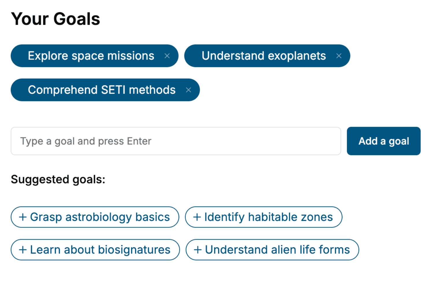
Specify your goals and choose sub-topics to include
- 3. Get your tailored book
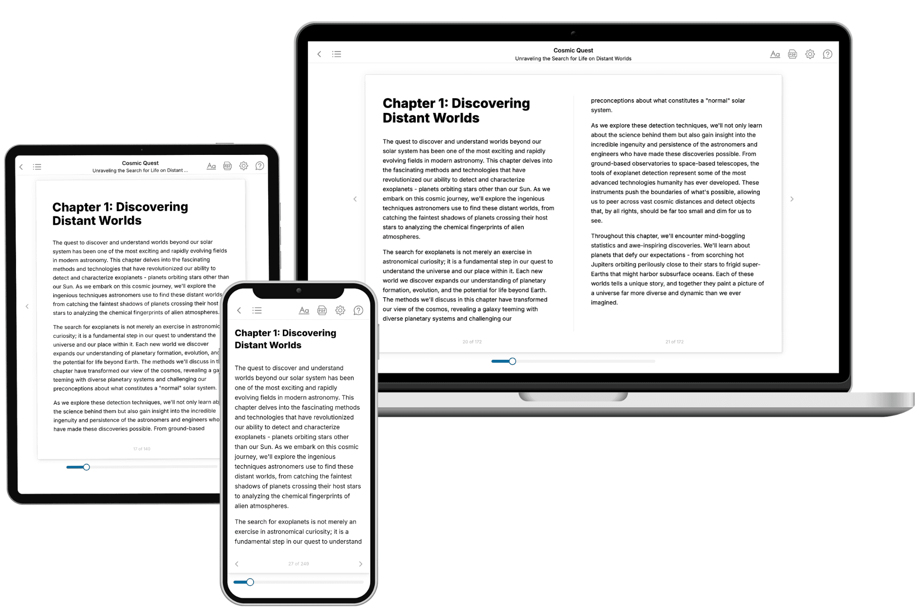
Your book is ready in 10 minutes. Read it online, download as EPUB or PDF, or send to Kindle.

Frequently asked questions
- What is TailoredRead?
TailoredRead creates complete, full-length nonfiction ebooks, written specifically for you. Tell us what you want to learn and why, and TailoredRead writes you a complete ebook on it, with charts, tables, and diagrams throughout. Read it online, on Kindle, or on any e-reader, or download it as a PDF or EPUB. Every book is written fresh the day you order it.
- How is the book tailored to me?
Before anything is written, you answer a few short questions about your goals, your background, and the topics you care about. Your book is then planned around your answers: written at your level, focused on your goals, and covering the topics you chose, instead of the one-size-fits-all book a store sells everyone.
- Will it read like generic AI content?
No. Your book isn't a chat transcript: it's planned chapter by chapter before a single word is written, told in a clear book voice, and explains ideas visually with charts, tables, and diagrams. After writing, every book goes through an editorial pass that polishes chapters so the book reads consistent from start to finish. Judge for yourself: browse thousands of sample books on our Explore Books page.
- How accurate is the content?
Our AI first checks whether your subject needs live research. Niche and fast-moving subjects are researched before writing, and the book plan and chapters are grounded in what it finds. After writing, every book goes through a quality pass that checks its facts and consistency, and researched books list their sources in a Notes section at the back.
- How long is the book?
You choose the length: Comprehensive (250-300 pages), Detailed (150-200 pages), Essential (70-100 pages), or Short (30-50 pages).
- Does the book include visuals?
Yes. Every book includes charts, tables, and diagrams: timelines, checklists, step-by-step guides, decision trees, comparison tables, matrices, and bar charts. A typical book includes dozens of these visual elements.
- Can I read it on Kindle? What formats can I download?
Yes. Every book can be read in our online reader or downloaded as a PDF or EPUB. The EPUB can be opened with Kindle, Apple Books, Kobo, and any e-reader. The Commercial and Author licenses also include an editable Word (DOCX) and Markdown download.
- How much does it cost?
About the price of a regular ebook. The exact price depends on your book's length and complexity, from $2 for the shortest books to $35 for the most comprehensive, and it's shown when you review your book, before you commit to anything. Each book is a one-time purchase, yours to keep, no subscription. Licenses that let you share, teach, or publish your book range from $35.99 to $49.99.
- Can I preview the book before purchasing?
Before you pay, you'll see what your book will cover: the title, a detailed description, the estimated length, and the complete chapter-by-chapter table of contents, so you know what you're getting before you decide. The full text itself is written after you purchase.
- How long does it take to create a book?
About 10 minutes. You can start reading the opening chapters while the rest is still being written.