The Urinary System Coloring Book
Learn Anatomy Through Interactive Illustrations and Detailed Exploration
Imagine having a complete visual map of your urinary system—every structure, every pathway, every connection—all at your fingertips.
191 pages8 chaptersDec 8, 2025Anatomy Coloring
About this book
This coloring book transforms abstract anatomical concepts into tangible, memorable learning experiences. By engaging with detailed illustrations and coloring them yourself, you'll develop an intuitive understanding of how your kidneys filter blood, how urine travels through your body, and how this remarkable system maintains your health. Whether you're a student preparing for exams, a healthcare professional seeking to refresh your knowledge, or simply curious about how your body works, this book combines the proven benefits of active learning with the satisfaction of creative expression. Each chapter builds systematically from basic structures to complex functions, with clear explanations accompanying every illustration. The act of coloring itself strengthens neural pathways, making anatomical knowledge stick in ways that passive reading never can. You'll finish this book not just knowing urinary anatomy—you'll truly understand it.
Table of contents
- IntroductionWhy Coloring Enhances Anatomical Learning, How to Use This Book, Overview of the Urinary SystemRead below
- Chapter 1: The Kidneys: Structure and LocationKidney Position and External Anatomy, Layers of the Kidney: Capsule to Medulla, Internal Regions: Cortex, Medulla, and Pyramids
- Chapter 2: The Nephron: Functional Unit of the KidneyNephron Structure and Components, The Renal Corpuscle: Glomerulus and Bowman's Capsule, The Renal Tubule: Proximal to Distal Segments, The Collecting Duct and Loop of Henle
- Chapter 3: Blood Supply and FiltrationRenal Artery and Capillary Networks, The Three-Step Filtration Process, Glomerular Filtration: Where It Begins
- Chapter 4: Urine Formation: Reabsorption and SecretionTubular Reabsorption in the Proximal Tubule, The Loop of Henle and Urine Concentration, Distal Tubule and Collecting Duct: Final Adjustments
- Chapter 5: The Ureters, Bladder, and UrethraUreters: Transporting Urine Downward, Bladder Structure and Wall Layers, The Urethra: Male and Female Differences
- Chapter 6: Innervation and Hormonal ControlSympathetic and Parasympathetic Innervation, ADH, Aldosterone, and Renin-Angiotensin System, The Micturition Reflex and Voluntary Control
- Chapter 7: Homeostasis and Electrolyte BalanceWater Balance and Osmotic Regulation, Sodium, Potassium, and Chloride Management, pH Regulation and Acid-Base Balance
- Chapter 8: Clinical Correlations and Common DisordersChronic Kidney Disease and Acute Injury, Urinary Tract Infections and Incontinence, Kidney Stones and Structural Abnormalities
- ConclusionIntegrating Your Anatomical Knowledge, Continuing Your Anatomy Journey
- Glossary of Terms
- Further Reading and Resources
Introduction
The urinary system stands as one of the body's most essential yet often overlooked networks. Every day, your kidneys filter approximately 180 liters of blood, producing about 1.5 liters of urine while maintaining the delicate chemical balance that keeps you alive. This remarkable system doesn't just eliminate waste—it regulates blood pressure, controls electrolyte levels, maintains pH balance, and even produces hormones that affect red blood cell production. Understanding how these organs work together requires more than memorizing names and locations. It demands a deep, spatial understanding of structures and their relationships to one another.
Traditional anatomy learning often relies on passive reading and two-dimensional diagrams that fail to engage multiple learning pathways simultaneously. Research in educational psychology consistently demonstrates that active learning techniques produce superior retention compared to passive methods. When you color an anatomical structure, you're not simply filling in spaces with pigment. You're tracing pathways with your hands, visualizing relationships with your eyes, and reinforcing connections in your brain. This multi-sensory approach creates stronger neural pathways and transforms abstract concepts into concrete, memorable knowledge that persists long after you close this book.
This coloring book takes you on a systematic journey through the urinary system, beginning with the kidneys' gross anatomy and progressing through microscopic structures, physiological processes, and clinical applications. Each chapter builds upon previous knowledge, creating a comprehensive understanding of how structure relates to function. Whether you're a nursing student preparing for exams, a medical professional refreshing your anatomical knowledge, or simply someone fascinated by how the human body maintains homeostasis, this book provides the tools you need to master urinary system anatomy. The combination of detailed illustrations, clear explanations, and hands-on coloring activities ensures that you'll not only learn the material but truly understand it at a level that supports clinical reasoning and practical application.
Why Coloring Enhances Anatomical Learning
The act of coloring anatomical structures engages your brain in ways that passive reading cannot match. When you select a color for the renal cortex and carefully fill in its boundaries, you're performing several cognitive tasks simultaneously. You're identifying the structure, distinguishing its borders from adjacent tissues, following its contours, and creating a visual memory that associates that specific color with that particular anatomical feature. This process activates multiple brain regions including visual processing centers, motor control areas, and memory formation pathways. Neuroscience research has shown that information processed through multiple sensory channels creates more robust and accessible memories than information received through a single channel[1].
Coloring also slows down the learning process in a beneficial way. Rather than quickly scanning a labeled diagram, you must spend time with each structure, examining its shape, size, and position relative to neighboring anatomy. This deliberate pace allows for deeper processing and reflection. As you color the nephron's proximal convoluted tubule, for example, you notice its coiled nature, its location in the cortex, and its connection to the glomerulus. These observations become embedded in your memory through the physical act of coloring, creating what educational psychologists call procedural memory—knowledge that's tied to actions and therefore more resistant to forgetting.
The kinesthetic component of coloring provides another powerful learning advantage. Your hands trace the pathways that blood follows through the renal circulation or the route that urine takes from the collecting ducts to the bladder. This physical tracing creates what's known as muscle memory, a form of learning that operates independently of conscious recall. Medical students have long used this technique when learning surgical procedures or physical examination techniques. By applying it to anatomical learning, you're leveraging the same neurological mechanisms that allow surgeons to navigate complex anatomy with precision and confidence.
Color coding creates visual associations that enhance recall during exams or clinical situations. When you consistently use red for arteries, blue for veins, and yellow for the renal pelvis, these color associations become mental shortcuts. During an exam, visualizing the colored diagram you created helps you recall not just the structure's name but its relationships, blood supply, and functional significance. This technique proves particularly valuable when learning complex three-dimensional relationships that are difficult to represent in traditional two-dimensional diagrams. The urinary system, with its intricate vascular networks and tubular structures, benefits enormously from this approach.
Research on active learning consistently demonstrates its superiority over passive methods. A comprehensive meta-analysis of 225 studies found that students in active learning environments performed better on exams and were less likely to fail compared to those in traditional lecture-based courses[2]. Coloring represents one of the most accessible forms of active learning, requiring no special equipment beyond colored pencils and providing immediate feedback as you see the anatomy take shape beneath your hands. This immediate visual feedback reinforces correct understanding and helps identify areas where your knowledge needs strengthening.
The creative aspect of coloring also reduces learning anxiety and makes studying more enjoyable. Many students approach anatomy with trepidation, overwhelmed by the volume of information and the precision required. Coloring transforms this potentially stressful activity into something more meditative and satisfying. As you complete each illustration, you experience a sense of accomplishment that motivates continued learning. This positive emotional association with the material enhances memory formation, as emotions and memory are closely linked in brain structures like the amygdala and hippocampus. When learning feels rewarding rather than burdensome, you're more likely to persist through challenging material and achieve mastery.
Finally, coloring creates a personalized study resource that's uniquely yours. The diagrams you color reflect your understanding, your color choices, and your learning journey. This personalization makes the book more valuable as a review tool because you're not just seeing generic illustrations—you're seeing your own work, which triggers memories of the learning process itself. When you review your colored diagrams before an exam, you remember not just the anatomy but also the insights you had while coloring, the questions you asked yourself, and the connections you made. This layered memory structure provides multiple retrieval pathways, making it easier to access the information when you need it most.
How to Use This Book
This book is designed for systematic progression through the urinary system, but your learning style and goals should guide how you use it. If you're a student following a course curriculum, align your coloring with your class schedule, using each chapter to reinforce lecture material and prepare for lab sessions. If you're a healthcare professional refreshing your knowledge, you might focus on chapters most relevant to your practice, spending extra time on clinical correlations. If you're learning independently out of curiosity, proceed at whatever pace feels comfortable, allowing yourself time to explore concepts thoroughly before moving forward. Regardless of your approach, consistency matters more than speed—regular, focused sessions produce better results than sporadic marathon study periods.
Before you begin coloring each chapter, read through the accompanying text completely. This initial reading provides context and helps you understand what you're about to illustrate. Pay attention to the relationships between structures, the directional terms used, and the functional significance of each anatomical feature. As you read, visualize the structures in three dimensions, imagining how they fit together in the living body. This mental preparation makes the coloring process more meaningful because you're not just filling in shapes—you're representing real anatomical structures that perform vital functions. Consider keeping a notebook alongside this book where you can jot down questions, observations, or connections you notice during your reading.
When you're ready to color, gather your materials and create a comfortable, well-lit workspace. Colored pencils work best for anatomical coloring because they allow for precision, layering, and shading. Choose a set with at least 12-24 colors to provide variety while maintaining consistency across chapters. Develop a personal color-coding system and stick with it throughout the book. For example, you might use:
- Red for arterial blood and oxygenated vessels
- Blue for venous blood and deoxygenated vessels
- Yellow for urine and collecting structures
- Green for the renal cortex
- Purple for the renal medulla
- Orange for the bladder wall layers
- Pink for epithelial linings
- Brown for connective tissue and capsules
As you color each structure, say its name aloud. This verbal reinforcement adds an auditory component to your visual and kinesthetic learning, further strengthening memory formation. If you're learning with a partner or study group, take turns quizzing each other on structure names, locations, and functions while coloring. Explain concepts to each other in your own words—teaching is one of the most effective learning strategies because it requires you to organize information clearly and identify gaps in your understanding. When you can explain why the loop of Henle dips into the medulla or how the juxtaglomerular apparatus regulates blood pressure, you've moved beyond memorization to genuine comprehension.
Don't rush through the illustrations. Take time to notice details like the relative sizes of structures, the thickness of vessel walls, or the coiling patterns of tubules. These details aren't arbitrary—they reflect functional adaptations. The thick muscular wall of the bladder, for instance, enables it to contract forcefully during urination. The extensive coiling of the proximal convoluted tubule increases surface area for reabsorption. As you color these features, think about why they exist and how they contribute to the system's overall function. This functional perspective transforms anatomy from a list of names into a coherent story about how the body maintains homeostasis.
After completing each chapter, test your knowledge before moving forward. Cover the labels on your colored diagrams and try to identify each structure from memory. Better yet, use a blank sheet of paper to draw the structures from memory, then compare your drawing to the colored illustration. This retrieval practice—actively recalling information rather than simply reviewing it—is one of the most powerful learning techniques available[3]. If you struggle to recall certain structures, that's valuable feedback indicating where you need additional review. Return to those sections, reread the text, and color the structures again with fresh attention to detail.
Use the clinical correlation sections to connect anatomical knowledge with real-world applications. Understanding that kidney stones often lodge at the ureteropelvic junction makes more sense when you've colored the ureters and noticed their narrow diameter. Recognizing that urinary tract infections ascend from the urethra to the bladder becomes clearer when you've traced that pathway with your colored pencils. These clinical connections not only make anatomy more interesting but also provide additional memory hooks. When you associate the trigone of the bladder with its clinical significance as a common site for bladder cancer, you're more likely to remember both the anatomy and its importance.
Finally, return to completed chapters periodically for review. Spaced repetition—reviewing material at increasing intervals—dramatically improves long-term retention. Schedule review sessions one day after completing a chapter, then one week later, then one month later. During these reviews, don't just look at your colored diagrams passively. Quiz yourself, draw structures from memory, or explain concepts aloud as if teaching someone else. This active review maintains and strengthens the neural pathways you've built, ensuring that your anatomical knowledge remains accessible when you need it for exams, clinical practice, or further learning.
Overview of the Urinary System
The urinary system consists of four main components working in coordinated sequence: the kidneys, ureters, bladder, and urethra. Each component performs specific functions that contribute to the system's overall mission of maintaining homeostasis through waste elimination and fluid regulation. The kidneys serve as the system's filtration and processing centers, continuously cleaning blood and producing urine. The ureters act as transport conduits, moving urine from the kidneys to the bladder through peristaltic contractions. The bladder functions as a storage reservoir, accommodating varying volumes of urine until voluntary elimination becomes convenient. The urethra provides the exit pathway, allowing urine to leave the body under both involuntary and voluntary control. Understanding how these components interact reveals the elegant efficiency of this vital system.
The kidneys are paired, bean-shaped organs located retroperitoneally on either side of the vertebral column, roughly between the T12 and L3 vertebrae. Each kidney measures approximately 10-12 centimeters in length, 5-7 centimeters in width, and 3 centimeters in thickness, though the right kidney typically sits slightly lower than the left due to the liver's position. Despite their relatively small size—each kidney weighs only about 150 grams—they receive approximately 20-25% of the cardiac output, processing roughly 1,200 milliliters of blood per minute. This extraordinary blood flow reflects the kidneys' critical role in continuously monitoring and adjusting blood composition. The kidneys don't simply filter waste; they actively regulate water balance, electrolyte concentrations, acid-base balance, and blood pressure while also producing hormones like erythropoietin and renin.
Within each kidney, approximately one million microscopic functional units called nephrons perform the actual work of filtration and urine formation. Each nephron consists of a renal corpuscle (containing the glomerulus and Bowman's capsule) and a renal tubule (including the proximal convoluted tubule, loop of Henle, and distal convoluted tubule). Blood enters the glomerulus under high pressure, forcing water and small solutes through the filtration membrane into Bowman's capsule. This filtrate then travels through the renal tubule, where approximately 99% of the filtered water and valuable solutes are reabsorbed back into the bloodstream. The remaining 1% becomes urine, containing waste products like urea, creatinine, and excess ions. This three-step process—filtration, reabsorption, and secretion—allows the kidneys to precisely control what stays in the body and what gets eliminated.
The ureters are muscular tubes approximately 25-30 centimeters long that connect the renal pelvis of each kidney to the bladder. Their walls contain three layers: an inner mucosa lined with transitional epithelium, a middle muscular layer (muscularis) with both longitudinal and circular smooth muscle fibers, and an outer adventitia of connective tissue. The muscular layer generates peristaltic waves—coordinated contractions that propel urine downward toward the bladder at a rate of about 1-5 contractions per minute. This active transport system works against gravity when you're upright and continues functioning regardless of body position. The ureters enter the bladder at an oblique angle, creating a valve-like mechanism that prevents urine from flowing backward (reflux) when the bladder contracts during urination. This anatomical arrangement protects the kidneys from potential infection or pressure damage.
The urinary bladder is a hollow, muscular organ that serves as a temporary storage reservoir for urine. When empty, it sits entirely within the pelvis, but as it fills, it expands upward into the abdominal cavity, potentially holding 400-600 milliliters comfortably, though maximum capacity can reach 800-1,000 milliliters. The bladder wall consists of four layers:
- The mucosa, lined with transitional epithelium that can stretch as the bladder fills
- The submucosa, containing connective tissue, blood vessels, and nerves
- The muscularis, composed of three layers of smooth muscle collectively called the detrusor muscle
- The adventitia (or serosa on the superior surface), providing an outer protective covering
The urethra is the final component, serving as the passageway through which urine exits the body. Significant anatomical differences exist between males and females. The female urethra measures only 3-4 centimeters in length and has a single function: urine transport. It travels from the bladder's neck to the external urethral orifice located anterior to the vaginal opening. This short length makes females more susceptible to urinary tract infections, as bacteria can more easily ascend from the external environment to the bladder. The male urethra, in contrast, measures approximately 20 centimeters in length and serves dual functions: urine transport and semen delivery. It consists of three regions—the prostatic urethra (passing through the prostate gland), the membranous urethra (passing through the urogenital diaphragm), and the spongy urethra (passing through the penis)—each with distinct anatomical characteristics.
The urinary system's primary functions extend far beyond simple waste elimination. Regulation of blood volume and pressure occurs through adjustments in water reabsorption and sodium retention, directly affecting the volume of blood circulating through your vessels. Regulation of blood osmolarity maintains the proper concentration of dissolved particles, preventing cells from swelling or shrinking due to osmotic imbalances. Regulation of electrolyte balance keeps sodium, potassium, calcium, chloride, and phosphate at optimal levels for nerve conduction, muscle contraction, and countless enzymatic reactions. Regulation of blood pH prevents dangerous acidosis or alkalosis by adjusting hydrogen ion secretion and bicarbonate reabsorption. Production of hormones includes erythropoietin (stimulating red blood cell production), renin (regulating blood pressure), and calcitriol (the active form of vitamin D). Excretion of metabolic wastes removes urea from protein metabolism, creatinine from muscle metabolism, uric acid from nucleic acid breakdown, and bilirubin from hemoglobin degradation.
Understanding the urinary system requires appreciating both its anatomical structures and their physiological functions. Structure and function are inseparable in anatomy—the shape, size, and arrangement of each component directly reflect its role in the system's overall mission. The extensive surface area of the glomerular capillaries facilitates efficient filtration. The length and countercurrent arrangement of the loop of Henle enables urine concentration. The muscular bladder wall allows for forceful contraction during urination. As you progress through this book, coloring each structure and learning its characteristics, you'll develop an integrated understanding of how anatomy enables physiology. This foundation will serve you whether you're preparing for exams, entering clinical practice, or simply satisfying your curiosity about how your body maintains the precise internal environment that keeps you healthy and alive.
Notes
Introduction
- 1. L. Shams and A. R. Seitz, "Benefits of multisensory learning," Trends in Cognitive Sciences, 2008
- 2. S. Freeman and others, "Active learning increases student performance in science, engineering, and mathematics," Proceedings of the National Academy of Sciences, 2014
- 3. H. L. Roediger and A. C. Butler, "The critical role of retrieval practice in long-term retention," Trends in Cognitive Sciences, 2011
This is the end of the free sample. The full book has 8 chapters.
Want this book written for you?
Tell us your goals and background, and get your own version of this book, tailored by AI to you.


Benefits of AI-tailored books
- Read one book, not ten:
- all the Anatomy Coloring knowledge you need, gathered into a single book.
- Save days of learning:
- tell us your goals, choose your topics, and the whole book is built around them.
- Learn effortlessly:
- Anatomy Coloring book written for your specific background and expertise.
- Understand at a glance:
- includes charts, tables, timelines, checklists, decision trees, and more.
- Stay current:
- written the day you order it, not years ago.
- Trust what you read:
- every book is checked for accuracy, with real research when your subject needs it.
Create your unique book in 3 steps
- 1. Select your focus
Select the focus of your Anatomy Coloring book and share your background
- 2. Personalize your book
Specify your goals and choose sub-topics to include
- 3. Get your tailored book
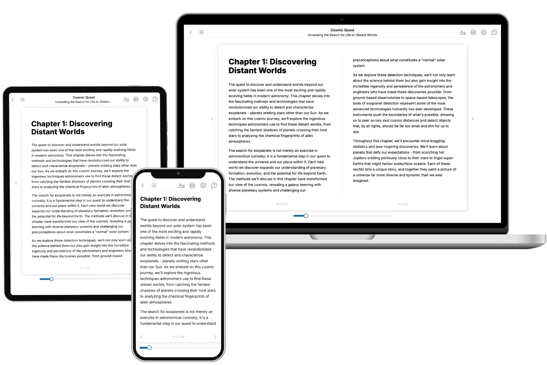
Your book is ready in 10 minutes. Read it online, download as EPUB or PDF, or send to Kindle.

Frequently asked questions
- What is TailoredRead?
TailoredRead creates complete, full-length nonfiction ebooks, written specifically for you. Tell us what you want to learn and why, and TailoredRead writes you a complete ebook on it, with charts, tables, and diagrams throughout. Read it online, on Kindle, or on any e-reader, or download it as a PDF or EPUB. Every book is written fresh the day you order it.
- How is the book tailored to me?
Before anything is written, you answer a few short questions about your goals, your background, and the topics you care about. Your book is then planned around your answers: written at your level, focused on your goals, and covering the topics you chose, instead of the one-size-fits-all book a store sells everyone.
- Will it read like generic AI content?
No. Your book isn't a chat transcript: it's planned chapter by chapter before a single word is written, told in a clear book voice, and explains ideas visually with charts, tables, and diagrams. After writing, every book goes through an editorial pass that polishes chapters so the book reads consistent from start to finish. Judge for yourself: browse thousands of sample books on our Explore Books page.
- How accurate is the content?
Our AI first checks whether your subject needs live research. Niche and fast-moving subjects are researched before writing, and the book plan and chapters are grounded in what it finds. After writing, every book goes through a quality pass that checks its facts and consistency, and researched books list their sources in a Notes section at the back.
- How long is the book?
You choose the length: Comprehensive (250-300 pages), Detailed (150-200 pages), Essential (70-100 pages), or Short (30-50 pages).
- Does the book include visuals?
Yes. Every book includes charts, tables, and diagrams: timelines, checklists, step-by-step guides, decision trees, comparison tables, matrices, and bar charts. A typical book includes dozens of these visual elements.
- Can I read it on Kindle? What formats can I download?
Yes. Every book can be read in our online reader or downloaded as a PDF or EPUB. The EPUB can be opened with Kindle, Apple Books, Kobo, and any e-reader. The Commercial and Author licenses also include an editable Word (DOCX) and Markdown download.
- How much does it cost?
About the price of a regular ebook. The exact price depends on your book's length and complexity, from $2 for the shortest books to $35 for the most comprehensive, and it's shown when you review your book, before you commit to anything. Each book is a one-time purchase, yours to keep, no subscription. Licenses that let you share, teach, or publish your book range from $35.99 to $49.99.
- Can I preview the book before purchasing?
Before you pay, you'll see what your book will cover: the title, a detailed description, the estimated length, and the complete chapter-by-chapter table of contents, so you know what you're getting before you decide. The full text itself is written after you purchase.
- How long does it take to create a book?
About 10 minutes. You can start reading the opening chapters while the rest is still being written.