Visual Evoked Potentials
Understanding How Your Brain Processes What You See
Imagine being able to see exactly how your brain responds to light and images in real time.
207 pages10 chaptersJun 2, 2026Clinical Neurophysiology
About this book
Visual Evoked Potentials make this possible by measuring the electrical activity your brain generates when processing visual information. This book demystifies VEPs—a powerful neurophysiological tool that bridges the gap between what you see and how your brain interprets it. Whether you're a student, clinician, researcher, or simply curious about visual neuroscience, you'll discover how VEPs work, why they matter in clinical diagnosis and research, and how they reveal the hidden mechanisms of visual perception. From the basic principles of neural electrical activity to advanced applications in detecting neurological disease, this guide walks you through the science with clarity and practical insight. Learn how VEPs differ from other vision tests, what the waveforms actually tell us, and why this technology continues to be essential in modern neurology and ophthalmology. By the end, you'll understand not just the "what" and "how" of VEPs, but the "why"—and how this knowledge applies to real-world clinical and research scenarios.
Table of contents
- IntroductionWhy Visual Evoked Potentials Matter, What You'll Learn in This Book, How to Use This GuideRead below
- Chapter 1: The Fundamentals of Visual PerceptionFrom Light to Brain: The Visual Pathway, Neural Basics: How Neurons Communicate, Electrical Activity in the Visual System
- Chapter 2: What Are Visual Evoked Potentials?Defining Visual Evoked Potentials, The Evolution of VEP Technology, Why VEPs Are Unique Among Vision Tests
- Chapter 3: The Neurophysiology Behind VEPsRetinal Processing and Signal Generation, Optic Nerve Transmission and Latency, Cortical Response and Waveform Generation
- Chapter 4: Types of Visual Evoked PotentialsPattern VEPs: Measuring Detailed Vision, Flash VEPs: Assessing Broad Visual Function, Sweep VEPs: Rapid Assessment Techniques
- Chapter 5: Recording and Measuring VEPsElectrode Placement and Setup, Stimulus Parameters and Protocols, Signal Acquisition and Averaging
- Chapter 6: Interpreting VEP WaveformsNormal VEP Components and Peaks, Understanding Amplitude and Latency, Recognizing Abnormal Patterns
- Chapter 7: Clinical Applications of VEPsDetecting Demyelinating Diseases, Assessing Optic Nerve Health, Evaluating Vision in Infants and Children
- Chapter 8: VEPs in Research and Advanced ApplicationsVEPs in Neuroscience Research, Studying Visual Attention and Cognition, Emerging Applications and Future Directions
- Chapter 9: VEPs and Other Diagnostic ToolsVEPs Versus Structural Neuroimaging, Complementary Vision Assessment Methods, Building a Comprehensive Diagnostic Picture
- Chapter 10: Practical Considerations and LimitationsTechnical Challenges and Artifacts, Patient Factors Affecting Results, Common Interpretation Pitfalls
- ConclusionKey Takeaways, The Future of VEP Technology
- Glossary
Introduction
Every time you open your eyes, your brain launches into a remarkable chain of electrical activity. Light enters through your pupils, strikes the retina, and within milliseconds, a cascade of neural signals races through your optic nerves toward the back of your brain. This process happens so automatically and so quickly that it feels effortless. But underneath that effortlessness lies an extraordinarily complex system of neural communication, one that scientists and clinicians have spent decades learning to measure, interpret, and apply in meaningful ways. Visual Evoked Potentials, or VEPs, are the tool that makes this hidden process visible.
A Visual Evoked Potential is simply the electrical response your brain generates when it processes a visual stimulus. When a clinician or researcher records a VEP, they are capturing the brain's reaction to something you see, whether that's a flashing light, a reversing checkerboard pattern, or a shifting grating. These responses are tiny, measured in millionths of a volt, but they carry a wealth of information about the speed and integrity of your visual pathways. The timing of the response, its size, and its shape all tell a story about what is happening between your eyes and your visual cortex.
What makes VEPs genuinely powerful is their objectivity. Unlike a standard eye chart test, which requires you to read letters aloud and cooperate fully, a VEP recording doesn't depend on your ability to communicate. Your brain generates the response automatically. This makes VEPs invaluable for assessing vision in newborns, young children, patients with cognitive impairments, and anyone who cannot participate in conventional vision testing. It also makes VEPs a sensitive early-warning system for neurological conditions like multiple sclerosis, where damage to the myelin sheath slows neural transmission long before a patient notices any symptoms. This combination of objectivity, sensitivity, and non-invasiveness is what has kept VEPs at the center of clinical neurology and vision science for more than half a century.
This book is designed to give you a thorough, accessible understanding of VEPs from the ground up. Whether you are a student encountering this topic for the first time in a neuroscience or clinical program, a clinician looking to deepen your understanding of a test you already use, or a researcher exploring the boundaries of what VEPs can reveal about visual cognition, this guide is built for you. The chapters move from foundational concepts to advanced applications, building knowledge progressively so that each new idea rests on a solid base of what came before.
Why Visual Evoked Potentials Matter
The visual system is the most extensively studied sensory system in the human brain, and for good reason. Roughly 30 percent of the human cerebral cortex is involved in processing visual information in some way.[1] Vision shapes how we navigate the world, recognize faces, read text, and interpret our environment. When something goes wrong in the visual system, the consequences can be profound. VEPs offer a direct, functional window into this system that no other single test can replicate.
Consider what happens in multiple sclerosis (MS), one of the most common neurological diseases affecting young adults. MS attacks the myelin sheath, the insulating layer that wraps around nerve fibers and allows them to conduct signals quickly. When myelin is damaged in the optic nerve, signals slow down. A VEP test can detect this slowing with remarkable precision, often identifying abnormalities before the patient has noticed any visual problems at all. Studies have shown that delayed VEP latency is present in up to 90 percent of MS patients with a history of optic neuritis, and in a significant proportion of those without any visual symptoms.[2] This diagnostic sensitivity is why VEPs remain a standard part of the MS workup in neurology clinics worldwide.
Beyond MS, VEPs matter across a wide range of clinical scenarios. They are used to evaluate optic nerve damage from glaucoma, trauma, or tumors. They help assess visual function in premature infants whose visual systems are still developing. They are used in the operating room to monitor the visual pathways during surgeries near the optic nerve or visual cortex, providing real-time feedback that can prevent accidental damage. In each of these contexts, VEPs provide information that is simply not available from structural imaging or standard behavioral tests alone.
From a research perspective, VEPs have opened doors to understanding how the brain processes not just light, but meaning. Researchers use VEPs to study how the brain responds to faces, words, colors, and motion. They use them to investigate how attention modulates visual processing, how the visual system develops in infancy, and how it changes with aging or disease. The same basic technology that a neurologist uses to diagnose optic neuritis is also used by cognitive neuroscientists to probe the timing of conscious visual awareness. This breadth of application is a testament to how fundamental and versatile VEP measurement really is.
It is also worth noting what VEPs represent in the broader landscape of neuroscience tools. Brain imaging techniques like MRI and CT scans are excellent at showing structure, revealing where a lesion is located or how large a tumor has grown. But they tell you relatively little about function, about whether the neural circuits in a given region are actually working. VEPs fill this gap. They measure function directly, capturing the electrical output of the visual system in real time. This functional perspective is irreplaceable, and it is why VEPs complement rather than compete with structural imaging in clinical practice.
Understanding VEPs also means understanding something fundamental about how the brain works. The principles behind VEP measurement, including neural signal generation, synaptic transmission, cortical organization, and signal averaging, are the same principles that underlie all of modern electrophysiology. Learning about VEPs is, in a very real sense, learning about the brain's electrical language. That knowledge has value far beyond any single clinical application.
Key insight: VEPs are not just a diagnostic test. They are a window into the functional architecture of the visual brain, offering information about speed, integrity, and organization that no other tool provides in quite the same way.
What You'll Learn in This Book
This book covers the full arc of VEP science, from the biological foundations of visual processing to the practical realities of clinical testing and the cutting edge of research applications. Each chapter builds on the last, so by the time you reach the advanced topics, you will have the conceptual framework needed to understand them fully. Here is a clear picture of what each part of the journey covers.
The first section of the book lays the groundwork. Chapter 1 covers the fundamentals of visual perception, tracing the path that visual information takes from the moment light enters your eye to the moment your visual cortex generates a response. You will learn how photoreceptors convert light into electrical signals, how those signals travel through the retina and optic nerve, and how the visual cortex is organized to process different aspects of what you see. This chapter also introduces the basics of neural communication, including action potentials, synaptic transmission, and the concept of electrical fields generated by populations of neurons.
Chapter 2 introduces VEPs directly, defining what they are, how they were discovered, and what makes them unique among vision assessment tools. You will learn about the history of VEP research, from the earliest flash-evoked responses recorded in the 1930s to the sophisticated pattern-reversal protocols used in modern clinics. This historical perspective helps explain why certain conventions in VEP recording exist and how the field has evolved in response to both technological advances and clinical needs.
Chapter 3 goes deeper into the neurophysiology, explaining exactly how the visual system generates the electrical signals that VEP recordings capture. This chapter covers retinal signal processing, the role of the optic nerve in transmitting signals to the brain, and the cortical mechanisms that produce the characteristic waveform peaks seen in a VEP recording. Understanding this neurophysiology is essential for interpreting what abnormal VEPs actually mean in clinical practice.
The middle section of the book focuses on the different types of VEPs and how they are recorded and interpreted. The key types covered include:
- Pattern VEPs (Chapter 4): The most commonly used type in clinical settings, generated by reversing checkerboard or grating patterns, and best suited for assessing detailed central vision.
- Flash VEPs (Chapter 4): Generated by brief flashes of light, useful when pattern stimuli cannot be used, such as in patients with very poor vision or in infants.
- Sweep VEPs (Chapter 4): A rapid technique that sweeps through a range of stimulus parameters to efficiently estimate visual acuity or contrast sensitivity.
Chapters 5 and 6 cover the practical side of VEP recording and interpretation. You will learn how electrodes are placed on the scalp, what stimulus parameters matter and why, how signals are acquired and averaged to extract the VEP from background brain noise, and how to read and interpret the resulting waveforms. These chapters include specific guidance on identifying the key components of a normal VEP, understanding what amplitude and latency values mean, and recognizing the patterns that indicate abnormality.
The final section of the book explores applications. Chapter 7 covers clinical uses in detail, including the diagnosis of demyelinating diseases, assessment of optic nerve health, and evaluation of vision in infants and children who cannot cooperate with standard tests. Chapter 8 moves into research territory, examining how VEPs are used to study visual attention, cognitive processing, and the developing visual system. Chapter 9 places VEPs in context alongside other diagnostic tools, explaining how they complement structural imaging and other vision tests. Chapter 10 addresses the practical challenges and limitations of VEP testing, including technical artifacts, patient factors that affect results, and common pitfalls in interpretation.
By the end of this book, you will be able to:
- Explain the neurophysiological basis of VEP generation from retina to cortex.
- Distinguish between the major types of VEPs and their appropriate clinical uses.
- Describe the key components of a normal VEP waveform and what deviations from normal indicate.
- Recognize the clinical conditions in which VEP testing provides critical diagnostic information.
- Understand how VEPs are used in neuroscience research and what questions they can and cannot answer.
- Appreciate the limitations of VEP testing and how to account for them in practice.
How to Use This Guide
This book is structured to work well whether you read it straight through from beginning to end or use it as a reference to explore specific topics. That said, if you are new to VEPs or to electrophysiology in general, reading the chapters in order is strongly recommended. The early chapters on visual pathways, neural communication, and VEP neurophysiology provide the conceptual vocabulary that makes the later chapters on interpretation and clinical application much easier to understand.
If you already have a solid background in neuroscience or clinical neurophysiology, you may find it useful to skim the first three chapters and focus your attention on the sections covering VEP types, recording protocols, waveform interpretation, and clinical applications. These chapters contain the most practically relevant material for someone who already understands the underlying biology. The glossary at the back of the book is also a useful quick-reference tool for terminology you encounter throughout the text.
Throughout the book, key terms are introduced in bold when they first appear and are defined in context. These terms are also collected in the glossary for easy reference. When a concept is particularly important or counterintuitive, it is highlighted in a blockquote or summary box to draw your attention. These callouts are worth pausing on, as they often capture the core insight of a section in a concise form.
Concrete examples and case scenarios appear throughout the text to ground abstract concepts in real-world situations. For instance, when discussing delayed VEP latency, the text walks through what this finding looks like in a patient with suspected optic neuritis, what the numbers mean, and how a clinician would interpret them in context. These examples are not just illustrations. They are meant to build the kind of practical pattern recognition that comes from seeing concepts applied to real cases.
A note on the level of technical detail: this book is written for readers with a foundational background in neuroscience or psychology. It assumes familiarity with basic concepts like neurons, action potentials, and the general organization of the brain, but it does not assume prior knowledge of electrophysiology or clinical neurophysiology. Where technical concepts are introduced, they are explained from the ground up with enough detail to be genuinely useful without becoming overwhelming. The goal is always clarity and practical understanding, not exhaustive technical coverage.
For those who want to go deeper on specific topics, footnotes throughout the text point to key primary sources and review articles. These references are drawn from the peer-reviewed literature in neurology, ophthalmology, and neuroscience, and they provide a starting point for further reading on any topic that captures your interest. The field of VEP research is active and evolving, and the references reflect both the foundational studies that established the field and more recent work that is pushing its boundaries.
Finally, approach this material with curiosity. VEPs sit at the intersection of physics, biology, clinical medicine, and cognitive science. They are a tool for measuring something that is, when you think about it, genuinely extraordinary: the electrical signature of the moment your brain sees the world. That is worth understanding deeply, and this book is designed to help you do exactly that.
A practical tip for getting the most from this book: After reading each chapter, take a moment to connect what you have just learned to the broader picture. Ask yourself how the new concept fits with what you already know, what questions it raises, and where you might encounter it in a clinical or research context. This kind of active engagement will help the material stick and make the later chapters feel like natural extensions of what came before.
This is the end of the free sample. The full book has 10 chapters.
Want this book written for you?
Tell us your goals and background, and get your own version of this book, tailored by AI to you.


Benefits of AI-tailored books
- Read one book, not ten:
- all the Clinical Neurophysiology knowledge you need, gathered into a single book.
- Save days of learning:
- tell us your goals, choose your topics, and the whole book is built around them.
- Learn effortlessly:
- Clinical Neurophysiology book written for your specific background and expertise.
- Understand at a glance:
- includes charts, tables, timelines, checklists, decision trees, and more.
- Stay current:
- written the day you order it, not years ago.
- Trust what you read:
- every book is checked for accuracy, with real research when your subject needs it.
Create your unique book in 3 steps
- 1. Select your focus
Select the focus of your Clinical Neurophysiology book and share your background
- 2. Personalize your book
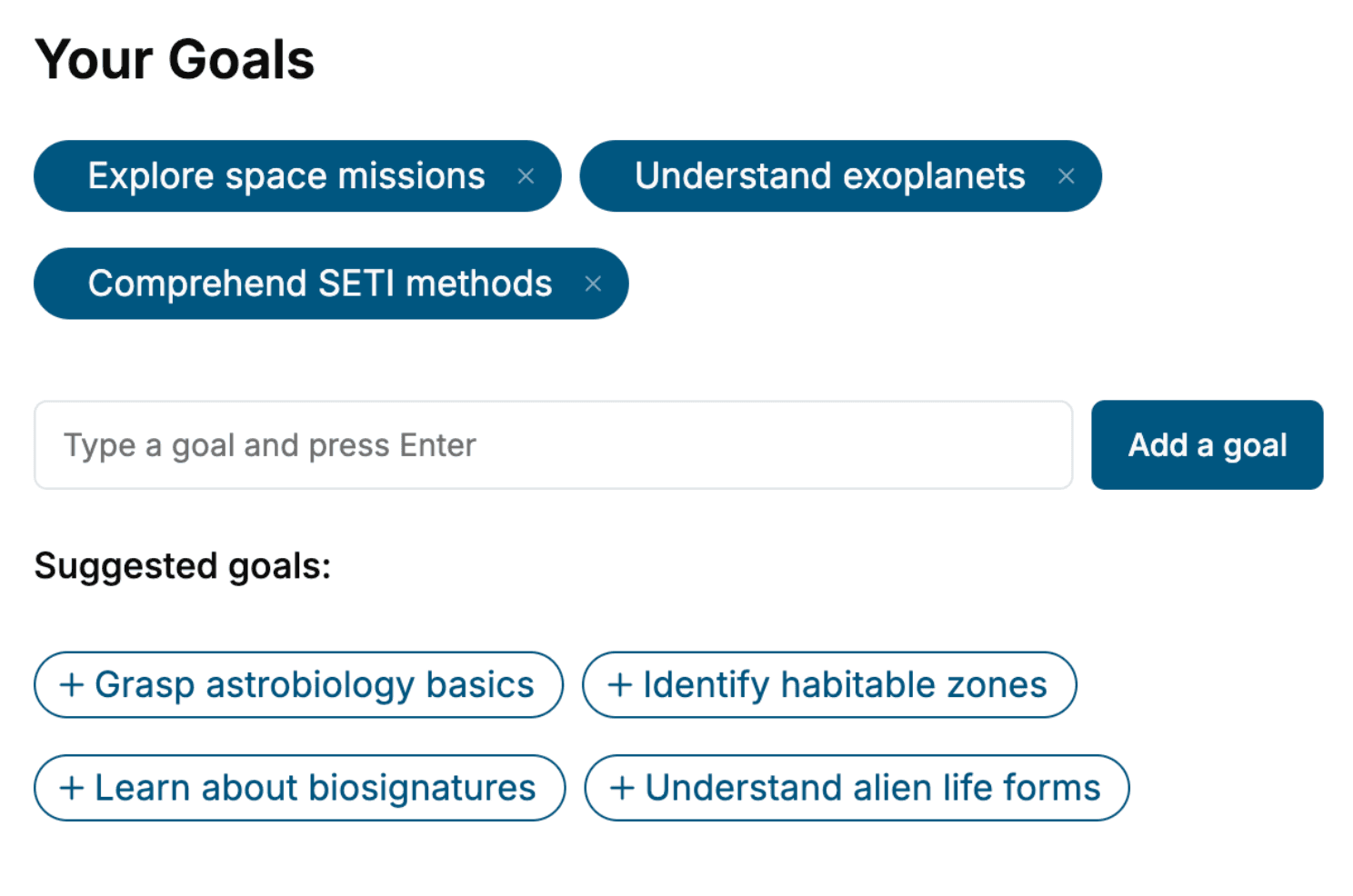
Specify your goals and choose sub-topics to include
- 3. Get your tailored book
Your book is ready in 10 minutes. Read it online, download as EPUB or PDF, or send to Kindle.


Frequently asked questions
- What is TailoredRead?
TailoredRead creates complete, full-length nonfiction ebooks, written specifically for you. Tell us what you want to learn and why, and TailoredRead writes you a complete ebook on it, with charts, tables, and diagrams throughout. Read it online, on Kindle, or on any e-reader, or download it as a PDF or EPUB. Every book is written fresh the day you order it.
- How is the book tailored to me?
Before anything is written, you answer a few short questions about your goals, your background, and the topics you care about. Your book is then planned around your answers: written at your level, focused on your goals, and covering the topics you chose, instead of the one-size-fits-all book a store sells everyone.
- Will it read like generic AI content?
No. Your book isn't a chat transcript: it's planned chapter by chapter before a single word is written, told in a clear book voice, and explains ideas visually with charts, tables, and diagrams. After writing, every book goes through an editorial pass that polishes chapters so the book reads consistent from start to finish. Judge for yourself: browse thousands of sample books on our Explore Books page.
- How accurate is the content?
Our AI first checks whether your subject needs live research. Niche and fast-moving subjects are researched before writing, and the book plan and chapters are grounded in what it finds. After writing, every book goes through a quality pass that checks its facts and consistency, and researched books list their sources in a Notes section at the back.
- How long is the book?
You choose the length: Comprehensive (250-300 pages), Detailed (150-200 pages), Essential (70-100 pages), or Short (30-50 pages).
- Does the book include visuals?
Yes. Every book includes charts, tables, and diagrams: timelines, checklists, step-by-step guides, decision trees, comparison tables, matrices, and bar charts. A typical book includes dozens of these visual elements.
- Can I read it on Kindle? What formats can I download?
Yes. Every book can be read in our online reader or downloaded as a PDF or EPUB. The EPUB can be opened with Kindle, Apple Books, Kobo, and any e-reader. The Commercial and Author licenses also include an editable Word (DOCX) and Markdown download.
- How much does it cost?
About the price of a regular ebook. The exact price depends on your book's length and complexity, from $2 for the shortest books to $35 for the most comprehensive, and it's shown when you review your book, before you commit to anything. Each book is a one-time purchase, yours to keep, no subscription. Licenses that let you share, teach, or publish your book range from $35.99 to $49.99.
- Can I preview the book before purchasing?
Before you pay, you'll see what your book will cover: the title, a detailed description, the estimated length, and the complete chapter-by-chapter table of contents, so you know what you're getting before you decide. The full text itself is written after you purchase.
- How long does it take to create a book?
About 10 minutes. You can start reading the opening chapters while the rest is still being written.