The Gastrointestinal System Coloring Book
Learn Anatomy Through Interactive Illustrations and Detailed Exploration
Understanding how your digestive system works requires more than reading text—it demands visual engagement and hands-on exploration.
222 pages10 chaptersApr 28, 2026Anatomy
About this book
Many learners struggle to retain complex anatomical details through passive study alone, missing the deeper connections between structures and their functions. This coloring book transforms your learning experience by combining detailed, scientifically accurate illustrations with interactive coloring activities that activate multiple learning pathways simultaneously. As you color each structure, you'll internalize the spatial relationships, layered organization, and functional connections within your gastrointestinal system. From the mouth to the colon, you'll explore every major organ and accessory structure, understanding not just where they are, but how they work together. Each illustration includes labeled anatomical features and accompanying explanations that deepen your comprehension. Whether you're a student preparing for exams, a healthcare professional seeking refresher knowledge, or an anatomy enthusiast, this book makes complex systems accessible and memorable through the proven power of visual learning and creative engagement.
Table of contents
- IntroductionWhy Visual Learning Works for Anatomy, How to Use This Coloring Book, Overview of the Gastrointestinal SystemRead below
- Chapter 1: The Mouth and Oral CavityTeeth Structure and Types, Tongue Anatomy and Function, Salivary Glands and Saliva Production
- Chapter 2: The Pharynx and EsophagusRegions of the Pharynx, Esophageal Structure and Peristalsis, The Swallowing Process
- Chapter 3: The StomachRegions and Curvatures of the Stomach, Stomach Wall Layers and Glands, Gastric Secretions and Digestion
- Chapter 4: The Small IntestineThe Duodenum and Pancreatic Duct, The Jejunum and Ileum, Villi and Microvilli for Absorption
- Chapter 5: The Large IntestineSections of the Colon, The Cecum and Appendix, The Rectum and Anal Canal
- Chapter 6: The Liver and Biliary SystemLiver Lobes and Internal Structure, Bile Production and Function, The Gallbladder and Bile Ducts
- Chapter 7: The PancreasPancreatic Structure and Regions, Exocrine Pancreas and Enzyme Production, Endocrine Pancreas and Islets of Langerhans
- Chapter 8: GI Tract Wall Layers and OrganizationThe Mucosa: Epithelium and Glands, The Submucosa and Muscularis Layers, The Serosa and Peritoneal Attachments
- Chapter 9: Blood Supply and Lymphatic DrainageArterial Supply to GI Organs, Venous Drainage and Portal Circulation, Lymphatic Vessels and Immune Function
- Chapter 10: Nervous System Control of DigestionThe Enteric Nervous System, Autonomic Nervous System Innervation, Hormonal Regulation of Digestion
- ConclusionIntegrating Your Anatomical Knowledge, Next Steps in Your Learning Journey
- Glossary of Terms
- Further Reading and Resources
Introduction
The human digestive system is one of the most intricate and fascinating networks in the body. From the moment food enters your mouth to the moment waste is eliminated, a precisely coordinated sequence of mechanical and chemical events unfolds across nearly 30 feet of interconnected organs. Understanding this system is not just an academic exercise. It is the foundation for comprehending how the body sustains itself, how nutrients reach every cell, and how disruptions in this process lead to disease. For students, clinicians, and curious learners alike, mastering gastrointestinal anatomy opens a window into the very mechanics of life.
Yet anatomy is notoriously difficult to learn through text alone. The gastrointestinal system involves dozens of structures layered on top of one another, folded into tight spaces, and connected by vessels, nerves, and ducts that weave through the abdominal cavity in complex patterns. Reading a description of the hepatopancreatic ampulla or the haustra of the colon is one thing. Seeing it, tracing it, and coloring it is something else entirely. That hands-on engagement transforms abstract information into spatial memory, making it far more likely to stick when you need it most, whether in an exam room, a clinical setting, or a conversation with a patient.
This coloring book was designed with that challenge in mind. Each chapter focuses on a specific region or functional component of the gastrointestinal system, pairing scientifically accurate illustrations with clear explanations and interactive coloring activities. The goal is not simply to label structures but to help you understand how those structures relate to one another and why their arrangement matters. As you work through each page, you will build a mental map of the digestive system that is grounded in visual experience rather than rote memorization. That kind of learning is deeper, more durable, and ultimately more useful.
Whether you are a first-year anatomy student preparing for practical exams, a nurse or physician assistant refreshing your foundational knowledge, or simply someone who wants to understand how the body works, this book meets you where you are. No prior expertise in anatomy is required beyond a basic familiarity with biological concepts. What is required is curiosity, a set of colored pencils or markers, and a willingness to engage actively with the material. The pages ahead will reward that engagement with a level of understanding that passive reading simply cannot provide.
Why Visual Learning Works for Anatomy
Human beings are fundamentally visual creatures. Roughly 65 percent of the population are visual learners, meaning they process and retain information most effectively when it is presented in a visual format.[1] The brain dedicates more neural real estate to visual processing than to any other sense, with approximately 30 percent of the cerebral cortex involved in interpreting what the eyes see. This is not a coincidence. It reflects millions of years of evolution during which survival depended on quickly recognizing patterns, shapes, and spatial relationships in the environment. Anatomy, which is fundamentally about shapes and spatial relationships, is therefore an ideal subject for visual learning methods.
Research in cognitive science supports this strongly. The dual coding theory, developed by psychologist Allan Paivio, proposes that the brain stores information in two separate but interconnected systems: one for verbal information and one for visual information.[2] When you learn something using both systems simultaneously, such as reading a label while also coloring the corresponding structure, you create two independent memory traces instead of one. This redundancy makes recall significantly more reliable. Studies have shown that students who use visual learning tools alongside text-based study outperform those who use text alone by a measurable margin in both short-term and long-term retention tasks.
Coloring, specifically, adds another powerful dimension: active engagement. Passive reading allows the mind to drift. Coloring demands sustained attention. You must look carefully at the structure, decide where its boundaries are, choose a color, and apply it deliberately. This process forces a level of focused observation that reading rarely achieves. In anatomy education, this matters enormously because many structures are distinguished by subtle differences in shape, position, or layering that are easy to overlook when simply reading a description.
There is also a strong motor memory component to coloring. The act of physically moving a pencil across a page engages the motor cortex and creates a kinesthetic memory trace alongside the visual one. This is the same principle that makes writing notes by hand more effective for retention than typing them.[3] When you color the layers of the stomach wall or trace the path of the bile duct, your hand is encoding that information in a way that reading alone never could. The result is a richer, more multi-dimensional memory that is easier to retrieve under pressure.
Color itself also serves as a powerful organizational tool in anatomy. When you consistently assign one color to arteries, another to veins, and another to nerves, you are building a visual coding system that mirrors the conventions used in professional anatomical atlases. This consistency trains your eye to recognize these structures quickly and accurately. Over time, the color associations become automatic, which is exactly the kind of fluency that clinical professionals need when interpreting imaging studies, surgical diagrams, or anatomical models.
Consider a practical example. A student studying the layers of the GI tract wall might read that the mucosa, submucosa, muscularis externa, and serosa are arranged concentrically from inside to outside. That sentence is easy to read and easy to forget. But when that same student colors each layer a different shade, tracing the boundary between the inner circular and outer longitudinal muscle layers with careful attention, the spatial reality of that arrangement becomes concrete and memorable. The next time they encounter a histological slide or a clinical question about where a peptic ulcer penetrates, they can visualize the answer rather than trying to reconstruct it from a verbal description.
This is why anatomy coloring books have been used in medical and health science education for decades. They are not a shortcut or a simplification. They are a pedagogically sound method for building the kind of deep, spatially grounded anatomical knowledge that supports clinical reasoning and professional practice. This book applies that method specifically to the gastrointestinal system, one of the most complex and clinically relevant systems in the body.
How to Use This Coloring Book
Getting the most out of this book requires a small amount of preparation and a consistent approach. The good news is that neither demands much time or expense. What matters most is that you engage actively with each page rather than simply reading through it. The following guidelines will help you build a productive coloring practice that maximizes your learning at every session.
Gather your materials before you begin. You will need a set of colored pencils or fine-tipped markers. Colored pencils are generally preferred because they allow for more precise shading and do not bleed through the page. Aim for a set with at least 12 to 20 colors so you have enough variety to distinguish different structures clearly. A pencil sharpener, an eraser, and a ruler for underlining key terms are also helpful. Some learners find it useful to keep a small notebook nearby for jotting down questions or observations as they work.
Before coloring any illustration, follow this three-step approach for each page:
- Read first. Read the accompanying text explanation fully before picking up a pencil. Understand what you are looking at, what each structure does, and how it relates to neighboring structures.
- Plan your colors. Decide which color you will use for each structure before you start. Write your color choices in the legend provided with each illustration. Consistency across chapters is valuable, so try to use the same color for the same type of structure throughout the book (for example, always using red for arteries and blue for veins).
- Color with intention. As you color each structure, say its name aloud or trace its outline with your finger first. This reinforces the connection between the visual form and the verbal label.
Each chapter in this book follows a consistent structure designed to support progressive learning. You will find an introductory explanation of the region or system being covered, followed by detailed illustrations with labeled structures, and then a set of review questions or self-check prompts at the end. Do not skip the review questions. They are designed to test whether you have internalized the material rather than simply colored it. If you cannot answer a question confidently, return to the illustration and the text before moving on.
A recommended coloring key to use consistently throughout this book is shown below. You are free to adapt it, but maintaining consistency will make your completed book a much more useful reference tool.
- Red or pink: Arteries and arterial structures
- Blue or purple: Veins and venous structures
- Yellow or gold: Nerves and neural structures
- Green: Lymphatic vessels and nodes
- Orange: Glands and secretory structures
- Light brown or tan: Connective tissue and supporting structures
- Light blue or gray: Luminal spaces and cavities
Pacing matters as much as method. Resist the temptation to rush through multiple chapters in a single sitting. The brain consolidates new information most effectively when learning is spaced out over time, a principle known as the spacing effect.[4] One chapter per study session, with a brief review of the previous chapter at the start of each new session, is an effective rhythm for most learners. This approach takes advantage of both the spacing effect and the retrieval practice effect, which shows that actively recalling information strengthens memory more than re-reading it.
This book can also be used effectively in group settings. Study groups can assign different colors to different members and compare their completed illustrations, discussing any discrepancies in how they interpreted the boundaries of a structure. Teaching a concept to another person is one of the most powerful ways to solidify your own understanding, and the visual nature of these illustrations makes them ideal conversation starters. If you are an educator using this book in a classroom or clinical training context, consider having students complete illustrations independently before comparing and discussing as a group.
Finally, treat your completed book as a living reference. Once you have finished a chapter, do not set it aside permanently. Return to it periodically, cover the labels, and test yourself on the structures. Use it alongside other resources such as anatomical atlases, physiology textbooks, or clinical case studies. The illustrations you have colored will serve as anchors for the more detailed information you encounter in those resources, helping you integrate new knowledge with the spatial framework you have already built.
Overview of the Gastrointestinal System
The gastrointestinal system, often called the digestive system, is a continuous muscular tube that runs from the mouth to the anus, supplemented by a set of accessory organs that contribute essential substances to the digestive process. Its primary job is to convert the food you eat into molecules small enough to be absorbed into the bloodstream and delivered to the cells of the body. This involves four fundamental processes: ingestion (taking food in), digestion (breaking it down mechanically and chemically), absorption (moving nutrients into the blood and lymph), and elimination (expelling undigested waste).
The main tube of the GI system, known as the alimentary canal or gastrointestinal tract, includes the following organs in sequence:
- The mouth (oral cavity), where mechanical digestion begins through chewing and chemical digestion begins through salivary enzymes
- The pharynx, the shared passageway for food and air at the back of the throat
- The esophagus, a muscular tube that propels food from the throat to the stomach
- The stomach, a J-shaped organ that churns food and secretes acid and enzymes to continue digestion
- The small intestine, a coiled tube approximately 20 feet long where most chemical digestion and nearly all nutrient absorption occur
- The large intestine, a wider tube approximately 5 feet long that absorbs water and electrolytes and compacts waste into feces
- The rectum and anal canal, which store and then expel feces from the body
Alongside this main tube, three major accessory organs contribute to digestion without food passing directly through them. The liver produces bile, a substance that emulsifies dietary fats to make them easier to digest. The gallbladder stores and concentrates bile between meals, releasing it into the small intestine when fatty food arrives. The pancreas produces a powerful mixture of digestive enzymes that break down carbohydrates, proteins, and fats, as well as bicarbonate that neutralizes the acidic contents arriving from the stomach. These three organs are connected to the small intestine by a system of ducts that deliver their secretions precisely where they are needed.
The wall of the GI tract is organized into four concentric layers that appear consistently from the esophagus to the rectum, though with regional variations that reflect the specific functions of each organ. From the inside out, these layers are the mucosa (the innermost lining that contacts food and absorbs nutrients), the submucosa (a connective tissue layer containing blood vessels, lymphatics, and nerves), the muscularis externa (smooth muscle layers responsible for the wave-like contractions that move food along), and the serosa or adventitia (the outermost covering). Understanding this four-layer organization is one of the most important conceptual frameworks in GI anatomy, and it will be revisited throughout this book.
Movement of food through the GI tract is driven by a process called peristalsis, a coordinated wave of muscular contraction and relaxation that propels contents forward. This movement is controlled by the enteric nervous system, a vast network of neurons embedded in the wall of the GI tract that operates largely independently of the brain. Sometimes called the "second brain," the enteric nervous system contains approximately 500 million neurons, more than the spinal cord, and it coordinates the timing and intensity of muscular contractions, glandular secretions, and blood flow throughout the digestive tract.[5]
The blood supply to the GI system is equally remarkable. The digestive organs receive blood from three major branches of the abdominal aorta: the celiac trunk, which supplies the stomach, liver, spleen, and upper duodenum; the superior mesenteric artery, which supplies the small intestine and the proximal large intestine; and the inferior mesenteric artery, which supplies the distal large intestine and rectum. Venous blood from the GI tract does not return directly to the heart. Instead, it drains into the portal vein, which carries nutrient-rich blood to the liver for processing before it enters the general circulation. This portal circulation is a critical feature of GI anatomy with major clinical implications.
Hormones also play a central role in coordinating digestion. Cells scattered throughout the lining of the GI tract secrete hormones in response to the presence of food, its chemical composition, and the degree of stretch in the organ walls. Key hormones include gastrin, which stimulates acid secretion in the stomach; secretin, which triggers bicarbonate release from the pancreas; and cholecystokinin (CCK), which stimulates the release of bile from the gallbladder and digestive enzymes from the pancreas. These hormonal signals travel through the bloodstream to reach their target organs, creating a sophisticated feedback system that adjusts digestive activity to match the type and quantity of food being processed.
As you work through the chapters of this book, you will explore each of these components in detail. The journey begins in the mouth and ends at the anal canal, following the path that food takes through the body. Along the way, you will encounter the accessory organs, the wall layers, the blood and lymphatic vessels, and the nervous and hormonal control systems that make the whole process work. Each chapter builds on the last, so by the time you reach the final pages, you will have assembled a comprehensive, visually grounded understanding of one of the body's most essential systems.
A useful way to think about the GI system is as a disassembly line. Just as a factory assembly line builds a product step by step, the digestive system breaks food down step by step, extracting what the body needs at each stage and passing the remainder along to the next. Every organ along the route has a specific job, and the system only works well when all the parts are functioning and communicating properly.
Keep this big picture in mind as you color each structure. Individual anatomical details are easier to remember when they are anchored to a clear understanding of the overall system and its purpose. The illustrations in this book are designed to show not just isolated structures but their relationships to neighboring organs, their positions within the abdominal cavity, and their connections to the vessels and nerves that support them. Use those relationships as your guide, and you will find that the details fall into place far more naturally than they would through memorization alone.
Notes
Introduction
- 1. R. M. Felder and L. K. Silverman, "Learning and Teaching Styles in Engineering Education," Engineering Education, 1988
- 2. A. Paivio, Mental Representations, Oxford University Press, 1986
- 3. P. A. Mueller and D. M. Oppenheimer, "The Pen Is Mightier Than the Keyboard," Psychological Science, 2014
- 4. N. J. Cepeda and others, "Distributed Practice in Verbal Recall Tasks," Psychological Bulletin, 2006
- 5. J. B. Furness, The Enteric Nervous System, Blackwell Publishing, 2006
This is the end of the free sample. The full book has 10 chapters.
Want this book written for you?
Tell us your goals and background, and get your own version of this book, tailored by AI to you.


Benefits of AI-tailored books
- Read one book, not ten:
- all the Anatomy knowledge you need, gathered into a single book.
- Save days of learning:
- tell us your goals, choose your topics, and the whole book is built around them.
- Learn effortlessly:
- Anatomy book written for your specific background and expertise.
- Understand at a glance:
- includes charts, tables, timelines, checklists, decision trees, and more.
- Stay current:
- written the day you order it, not years ago.
- Trust what you read:
- every book is checked for accuracy, with real research when your subject needs it.
Create your unique book in 3 steps
- 1. Select your focus
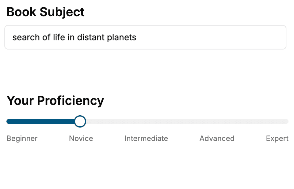
Select the focus of your Anatomy book and share your background
- 2. Personalize your book
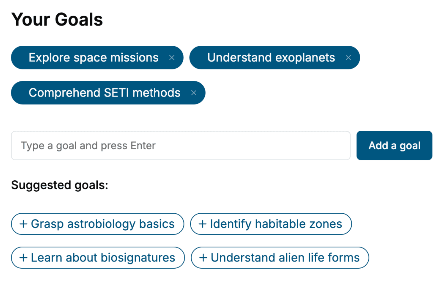
Specify your goals and choose sub-topics to include
- 3. Get your tailored book
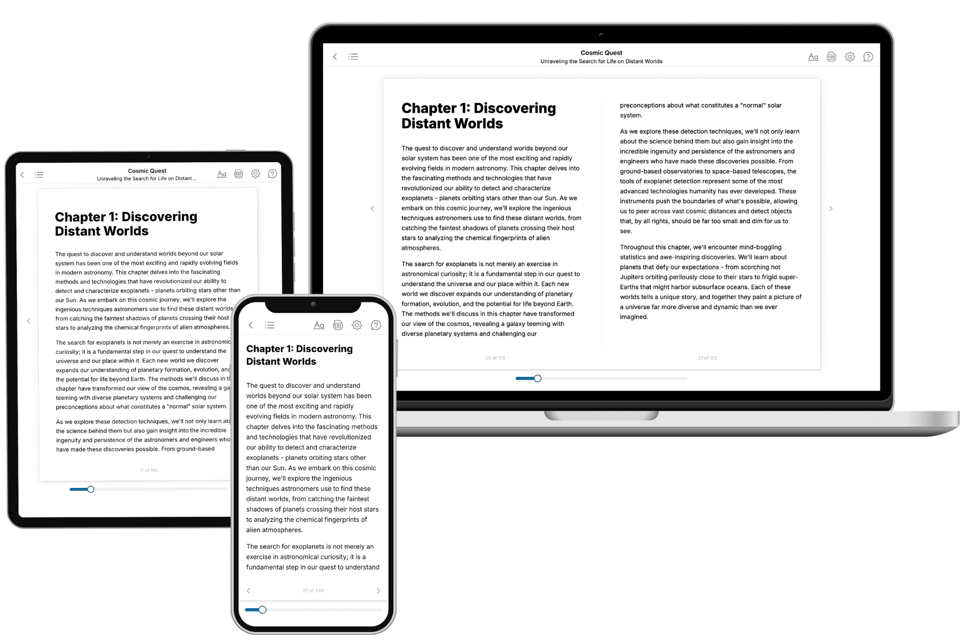
Your book is ready in 10 minutes. Read it online, download as EPUB or PDF, or send to Kindle.
Frequently asked questions
- What is TailoredRead?
TailoredRead creates complete, full-length nonfiction ebooks, written specifically for you. Tell us what you want to learn and why, and TailoredRead writes you a complete ebook on it, with charts, tables, and diagrams throughout. Read it online, on Kindle, or on any e-reader, or download it as a PDF or EPUB. Every book is written fresh the day you order it.
- How is the book tailored to me?
Before anything is written, you answer a few short questions about your goals, your background, and the topics you care about. Your book is then planned around your answers: written at your level, focused on your goals, and covering the topics you chose, instead of the one-size-fits-all book a store sells everyone.
- Will it read like generic AI content?
No. Your book isn't a chat transcript: it's planned chapter by chapter before a single word is written, told in a clear book voice, and explains ideas visually with charts, tables, and diagrams. After writing, every book goes through an editorial pass that polishes chapters so the book reads consistent from start to finish. Judge for yourself: browse thousands of sample books on our Explore Books page.
- How accurate is the content?
Our AI first checks whether your subject needs live research. Niche and fast-moving subjects are researched before writing, and the book plan and chapters are grounded in what it finds. After writing, every book goes through a quality pass that checks its facts and consistency, and researched books list their sources in a Notes section at the back.
- How long is the book?
You choose the length: Comprehensive (250-300 pages), Detailed (150-200 pages), Essential (70-100 pages), or Short (30-50 pages).
- Does the book include visuals?
Yes. Every book includes charts, tables, and diagrams: timelines, checklists, step-by-step guides, decision trees, comparison tables, matrices, and bar charts. A typical book includes dozens of these visual elements.
- Can I read it on Kindle? What formats can I download?
Yes. Every book can be read in our online reader or downloaded as a PDF or EPUB. The EPUB can be opened with Kindle, Apple Books, Kobo, and any e-reader. The Commercial and Author licenses also include an editable Word (DOCX) and Markdown download.
- How much does it cost?
About the price of a regular ebook. The exact price depends on your book's length and complexity, from $2 for the shortest books to $35 for the most comprehensive, and it's shown when you review your book, before you commit to anything. Each book is a one-time purchase, yours to keep, no subscription. Licenses that let you share, teach, or publish your book range from $35.99 to $49.99.
- Can I preview the book before purchasing?
Before you pay, you'll see what your book will cover: the title, a detailed description, the estimated length, and the complete chapter-by-chapter table of contents, so you know what you're getting before you decide. The full text itself is written after you purchase.
- How long does it take to create a book?
About 10 minutes. You can start reading the opening chapters while the rest is still being written.